
Abstract
Solutions of Pluronic F-127 in ethylene glycol were prepared at different concentrations (10–25%, w/w), and the behaviour of the samples during heating to 150°C was observed. During heating, the solubility of Pluronic in ethylene glycol increased, but no gel formation was observed. However, gel formation in the sample containing 25% Pluronic F-127 was observed near 50°C during cooling, and all the samples become solid upon cooling to 25°C. Fourier transform infrared spectroscopy, differential scanning calorimetry and optical microscopy were performed to analyse the gel formed by 25% Pluronic F-127 in ethylene glycol, and it was found that the gel formation in ethylene glycol was different from the gel formation of Pluronic in water as the latter gels are crystalline in nature whereas the former are amorphous. Hydrogen bonding was considered to be the main factor behind the gel formation.
Introduction
The support material plays an important role during the layer by layer manufacturing of three-dimensional parts via some of the commercially available additive manufacturing (AM) processes. A support material not only facilitates the removal of the part from the build platform after its completion but also enables manufacturing of intricate features, such as downward facing curved surfaces, overhanging features, holes and cavities. In addition, the support material provides resistance against the tendency of a model to deform due to applied forces during build.1, 2
Most of the jetting based AM processes, such as multijet modelling, PolyJet and Solidscape 3D printing, make use of support material during manufacture of parts, and these support materials are jetted in a similar way to the build material. Jetting of caprolactam is a novel AM process that is currently under research in our laboratories. The aim of this process is to utilise inkjet heads to deposit liquid droplets of caprolactam, which would polymerise into polyamide 6 after being jetted. Details of this process have been discussed elsewhere.3–5
A suitable support material is therefore required for the jetting of caprolactam. As with the jetting based AM processes mentioned above, the support material will be jetted using inkjet heads and thus should be liquid with low enough viscosity at or below 80°C, which is the maximum operating temperature for the inkjet head being used in this project. The selected material for the research is Pluronic F-127, a block co-polymer of polyethylene oxide (PEO) and polypropylene oxide (PPO), due to its reverse thermal gel formation abilities in water.
Pluronic (BASF) is a trade name of a family of more than 30 block co-polymers. These are triblock co-polymers and have a structure consisting of PEO–PPO–PEO (PEOa–PPOb–PEOa), where a and b represent the degree of polymerisation of each block (i.e. number average chain length). The chemical structure of Pluronics is as follows
Applications of Pluronics mainly involve water as solvent, but for applications for which the presence of water is unacceptable37, 38 and/or to explain the hydrophobic effect causing the self-aggregation of Pluronics in different solvents, non-aqueous polar solvents have been studied recently.40, 44–51 The study of non-aqueous solvents for Pluronics is considered as of ‘fundamental and practical interest’.45 Furthermore, in the present work, water presents difficulties as it inhibits the polymerisation of caprolactam into polyamide 6, so a novel combination of Pluronic F-127 and ethylene glycol was investigated. Ethylene glycol is a solvent that could be a possible alternative to water in applications requiring a non-aqueous solvent. This work is therefore intended to replace water with ethylene glycol as a non-aqueous solvent for Pluronics. Pluronic F-127 was selected as it is the most commonly studied Pluronic polymer.52
Materials
Pluronic F-127 (Mw = 12 600, PEO content = 70% of total molecular weight) was purchased from BASF, and ethylene glycol was purchased from Fisher Scientific; both were used as received without any further purification. Four different Pluronic in ethylene glycol samples, with compositions ranging from 10 to 25% (w/w) in increments of 5%, were prepared by weighing out the appropriate quantities of F-127 and ethylene glycol. Mixing was performed using a magnetic stirrer, and each sample was stirred for ∼24 h to allow complete dissolution/dispersion.
Experimental
Sample heating
The samples were heated, and a tube inversion method was used to identify gel formation. Approximately 6 g of each different composition was placed in a glass tube (inner diameter 23 mm), and the four samples were heated from 25 to 150°C in order to observe the gel formation. The tubes were inverted after every 5°C rise in temperature up to 50°C and after every 10°C rise in temperature up to 150°C. No movement of meniscus upon inverting the tube was identified as the formation of gel state. After heating to 150°C, the samples were allowed to cool back to the initial temperature (i.e. 25°C), and observations were also made during the cooling of the samples.
Fourier transform infrared spectroscopy
Fourier transform infrared spectroscopy of ethylene glycol, Pluronic F-127 and the four samples before heating and after heating was performed. A Shimadzu FTIR-8400S spectrometer was used to analyse all the samples with a standard deuterated L-alanine doped triglycine sulphate detector. The spectral resolution was 4 cm−1, and the IR spectra were recorded by scanning 64 times for each measurement. Apart from Pluronic F-127, which was in the form of flakes, all the other samples were sandwiched between two round (25 mm diameter×4 mm thick) NaCl crystal windows. The IR spectrum for Pluronic F-127 was obtained using attenuated total reflectance with a diamond crystal.
Differential scanning calorimetry
Differential scanning calorimetry was performed on Pluronic F-127, 25% Pluronic F-127, in ethylene glycol sample before and after heating in order to determine the melting and crystallisation temperatures upon cooling for Pluronic F-127 and to determine whether or not crystallisation takes place in Pluronic in ethylene glycol samples. The equipment used for the DSC was a DSC 2010 by TA Instruments. All of the samples were prepared in aluminium sample pans with lids and placed in the heating chamber. Nitrogen gas was supplied at a rate of 50 cm3 min−1 to the heating chamber. The samples were heated from 20 to 150°C and cooled back to 10°C using heating/cooling rates of 5°C min−1. Thermal equilibrium was maintained at the maximum temperature for 1 min for all the samples.
Hot stage microscopy
The hot stage microscopic examinations of Pluronic F-127 powder and Pluronic F-127 in ethylene glycol sample with the highest concentration (i.e. 25%) were performed using a Leica DM/LM microscope with an objective lens of 10× magnification. A total magnification of 100× was used to observe the two samples. A Mettler Toledo FP90 central processor was used to control the temperature during microscopic examination of the samples, and they were mounted on the microscope slide using a Mettler Toledo FP82HT hot stage. Three samples each of Pluronic F-127 powder and Pluronic F-127 in ethylene glycol solution were prepared, and all the samples were heated from 25 to 100°C using a heating rate of 5°C min−1 and then kept at 100°C for 1 min. The samples were cooled at natural cooling rate. The images were captured using a JVC colour video camera at 1 frame/s. A video file was recorded for each sample, and the images at the desired temperatures were obtained from the video files using video file playback, editing and image capturing software (Studio Player version 3.0). The microscope was set up for transmitted light, and crossed polarisers were used to observe the samples for any change in the crystalline state during heating and cooling between 25 and 100°C.
Tyndall test
The Tyndall test is a commonly employed qualitative test for the confirmation of particle suspension present in a solution. The test is a qualitative tool to detect the presence of very small particles suspended in a clear medium (i.e. liquid). Light from a laser source is passed through the sample. The presence of particles in a sample causes the scattering of light so that the laser beam becomes visible through the sample, whereas in the absence of any particle, the light will not be scattered, and the laser beam will not be visible. The increase in the particle concentration results in a relatively brighter beam than a low concentration of particles. This test was performed on 15, 20 and 25% samples at 60 and 120°C to confirm the presence of particles (i.e. micelles) at these temperatures using a JDS Uniphase He–Ne laser. The wavelength of the laser was 633 nm, and the power was 4 mW. The samples were heated using an oil bath to the desired temperature and kept at the same temperature for ∼30 min to ensure thermal equilibrium. The laser was passed through in each sample, and the images were captured using a Sony Cyber-shot (DSC-S950) camera.
Results
Sample heating/cooling
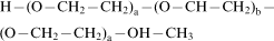
All four samples were turbid at room temperature (Fig. 1a), which reflected the insolubility of Pluronic F-127 in ethylene glycol. As the temperature was increased, the solutions started turning clear at ∼35°C, and all the four samples were completely clear (transparent) solutions at 50°C. No change was observed in either the colour or the meniscus movement of the solutions upon increasing the temperature up to 150°C (Fig. 1b). After reaching the highest temperature (i.e. 150°C), the solutions were allowed to cool to room temperature. As the solutions were cooled, the 25% solution formed a clear, soft gel near 50°C, whereas the other three samples were liquid at this temperature (Fig. 1c). Further cooling to near 25°C resulted in the appearance of turbidity in all the four samples, which resulted in a white coloured, soft (wax-like) solid at 25°C for all the four samples (Fig. 1d). In order to observe the gel melting temperature, the 25% sample was reheated. The sample at the start of reheating was a white wax-like solid. As the temperature was increased, it started turning turbid near 30°C and was a clear gel near 40°C. The gel melting was observed near 50°C.
Pluronic F-127 in ethylene glycol samples at a 20°C, b 150°C, c 50°C, during cooling and d 25°C, cooled back
Fourier transform infrared spectroscopy
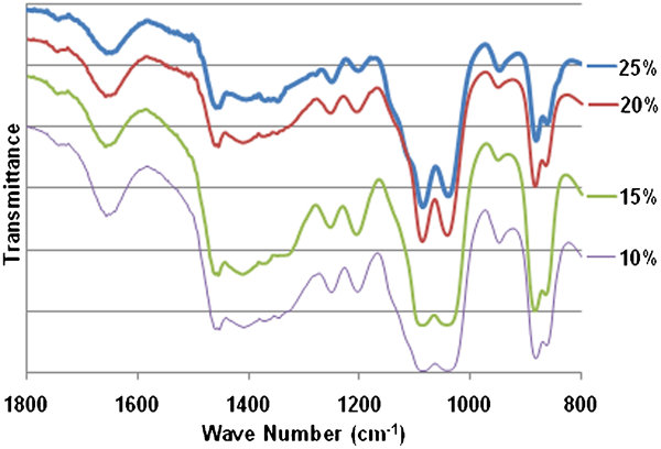
Figures 2–4 show the IR spectra (transmittance) obtained for the samples, and the results are summarised in Table 1. The infrared spectra of F-127 and ethylene glycol are presented in Fig. 2, whereas the spectra for F-127 in ethylene glycol liquid and solid (i.e. after heating) samples between 800 and 1800 cm−1 are presented in Figs. 3 and 4 respectively.
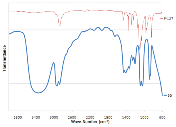
Fourier transform infrared spectra of Pluronic F-127 and ethylene glycol

Fourier transform infrared spectra of Pluronic in ethylene glycol solutions (liquid, before heating)
Fourier transform infrared spectra of Pluronic in ethylene glycol solutions (solid, after heating)
Peaks obtained for different samples used in FTIR spectroscopy
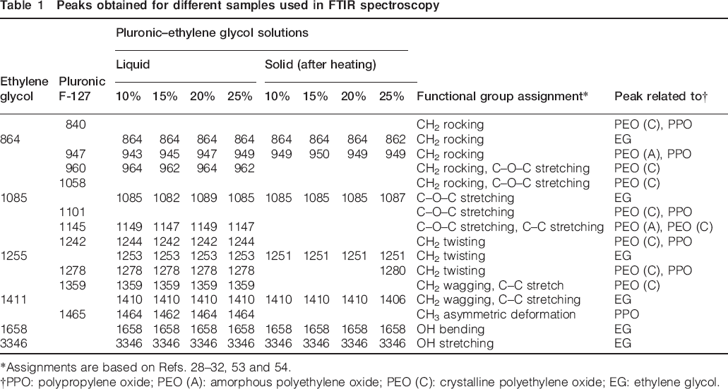
*Assignments are based on Refs. 28–32, 53 and 54.
†PPO: polypropylene oxide; PEO (A): amorphous polyethylene oxide; PEO (C): crystalline polyethylene oxide; EG: ethylene glycol.
Differential scanning calorimetry
Figure 5 shows the DSC curves for Pluronic F-127 and the 25% liquid and solid samples. The melting peak for F-127 was obtained at 59°C, and an exotherm peak due to crystallisation upon cooling was obtained at 35°C. For the 25% F-127 in ethylene glycol liquid sample, sharp melting endothermic and exothermic peaks were observed at 43 and 16°C respectively. The DSC curve for the 25% F-127 in ethylene glycol solid sample shows a melting endotherm with peak at 39°C and a crystallisation peak at 10°C. These crystallisation peaks obtained by the DSC are below the room temperature, so after cooling at 5°C min−1 to room temperature, Pluronic F-127 would not have crystallised.
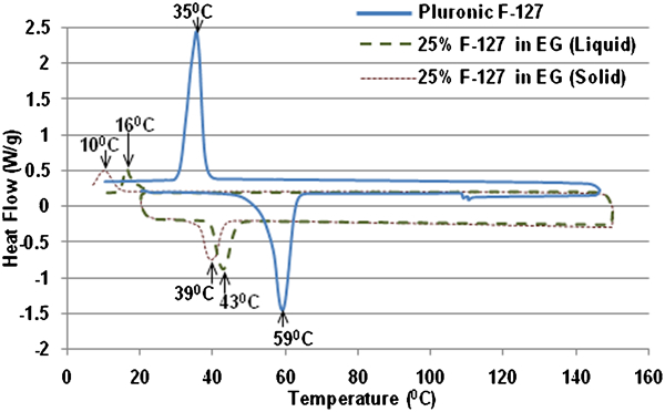
Differential scanning calorimetry curve for Pluronic F-127 and 25% liquid and solid samples
Hot stage microscopy
Figure 6a and b depicts the micrographs at 25 and 55°C, showing the presence of a crystalline structure at these temperatures, while Fig. 5c shows the melting of Pluronic F-127 near 58°C. Figure 6d–f shows the onset of crystallisation of F-127 starting at 38°C until complete crystallisation occurs at 25°C. Similarly, Fig. 7 shows the micrographs obtained at different temperatures during the heating of 25% F-127 in ethylene glycol, indicating the presence of a crystalline structure at 25 and 40°C (Fig. 7a and b respectively) and the onset and completion of dissolution at 43 and 46°C (Fig. 7c and d respectively). During cooling, the crystallisation starts near 28°C (Fig. 8b) and 25°C, and the sample (i.e. solid) is completely crystallised (Fig. 8c and d). As the sample cooling was performed under natural cooling rate, the recrystallisation values are consistent with the sample heating/cooling and are higher than those obtained during DSC.
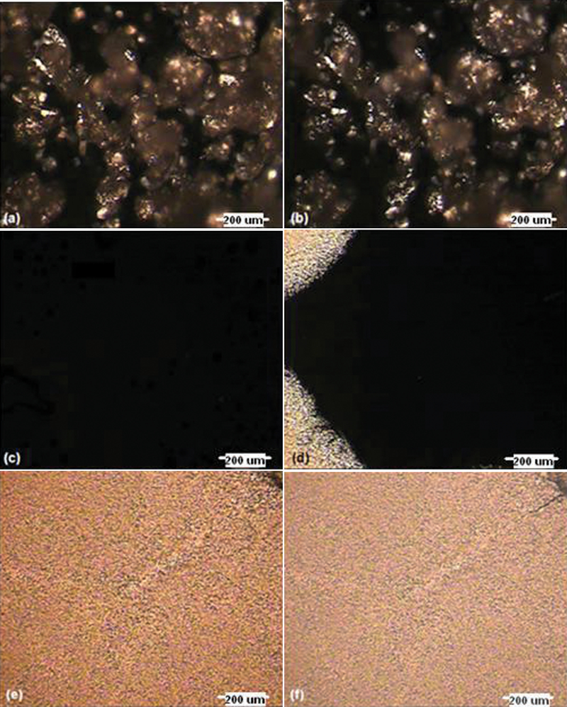
Micrographs of Pluronic F-127 at a 25°C, b 55°C, c 58°C, d 38°C, during cooling, e 36°C, during cooling and f 25°C, after cooling
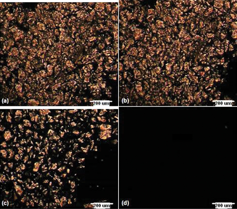
F-127 (25%) in ethylene glycol sample during heating at a 25°C, b 40°C, c 43°C and d 46°C
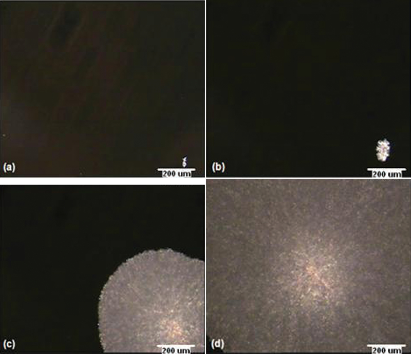
F-127 (25%) in ethylene glycol sample during cooling at a 30°C, b 28°C, c 25°C and d 24°C
Tyndall test
The Tyndall test results are presented in Fig. 9. The presence of particles (micelles) in the clear solutions at 60 and 120°C is confirmed by the observation of the visibility of the laser beam through the clear samples. In addition, it must be noted that compared with the 15 and 20% samples, the 25% sample presents a very clearly visible (brighter) beam indicating the possible presence of micelles.
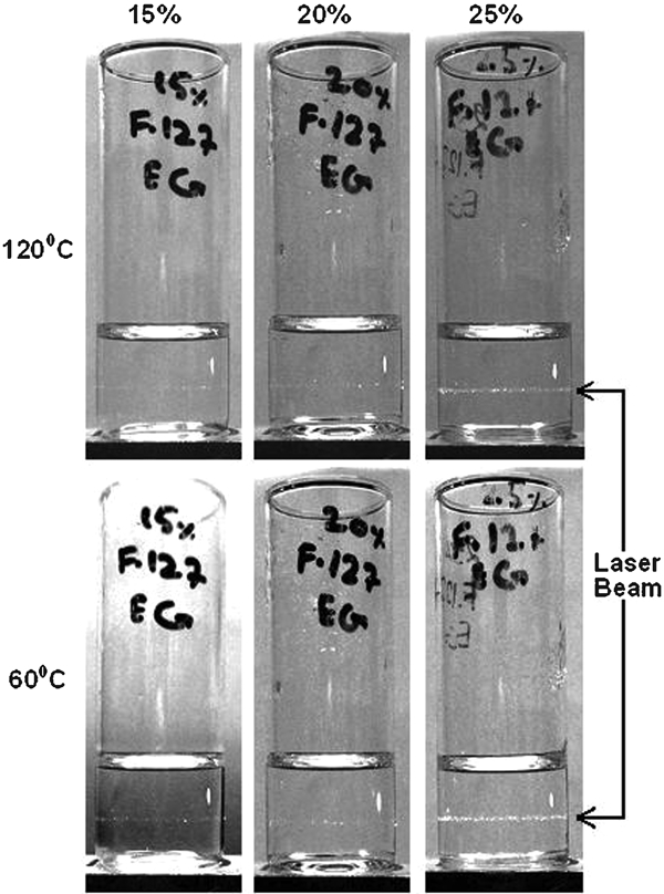
Tyndall test of F-127 in ethylene glycol solutions at 60 and 120°C
Discussion
Table 1 summarises the FTIR frequencies obtained for ethylene glycol, Pluronic F-127 and the four Pluronic in ethylene glycol samples in the liquid state (i.e. before heating) and in the solid state (i.e. after heating) along with the functional groups assigned to these frequencies. The assignments were based on the literature.28–32, 53, 54 It is well known that Pluronic is comprised of blocks of PEO and PPO with a PPO chain centred between two PEO chains, where the PEO is present in both amorphous and crystalline forms, and the PPO is preset in the amorphous form.28–32 Therefore, in Table 1, the FTIR frequencies achieved for all the samples have been related with the structural components of the composition (i.e. ethylene glycol, PPO and PEO). Polyethylene oxide peaks were also assigned to crystalline and amorphous PEOs. The bands at 840, 947, 960, 1058, 1101, 1145, 1242, 1278, 1342, 1359 and 1465 cm−1 are the characteristic peaks related to the PEO structure in Pluronic F-127. Similarly, the bands due to PPO include 840, 947, 1101, 1242, 1278 and 1373 cm−1. Ethylene glycol gives rise to bands at 864, 1085, 1255, 1411, 1658 and 3346 cm−1. It is worth noting here that some of these bands, such as 840, 947, 1101, 1242 and 1278 cm−1, can be associated with both PPO and PEO parts of Pluronic since both contain similar groups (e.g. CH2 and C–O). Compared to the bands related to the PPO, a large number of bands in the Pluronic F-127 spectra are related to the PEO, which is due to the relatively high proportion of PEO chains in the F-127 molecule (i.e. 70% PEO and 30% PPO). From the data in Table 1, it is clear that some of the peaks (840, 1058 and 1101 cm−1) associated with PEO disappear in the liquid samples, whereas the remaining bands associated with PEO (960, 1145, 1242, 1278, 1359 and 1465 cm−1) also disappear in the solid samples (i.e. after heating). The bands at 840, 1101 and 1270 cm−1, associated with PPO, disappear in all the samples, whereas the PPO bands at 960, 1145, 1242 and 1278 cm−1 disappear in the solid samples. All the bands associated with ethylene glycol appear in all the samples. The behaviour of solutions upon heating can thus be explained on the basis of these results along with the microscopic images and DSC of Pluronic F-127 and 25% Pluronic in ethylene glycol solution as follows.
Loss of turbidity between 35 and 50°C
When Pluronic F-127 is mixed with ethylene glycol, both PEO and PPO present in Pluronic F-127 are insoluble, as indicated by presence of crystals in the 25% solution at 25 and 40°C (Fig. 7a and b), resulting in turbid liquids at this temperature. A straightforward reason for the disappearance of turbidity could be the melting of Pluronic F-127 near 50°C. The melting temperature of F-127 (as quoted in the data sheet supplied by manufacturer) is 53–57°C. This melting point is also confirmed by both the microscopic image of F-127 at 58°C (Fig. 6c) and the DSC of F-127 (Fig. 5). Therefore, the melting of F-127 can be considered as a reason for the increased solubility and thus appearance of clear solutions near 50°C. However, as suggested by the disappearance of bands at 840, 1058 and 1101 cm−1, which are associated with the crystalline PEO,30–32 it can be said that some of the crystalline PEO has dissolved in ethylene glycol. The reason for the disappearance of peaks associated with PPO (i.e. 840, 1101 and 1270 cm−1) can be considered to be due to the masking of these peaks under the relatively broader peaks associated with ethylene glycol at 864, 1085 and 1255 cm−1, so it can be said that PPO is insoluble and PEO is partially soluble at room temperature in ethylene glycol, resulting in turbid liquids. The remaining PEO bands at 960, 1145, 1242, 1278, 1359 and 1465 cm−1, which were present in the liquid samples, disappeared in the solid samples, indicating increased solubility of PEO as a result of heating to a higher temperature. Some of these bands (i.e. 960, 1145, 1242 and 1278 cm−1) are also associated with PPO, so it can also be stated that the PPO solubility has increased with increasing temperature. The micrographs of 25% F-127 in ethylene glycol at 43 and 46°C (Fig. 7c and d) clearly suggest complete dissolution above 40°C. This increased solubility of both PEO and PPO can be considered the main reason behind the disappearance of turbidity and appearance of clear solutions near 50°C. In addition, the observation of a visible laser beam through these clear liquids at high temperatures (i.e. at 60 and 120°C) confirmed the presence of particles in the liquid phase. These particles can be due to micelles that are formed by the difference in solubility of the PEO and PPO chains.
Gel formation by 25% sample on cooling near 50°C
As the samples are cooled down, the highest concentration solution (i.e. 25%) converts from a viscous liquid into a soft gel structure near 50°C. As suggested by the FTIR spectra, the bands associated with crystalline PEO at 960, 1145, 1242, 1278, 1359 and 1465 cm−1 disappeared in all the samples after they were heated and cooled back to a solid state. The disappearance of these bands can be caused by a change in the crystalline structure and/or the conformational structure of the PEO.31 Furthermore, the micrographs at 30 and 28°C (Fig. 8a and b) and the DSC curves of both liquid and solid 25% F-127 in ethylene glycol samples (Fig. 5) clearly showed the absence of any crystallisation between 30 and 50°C. The absence of FTIR bands associated with crystalline PEO at 960, 1145, 1242, 1278, 1359 and 1465 cm−1 can thus be considered to be due to a change in the conformational structure of crystalline PEO, as a change in the conformation of PEO from trans–gauche–trans in the crystalline state to gauche–gauche–trans upon melting into the amorphous state has been observed previously.31, 55 Figures 10 and 11 respectively show the trans and gauche conformations of OCCO backbone within PEO. From the FTIR results, it is clear that the band near 1242 cm−1 disappeared in the solid samples, and the intensity of the band near 1251 cm−1 is higher than that of the liquid samples (Figs. 3 and 4). The bands near 1240 and 1280 cm−1 were assigned to the trans conformation, and the band near 1250 cm−1 was assigned to gauche conformation;55, 56 therefore, the disappearance of bands near 1240 and 1280 cm−1 and the increased intensity of 1250 cm−1 band also present evidence of an increased amount of gauche conformation.
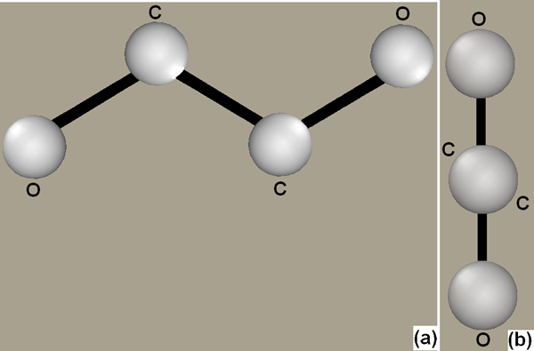
a transconformation of OCCO group within PEO and b viewed along C–C bond
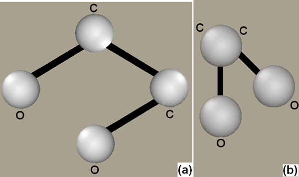
a gauche conformation of OCCO group within PEO and b viewed along C–C bond
As the conformation changes in a polymer dissolved in a solvent, this can also result in the formation of the gel phase.57 The gel formation shown by 25% F-127 in ethylene glycol could thus be caused by this conformational change. Both PEO and ethylene glycol have strong hydrogen bonding tendencies; therefore, the strong hydrogen bonding between the PEO chains and ethylene glycol as compared to hydrogen bonding between the less polar PPO and ethylene glycol resulting in increased polymer to polymer interaction can also be a cause of this gel formation.58 Ether oxygen present within the PEO is the hydrogen bonding site present in these systems. The intensity of the band at 1658 cm−1 decreased in the solid samples. This band has been assigned to OH bending vibration, and it has been reported that an increase in hydrogen bonded OH bending as opposed to free OH bending results in the increased intensity of this band.59 In addition, it has been suggested previously that bands related to ether (C–O–C) stretching and CH2 rocking vibrations are most likely to show changes due to hydrogen bonding effects.56, 60, 61 Thus, the disappearance of bands near 840, 960, 1058, 1101 and 1145 cm−1 along with the reduced intensity of the 1658 cm−1 band clearly presents evidence of hydrogen bonding in the solid samples. Furthermore, the observation of the same solution–gel and gel–solution transformation temperature (i.e. near 50°C) along with evidence of the lack of crystallinity demonstrated by micrographs and DSC results (Figs. 5 and 8a and b) and the conformational change represented by the disappearance of PEO peaks suggest that this gel phase could be an amorphous gel.57, 62 The gel phase formed by Pluronic in ethylene glycol is therefore different from the gels formed by Pluronic in water in that Pluronic in water gels has been shown to be crystalline in nature, with the packing of micelles in the liquid crystalline state as the main cause of the gel formation,10, 12, 13, 14, 20 whereas the gel formed by Pluronic in ethylene glycol is amorphous, and PEO conformation change could be the main contributing factor towards the gel formation. In addition, aqueous Pluronic gels return to their initial state (i.e. clear solution) upon cooling, whereas Pluronic F-127 in ethylene glycol samples did not return to the initial state of a turbid mixture, rather a white wax-like solid structure was achieved.
Since the lower concentrations (i.e. 10–20%) do not form the gel at any temperature, it can be said that the polymer–polymer interactions resulting in gel formation only take place at a critical concentration of 25% or higher.
Formation of soft wax-like solids
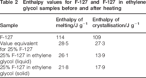
Further cooling results in the appearance of turbidity in all the samples near 25°C. This could be caused by recrystallisation of Pluronic F-127. Differential scanning calorimetry results (Fig. 5) show that Pluronic F-127 recrystallises in a less perfect (lower melting) form at subambient temperatures. However, as the solution was cooled to 25°C, a very slow crystal formation was observed (Fig. 8c and d). This is also confirmed by microscopic images (Fig. 8a and b); as near 28°C, only a very small amount of crystallised structure can be seen, indicating the absence of crystalline PEO. Recrystallisation was observed near room temperature during the microscopy and bulk heating/cooling experiments since cooling during both of these experiments was performed more slowly, whereas during DSC, a faster cooling rate was used (i.e. 5°C min−1). The FTIR spectra of samples after they were cooled (i.e. solid) clearly showed that most of the peaks associated with crystalline PEO had disappeared. Since the conformational structure of PEO has changed, the appearance of the white, wax-like solid state upon cooling back to near 25°C can be considered as the recrystallisation of the PEO with a changed conformational structure. Enthalpy values during heating and cooling and F-127 and 25% F-127 in ethylene glycol before and after heating obtained from the DSC curves are presented in Table 2. The change in enthalpy for F-127 during heating (114 J g−1) and cooling (109 J g−1) is nearly the same. On the other hand, the change in enthalpy for 25% F-127 in ethylene glycol during cooling (13·9 J g−1) is significantly reduced compared to that during heating (26·1 J g−1). Comparing the enthalpy values of 25% F-127 in ethylene glycol to corrected values for 25% of F-127 also highlights that the change in enthalpy for 25% F-127 in ethylene glycol during heating is nearly the same as the corrected value for 25% of F-127, whereas the change in enthalpy during cooling is significantly lower than the corrected value during cooling. This reduced value of enthalpy during cooling of 25% F-127 in ethylene glycol suggests that ethylene glycol becomes entrapped between the solidified PEO and PPO structures on a macrolevel with some of the PEO dissolved in ethylene glycol and/or present in the form of gel, and thus, a white wax-like solid structure appears. When these solids are subjected to compression (i.e. pressure applied by hands), the ethylene glycol entrapped between solidified PPO and PEO is released out. The presence of all the bands associated with ethylene glycol in both liquid and solid samples also confirmed the presence of ethylene glycol in the resulting solid structure.
Enthalpy values for F-127 and F-127 in ethylene glycol samples before and after heating
Conclusions
Ethylene glycol was investigated as a non-aqueous solvent for Pluronic F-127, and samples of concentrations ranging from 10 to 25% (w/w) F-127 in ethylene glycol were prepared and analysed for gel formation. The gel formation in 25% F-127 in ethylene glycol was observed during the cooling of the sample, and the solution to gel transformation took place near 50°C. Reheating the sample also resulted in a sol–gel transition near 50°C. The FTIR, DSC and hot stage microscopy on F-127 and 25% F127 in ethylene glycol before and after heating were performed, and the results were presented. On the basis of the results obtained, it can be concluded that the gel formed by Pluronic F-127 in ethylene glycol is caused by hydrogen bonding as opposed to aqueous Pluronic gels, which are due to the arrangement of micelles into lyotropic liquid crystalline phases. Furthermore, the gel formed by F-127 in ethylene glycol is an amorphous gel phase as compared to the lyotropic liquid crystalline gels formed by Pluronics in water. It can further be concluded, based on the results, that the gel formation by Pluronic F-127 in ethylene glycol can only be observed for a concentration of 25% (w/w) or higher. The results further suggest that although a gel was not obtained at higher temperatures, if used for lower temperature build environments (i.e. 25°C), the compositions investigated can provide a possible material to act as support, which can be very easily removed due to its water solubility.
Footnotes
Acknowledgements
The authors acknowledge the NED University of Engineering and Technology (Pakistan) for providing funds, in the form of a study scholarship, to carry out this research as part of the PhD of one of the authors.