
Abstract
Layered organic/inorganic nanocomposites have been widely investigated in recent years. In this work, for the first time, the authors prepared laminated hydroxyapatite (HAp)/poly(methyl metacrylate) (PMMA) nanocomposites via one-step intercalative bulk polymerization. X-ray diffraction (XRD) and scanning electron microscopy (SEM) results revealed that different morphologies of PMMA/HAp nanocomposites were obtained by varying HAp contents. Differential scanning calorimetry (DSC) and tensile strength analysis indicated that both thermal stability and tensile strength were enhanced with incorporation of laminated HAp. The results reported here can be expanded to other HAp polymer systems, thus paving a new way of designing and fabricating biomimetic nanocomposites for biomedical applications.
Introduction
Hydroxyapatite (HAp) is commonly used as a filler to replace amputated bone or as a coating to promote bone ingrowth because of its excellent biocompatibility, bioactivity, osteoconductivity1 and its similarity in chemical composition to apatite in the human skeletal system.2 However, the sintered HAp material is brittle and poor in strength which makes it liable to fracture when subjected to clinical applications under load bearing conditions.3 As a result, HAp/polymer composites were synthesised in order to overcome the shortcomings of pure HAp. Poly(methylmethacrylate) (PMMA) is commonly used as bone cement due to its ability to conform to the shape of its surroundings, thus allowing an even distribution of implant loads and forming a strong mechanical bond with the implants. However, a big disadvantage to this bone cement is its poor bioactivity4 and osteoconductivity.5 With the aim to combine the advantages of HAp and PMMA, the PMMA/HAp composites have been vigorously investigated in recent years.6–10 Dalby et al.11 prepared a PMMA/HAp composite with improved bioactivity and better cell anchorage. Itokawa et al.5 developed a PMMA/HAp composite by mixing HAp granules with PMMA and the resulted cranioplasty plate material was expected to possess both the good osteoconductivity of HAp and the high strength of PMMA. However, a critical issue in preparing PMMA/HAp composite is the homogeneous dispersion of HAp in PMMA matrix. Aggregation is inevitable in most cases, particularly when nanosized HAp particles are used. As a matter of fact, this critical problem has been solved in the case of clay nanocomposites by intercalation method.12 In our previous work,13 a lamellar HAp was prepared by surfactant template synthesis technique, which makes it possible to fabricate laminated HAp/PMMA composites by intercalation. In this work, for the first time, laminated HAp/PMMA nanocomposites were obtained by intercalative bulk polymerisation.
Experimental
Laminated HAp was prepared as reported in our previous work.13 The laminated HAp/PMMA nanocomposites were prepared by one-step intercalative bulk polymerisation. Under magnetic stirring, 20 mL methyl metacrylate (MMA) was mixed with 0·06 g azobisisobutyronitrile (AIBN). Then, different proportions of laminated HAp (5 and 10%) were added respectively and stirred for 6 h at room temperature, following by prepolymerisation at 80°C for ∼40 min. At last, the reaction mixtures were heated in the oven and let the polymerisation finish at 80°C, thus obtaining HAp/PMMA nanocomposites with different HAp weight ratios, i.e. 5 and 10%, which are denoted as 5HAp/PMMA and 10HAp/PMMA respectively.
Results and discussion
X-ray diffraction analysis
X-ray diffraction is a classical technique for determining the gallery height (d spacing) of clay. Herein, XRD was used for the preliminary confirmation of laminated structure of HAp and composites. Fig. 1a gives the small XRD patterns of HAp and HAp/PMMA nanocomposites obtained in this work. It is observed that HAp showes three intense diffraction peaks at 2θ value of 2·8, 5·6 and 8·5°, corresponding to d spacing of d100 = 3·1 nm, d200 = 1·5 nm and d300 = 1·0 nm respectively. The three intense peaks indicates the formation of a long range ordered laminated structure of HAp.13 Compared with HAp, the 10HAp/PMMA composite shows a diffraction peak at 2θ value of 1·2° corresponding to a d spacing of 7·2 nm. The increase in d spacing of the composites is due to the entry of PMMA into gallery of HAp. As for 5HAp/PMMA composite, the diffraction peak disappears in the 2θ range from 1 to 10°, indicating that the PMMA has broken the ordered laminated structure of HAp totally and formed exfoliated structure. Similar results have been obtained in the PMMA/MMT composites 14 , 15 and laminated HAp/gelatin or chitosan composites in our previous work.16–18 The wide angle diffraction peaks of different samples are shown in Fig. 1b. It is found that the HAp characteristic peaks of PMMA/HAp nanocomposites matched well with the pure HAp.
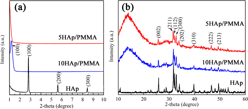
a small and b wide angle XRD patterns of various samples
Scanning electron miscrosocpy observation
The SEM images of HAp/PMMA nanocomposites are shown in Fig. 2. Figure 2a shows the low magnification structure of cross-section of HAp/PMMA composite sample prepared in liquid nitrogen. It is observed that the 10HAp/PMMA (Fig. 2b) possesses intercalated structure, and the layer spacing is between 7 and 8 nm (inset of Fig. 2b), which is consistent with the XRD results. With decreasing HAp content, structure of 5HAp/PMMA composite is shown in Fig. 2c. Some ordered lamellar HAp groups are separated into single HAp lamellae because of the relative increase in PMMA content. The EDS analysis result (Fig. 2d) suggests that HAp/PMMA composite contains Ca and P, which confirms the existence of HAp in the composite.
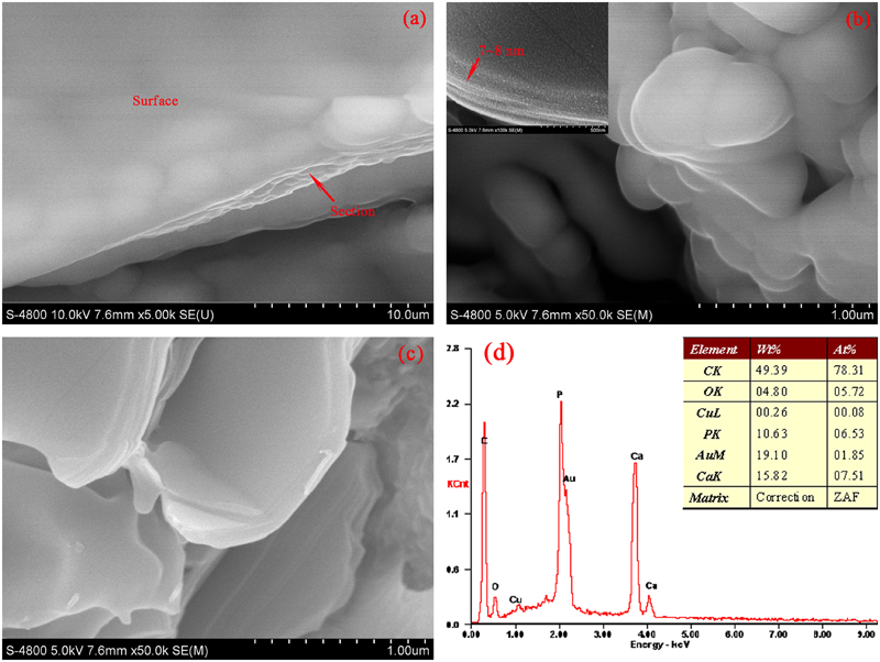
Images (SEM) of low magnificated structure of a HAp/PMMA sample, b 10HAp/PMMA composite, c 5HAp/PMMA composite and d EDS pattern
Differential scanning calorimetry analysis
Differential scanning calorimetry traces of PMMA and PMMA/HAp composites are shown in Fig. 3, which indicates that the composites show higher Tg than PMMA. The increment of Tg in the composites can be attributed to that the HAp lamellae provide large surface area for interacting with PMMA matrix, which lead to the restricted segmental motions near the organic/inorganic interfaces. Similar results were obtained in the investigations of clay/PMMA composites.19 It is believed that PMMA chains are strongly bound to the HAp lamellae either through intercalation or exfoliation of the lamellae and consequently the thermal stability of PMMA is improved.20
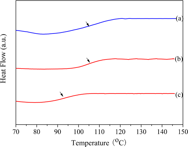
Differential scanning calorimetry curves of a 10HAp/PMMA composite, b 5HAp/PMMA composite and c pure PMMA
Tensile strength analysis
Figure 4 shows the tensile strength values of pure PMMA and HAp/PMMA nanocomposites with various HAp contents. It is detected that 5HAp/PMMA nanocomposite shows distinct increased tensile strength than pure PMMA. This is attributed to that HAp lamellae can distribute the load uniformly, thus bring down the possibility of breaking. However, the tensile strength decreases when the HAp content is 10%, which may be due to the aggregation of HAp lamellae. This trend is in agreement with the results of PMMA/MMT composites.21
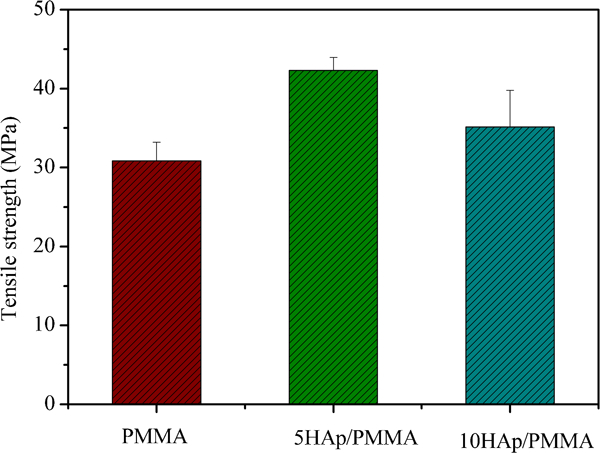
Tensile strength values of pure PMMA and HAp/PMMA nanocomposites
Conclusions
Via one-step intercalative bulk polymerisation, the laminated HAp/PMMA nanocomposites were prepared successfully in this work. X-ray diffraction and SEM results revealed that different morphologies, i.e. intercalated and exfoliated structures were obtained in 10HAp/PMMA and 5HAp/PMMA respectively. The enhancement of thermal stability of PMMA/HAp composites was confirmed by DSC analysis and it is believed that the dynamic movements of PMMA chains were blocked by the HAp lamellae. Tensile strength analysis confirmed that the HAp/PMMA nanocomposites show distinct increased tensile strength than pure PMMA.
Footnotes
Acknowledgements
This work was supported by the Hebei Province Science and Technology Project (12211203), Tangshan Municipal Science and Technology Project (12110211b), Doctor Research Foundation of Hebei United University (grant no. 35395601) and the National Natural Science Foundation of China (grant no. 51172158).
This article is part of a special issue on the 2nd International Conference on Engineering Against Fracture