
Abstract
Polyaniline (PANI) has been synthesised using a combined ultrasonic interfacial polymerisation method. Both the morphological structure and chemical compositions were analysed using field emission scanning electron microscopy (FESEM), X-ray diffraction (XRD) and Fourier transmission infrared radiation (FTIR). Results from FESEM showed that a granular morphology of PANI with a partial amount of nanofibres was obtained. Results from XRD results revealed the formation of partially crystalline PANI. Analysis by FTIR also confirmed the formation of PANI. This paper aims to study synthesis of PANI with nanofibre-granular morphology.
Introduction
Conductive polymers are an attractive group of materials with unique electrical and optical properties. Among conductive polymers, polyaniline (PANI) has gained more attentions due to its low cost monomer, ease of synthesis, unique redox properties, good electrical properties and high environmental stability. Various applications such as diodes,1 transistors,2 sensors,3 fuel cells4 and anticorrosive5 and self-cleaning coatings6 benefit from PANI. Generally, chemical synthesis methods are used to fabricate PANI in large quantities. Chemical synthesis methods resulted in PANI particles with different morphologies. In conventional methods, the synthesised PANI has granular morphology. However, using methods such as hard templates,7 soft template,8 interfacial polymerisation,9 seeding polymerisation,10 electrospinning11 and ultrasonic irradiation,12 the PANI nanofibres could be obtained. In this study, we aim to fabricate PANI with partial nanofibres at granular matrix. For this purpose, the ultrasonic interfacial polymerisation method has been used. Based on the literature, no study has been carried out before to fabricate PANI with a novel morphology through ultrasonic interfacial polymerisation method. Thus, this is the first work to investigate the production of PANI with partial nanofibre granular matrix through ultrasonic interfacial polymerisation method.
Experimental
Materials
The aniline monomer, ferric chloride, chloroform and hydrochloric acid were purchased from Sigma-Aldrich Company. All the chemicals were of analytical grade and used as received.
Synthesis of PANI with novel morphology
The method for synthesis of PANI was as follows. First, two separate solutions were prepared: for solution I, 4 mmol ferric chloride was dissolved in 20 mL hydrochloric acid (1M) by magnetic stirrer at room temperature; for solution II, 4 mmol aniline was added to 20 mL chloroform under magnetic stirring. Solution I was then added to solution II under ultrasonic irradiation, and the reaction was left at ambient temperature for 30 min, 2 and 4 h. The resulting dark green PANI precipitate was washed with distilled water and acetone several times to remove unreacted aniline monomers and chloroform. Finally, the precipitate was dried at ambient temperature for one week.
Characterisation
The morphology of the PANI particles was examined using a FESEM (Zeiss-LEO Model 1530). The X-ray diffraction (XRD) analysis of the PANI was on Ultima III X-ray diffractometer. The Fourier transmission infrared (FTIR) analysis was performed by using a FTIR (VERTEX70 infrared spectrometer).
Results and discussion
Morphological study
The morphology of the PANI was characterised by FESEM. As shown in Fig. 1, moderate amount of PANI nanofibres with mean diameter of 20–30 nm was formed on granular PANI. The FESEM results obtained in this study are in good agreement with the morphology of PANI synthesised by Zhou et al., in which they have also gained granular PANI–partial nanofibre network.13 As shown in Fig. 1a, the nanofibres with mean diameter of 20–30 nm and length of sub-micrometres were formed on the PANI matrix. By increasing the ultrasonic irradiation time, also nanofibres were formed on the granular PANI. As shown in Fig. 1b, the nanofibres with average diameter of 20–28 nm and length of sub-micrometres were formed. Figure 1c also shows that nanofibres with mean diameter of 18–22 nm were synthesised. It is believed that the morphology of as fabricated PANI exhibits the coexistence of nanoparticle agglomerates and nanofibres.
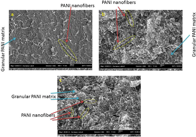
Granular polyaniline with partially nanofibre synthesised through ultrasonic interfacial polymerisation method at a 30 min, b 2 h and c 4 h
FTIR spectroscopy
Figure 2 shows the FTIR spectra of the synthesised PANI under ultrasonic irradiation time of 2 h. The main peaks at around 1569 and 1488 cm−1 can be assigned to the stretching vibrations of quinone and benzene rings respectively. The peaks at 1301 and 1243 cm−1 correspond to the C–N stretching vibration. The in plane bending of C–H is reflected in the 1110 cm−1 peak. The peak at 802 cm−1 is attributed to the out of plane bending of C–H. These assignments confirm the formation of PANI by an ultrasonic interfacial polymerisation method.
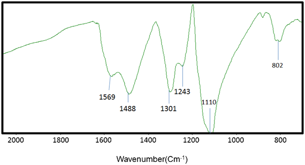
FTIR spectra of PANI synthesised through ultrasonic-interfacial polymerization under ultrasonicirradiation of 2 h
XRD analysis
Figure 3 shows the XRD spectrum of the synthesised PANI by ultrasonic interfacial polymerisation method. It has been reported that the ratio of half-width to height of an XRD peak reflects the order of the polymer crystallinity. Rahy and Yang showed that the metallic PANI has an XRD pattern consisting of three peaks at 15, 20 and 25.14 As shown in Fig. 3, three main peaks appeared at around 2θ = 15, 20 and 25°. Thus, it has been found that the synthesised PANI is partially crystalline.
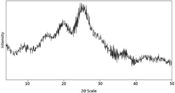
XRD spectra of the synthesised PANI under ultrasonicirradiation time of 2 h
Conclusions
In summary, PANI has been fabricated by a combination of interfacial polymerisation and ultrasonic irradiation method. The morphology of as fabricated PANI exhibits the coexistence of nanoparticle agglomerates and nanofibres. It is proposed that the ultrasonic irradiation combined with interfacial polymerisation could effectively shape the PANI morphology. The XRD results also showed that the fabricated PANI is partially crystalline. This study may provide good opportunities for introducing a simple method to fabricate PANI with granular-nanofiber morphology.