
Abstract
Chitosan (CS) has been proposed for the use of electrically modulated drug delivery. However, the gel fatigue makes CS hydrogel difficult to achieve precise and prolonged drug release. In this study, laminated hydroxyapatite (HAp)/CS composite hydrogels were prepared via solution intercalation method. Cyclic electrostimulation test revealed that the fatigue of neat CS was significantly improved by incorporation of laminated HAp. It is detected by transmission electron microscopy that laminated HAp distributed disorderly in the CS matrix, and the dimension of the HAp lamella is ∼150 nm. Furthermore, the in vitro cytotoxicity of 2HAp/CS was evaluated by optical microscopy, scanning electron microscopy and [3-(4,5-dimethylthiazol-2-yl)-2,5-diphenyltetrazolium bromide) assay. The prepared composite hydrogels exhibit improved swelling fatigue property and good biocompatibility, which will further provide a wide range of potential application in the drug delivery.
Introduction
In order to reduce the patient expenses and risks brought by toxicity, controlled drug delivery system drawn extensive attention attributed to its high drug efficacy, specificity, tolerability and therapeutic index of corresponding drugs.1–3 The electrostimulus responsive hydrogel is one kind of drug delivery system which can realise controlled drug release through the swelling, shrinkage and other behaviours as external electric field changes. In general, an important goal of electric controlled drug delivery is to obtain a constant and precise drug release during a prolonged time. However, it is a common problem of all hydrogels that the responsivity and reversibility will deteriorate after several on–off switching electrostimulation operations. This unstable release phenomenon is defined as electrostimulus responsive fatigue, which makes it difficult to precisely control the drug release by electrostimulation.
In the past decades, a wide range of synthetic polymers, such as polyvinyl alcohol 4 and polydimethyl amine acrylamide, 5 have been applied as electrostimulus responsive drug carriers. Although these synthetic polymers show inspired properties, the relative poor biocompatible and biodegradable of synthetic polymer greatly restrict their application as drug carriers. Compared with these synthetic polymers, chitosan (CS) possesses larger potential in the use of drug carrier because of its excellent biocompatibility and biodegradability. 6 CS is a cationic biopolymer and has been proposed for application as electrically modulated drug delivery. 7 However, a significant drawback of mere CS is that the responsivity and reversibility often deteriorates with increasing number of on–off electrostimulus operation cycles, which makes CS hydrogel difficult to achieve precise and prolonged drug release. 8 In order to overcome the drawback of mere CS, laminated inorganic material such as clay was incorporated, and thus, the electrostimulus responsive fatigue against cyclic electric stimulations was significantly improved. 8 Unfortunately, there is no general report on the safety for application of clay as biomaterial. Compared with clay, hydroxyapatite (HAp) exhibits excellent biocompatibility and osteoconductivity attributed to the similarity of its chemical composition to mineral component of natural bone tissues. 9 Therefore, this work presents the fabrication of laminated HAp/CS composite hydrogels. Laminated HAp was synthesised by a surfactant template technique as described in our previous study. 10 Then, solution intercalation method was applied in the preparation of laminated HAp/CS composite hydrogels.
Materials and methods
Materials
The materials used in the present work included CS, acetic acid, calcium nitrate [Ca(NO3)2.4H2O], sodium hydroxide (NaOH), sodium dodecyl sulphonate (C12H25SO3Na), ammonium hydrogen phosphate [(NH4)2HPO4], ethanol and deionised water. All chemicals used were of analytical grade and were used without further purification.
Preparation of laminated HAp
Sodium dodecyl sulphonate (0·5 g) with 15 mL deionised water and 30 mL ethanol was mixed under magnetic stirring (2000 rev min−1). The mixture was then heated to 60°C in which 15 mL 3·3M Ca(NO3)2.4H2O was added, followed by adding 30 mL 1M (NH4)2HPO4 and 30 mL ethanol. Then, 20 mL 5M NaOH solution and 20 mL ethanol were added. The mixture was refluxed at 83°C for 14 h. Then, the precipitate was deposited at room temperature for 12 days. After being centrifuged and immersed in deionised water for 3 days, the precipitate was rinsed several times in deionised water and centrifuged again, followed by drying at 60°C for 2 h.
Preparation of HAp/CS composite hydrogels
In a typical procedure, 0·01 g of prepared laminated HAp was added into 50 mL deionised water, and intermittent ultrasonic treatment was carried out for well dispersion. Chitosan (1·0 g) was dissolved with 40 mL 1% (wt-%) acetic solution and was poured into a 250 mL flask. Then, HAp suspension was added dropwise into the flask under vigorous stirring, followed by reacting for 6 h at 60°C. The mixture was poured into Petri dishes and dried at 37°C for 24 h. Finally, the CS/HAp composite hydrogel was obtained by washing with 1%NaOH solution and deionised water thoroughly. The same procedure was repeated twice by changing the amount of HAp (0·02 and 0·03 g), and the CS/HAp composite hydrogels with different HAp contents (1, 2 and 3%) were obtained, which are denoted as 1HAp/CS, 2HAp/CS and 3HAp/CS respectively.
Characterisation
Cyclic electrostimulation investigation
CS, 1HAp/CS, 2HAp/CS and 3HAp/CS films were first cut into circular plates (radius ∼1 cm). The average thickness of the obtained dried films was ∼0·30 mm. Then, the films were pre-equilibrated and swollen in phosphate buffered saline (PBS). Two platinum electrodes (diameter: 30 mm) were kept in contact with the opposite surface of the film. A repeated on–off electrostimulus operation was applied on the swollen films with a 25 min interval (5 min for electrical stimulation and 20 min for swelling). The released water from the hybrid films was removed using filter paper, and the weight change of the swollen hybrid films was checked. All results reflect the average figure of five measurements. The difference between each measurement is <5%.
TEM observation
Transmission electron microscopy (TEM) experiment was performed on an FEI Tecnai G2 F2 TEM operating at 200 kV accelerating voltage. Samples were ultrasonically dispersed by ethanol. Then, the suspension was dropped on the copper grids and dried in air.
Biocompatibility test
Before the test, the composite hydrogels were washed with high purity water for several times and sterilised by immersing in 70 wt-% ethanol aqueous solution for 12 h and then subjected to UV irradiation for 12 h. The hydrogels were then seeded in a 24-well cell culture plate, and the blank wells were used as the control group. The frozen New Zealand rabbit cartilage cells were recovered and digested by adding 0·25% trypsin solution. Then, the cell suspension was centrifuged for 3 min at a rotational speed of 1000 rev min−1 and was adjusted to 2·0×104 mL−1 by adding fresh serum Dulbecco's modified Eagle's medium containing 10% fetal bovine. The obtained cell suspension was inoculated to the 24-well cell culture plate by adding 1 mL to each well and incubated at 37°C in a humidified atmosphere of 5% CO2 in air. The growth of cells on the material was recorded by Olympus inverted microscope and IPP image analysis system after 1, 3, 5 and 7 days respectively.
The morphology of the cells on the films was examined by environmental scanning electron microscopy (ESEM). The samples were washed with PBS three times and fixed with 4% paraformaldehyde at 4°C for 12 h. After washing with PBS for more than three times, the films were dehydrated through a graded series of aqueous ethanol soaks (30–100%). After the sample was dried and subjected to a surface spray with gold, ESEM was employed to investigate the microscopic morphology of cells on the surface of composite hydrogels.
The 3-(4,5-dimethylthiazol-2-yl)-2,5-diphenyltetrazolium bromide (MTT) assay was used as a measure of relative cell viability according to the method of Mosmann. 11 The material free blank plates were used as control groups. The obtained cell suspension was inoculated to the 24-well cell culture plate by adding 500 μL for each well and incubated at 37°C in a humidified atmosphere of 5% CO2 in air. The culture solution is carefully sucked out after l, 3, 5 and 7 days respectively, followed by washing twice with Hanks’ balanced salt solution and adding 0·5 mL serum free medium to each well. MTT (50 μL) was added to each well in the dark conditions and drained after cultured at 37°C for 4 h. Then, 450 μL dimethyl sulphoxide was added and shaken in a shaker for 5 min, followed by adding to 96-well plates immediately (150 μL/well). The absorbance value of each well was measured at 490 nm using a microplate reader, and all material extracts were tested for a minimum of three separate experiments with comparable results.
Results and discussion
Swelling fatigue performance of hydrogels
In this study, the fatigue performance of hydrogels was defined as the ability of the hybrid to structurally return to the initial swelling state under cyclic electric stimulation. Figure 1 shows the weight changes of the pure CS hydrogel and the CS/HAp composite hydrogels under consecutive on–off operations. It can be seen in Fig. 1a that the pure CS hydrogel swelling equilibrium rate is apparently decreased when the on–off electrostimulus operation exceeds five cycles, which indicates that the structure return ability of CS is destroyed by cyclic electric stimulation. It is documented that the contractile deformation and volume recovery of CS hydrogel are associated with viscoelasticity, which is controlled by cross-linking density. 12 Here, lamellar HAp was incorporated as a physical cross-linking agent. Compared with mere CS hydrogel, the swelling fatigue property of CS/HAp composite hydrogels was improved significantly by the incorporation of lamellar HAp. Among the three composite hydrogels, 2CS/HAp (Fig. 1c) shows the best anti-fatigue performance, in which the swelling and deswelling behaviour remained identical and steady even after more than 20 on–off electrostimulus cycles. It may be attributed to that the cross-linking density is improved by incorporation of lamellar HAp. Compared with 1HAp/CS, 2HAp/CS shows better antifatigue performance because the higher cross-linking density is capable of bearing more on–off switching electrostimulus operations. However, 3HAp/CS shows less stability than 2HAp/CS. This is because the relative large content of HAp deteriorated the crystallinity of CS. The deterioration of crystallinity weakened the strength of the nanohydrogels and thus reduced the responsiveness after cyclic on–off operations. This trend is consistent with CS/MMT system in the study of Liu et al.8, 12 Therefore, the optimal range of HAp content in the nanohydrogels is 2 wt-%, in which the level hydrogel possesses optimal cross-linking density to keep the best antifatigue performance.
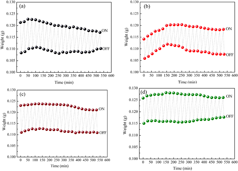
Weight changes of a pure CS, b 1CS/HA, c 2CS/HAp and d 3CS/HAp composite hydrogels under cyclic on–off switching of 5 V electrostimulation
Microstructure observation
TEM was used to detect the microstructure of 2HAp/CS composite hydrogel. It can be seen in Fig. 2a that the laminated HAp distributed disorderly in the CS matrix, which corresponds to our previous work. 10 The crystalline nature of the HAp lamella could be confirmed by the selected area diffraction pattern with the diffraction rings corresponding to (002) and (300) reflection of HAp (inset of Fig. 2a). Figure 2b shows the TEM image of one single HAp lamella, and it can be measured that the dimension of the HAp lamella is about 150–200 nm.
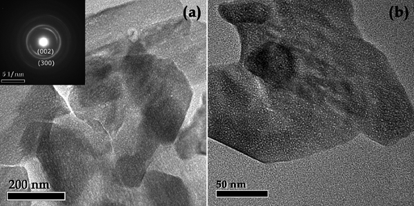
Images (TEM) of a distributed HAp and b magnified image of single HAp lamella in 2CS/HAp: inset of a is reflection of HAp in 2CS/HAp
Biocompatibility evaluation
Figure 3 shows the cell morphology of the control and experimental groups under different incubation time (1–7 days). It is indicated that the cells exhibit adherent growth and spread well on the material surface through cytoskeletal processes after 1 day culture. Three days later, cells proliferate and present fusiform shape. After 5 and 7 days culture, the cells show large scale proliferation and stratified clustered growth. It is identified that cell morphology in the experimental group was not significantly different from that in the negative control group at the different stages of the cultivation, indicating that the material is not obvious toxicity.
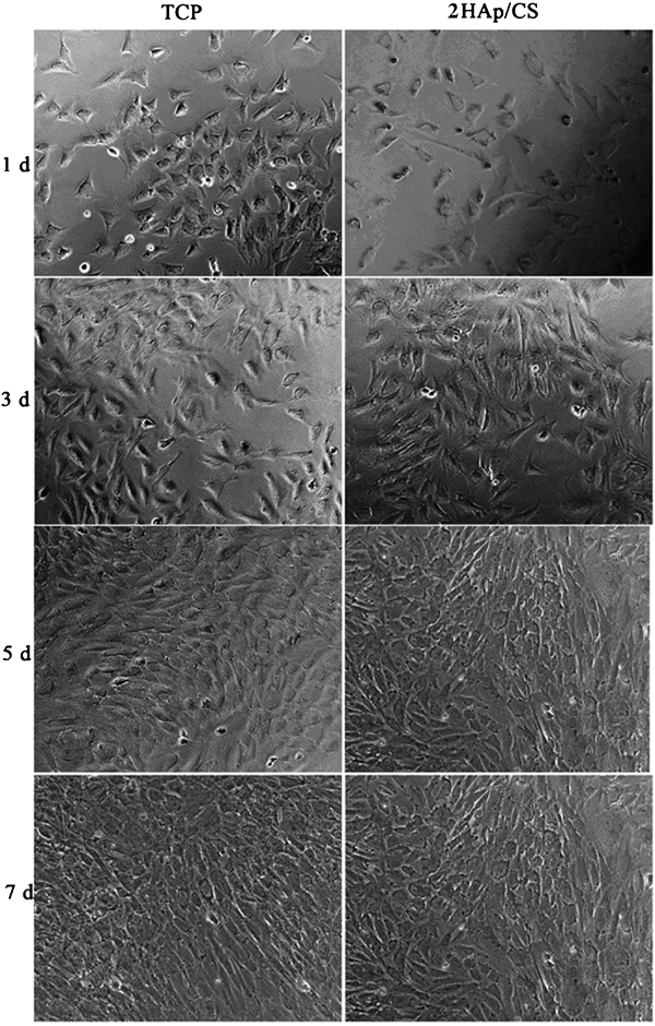
Optical micrographs of cartilage cells on TCP (left) and 2HAp/CS (right) with culture times for 1, 3, 5 and 7 days respectively
Scanning electron microscopy was used in this experiment in order to observe the cell growth morphology on the surface of the 2HAp/CS. As can be seen in Fig. 4, cartilage cells show typical cell adhesion morphology, and the cell pseudopodia stretch to several directions to form a good bond with the material matrix. The cell morphology obtained in the present experiment is similar to that in the study of Lu et al. 13 and further indicates a good biocompatibility of the material.
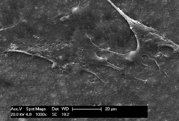
Microscopic morphology of cartilage cells on surface of 2HAp/CS
MTT assay was used in this study in order to quantitatively analyse the cytotoxicity and the proliferation of chondrocytes on 2HAp/CS. Figure 5 shows the results of MTT test, and hence, a calculated cell relative proliferation rate of experimental group is >80% at each stage of culture, indicating that 2HAp/CS is nontoxic in each time period. This is consistent with the cell morphology test.
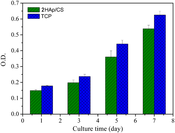
Proliferation of cartilage cells in TCP control group and 2HAp/CS group
Conclusions
Laminated HAp/CS composite hydrogels were successfully prepared by means of solution intercalation in this study. Compared with mere CS, composite hydrogels show improved swelling fatigue property, and the swelling/deswelling behaviour of 2CS/HAp remained identical and steady even after >20 on–off cycles. In vitro cytotoxicity was evaluated using MTT assay, and it was demonstrated that 2HAp/CS composite hydrogel has good biocompatibility.
Footnotes
Acknowledgements
This work is supported by the Cultivate Foundation of Hebei United University (Grant No. SP201305), the Doctor Research Foundation of Hebei United University (Grant No. 35395601), the National Natural Science Foundation of China (Grant No. 51172158), the Science and Technology Support Program of Tianjin (Grant No. 11ZCKFSY01700) and the Natural Science Foundation of Hebei Province (Grant No. H2013209192).