
Abstract
In this study, nanocrystalline Ni0·64Zn0·36Fe2O4 powders were prepared using a planetary ball mill. The evolution of the microstructure and magnetic properties during the milling were studied by X-ray diffraction technique, scanning electron microscopy, transmission electron microscopy and vibrating sample magnetometre. It is revealed from the results of the phase analysis that nanocrystalline Ni0·64Zn0·36Fe2O4 ferrite with average crystallite size of 6·18 nm and non-uniform lattice strain of 0·33% has been formed after 60 h of milling time. A progressive increase of saturation magnetisation and a dramatic decrease in coercivity were also observed with increasing milling time.
Introduction
The Ni–Zn ferrite (Ni1-xZnxFe2O4) is the most versatile ferrite from the viewpoint of its large number of applications, due to its high value of magnetic permeability, high electrical resistivity, high Curie temperature and low power loss at high frequencies.1,2 Ni0·64Zn0·36Fe2O4 ferrite is one of the Ni–Zn ferrite compositions, well known for high electrical resistivity (ρ≅6200 Ω cm)3 and high saturation magnetisation (Ms≅78 emu g−1).4
Recently, more and more attentions have been paid to the magnetic nanostructured materials due to their unique magnetic properties compared to those obtained for the same conventional microstructured materials.5 Several methods, such as coprecipitation,6 reverse micelle,7 sol–gel,8 autocombustion synthesis9 and ball milling10– 12 are used to synthesise nanostructured Ni–Zn ferrite. Among these methods, ball milling method is a relatively simple technique, which allows the preparation of nanostructured Ni–Zn ferrite at a relatively low temperature.10– 12 Microstructure characterisation and phase transformation kinetics of Ni0·5Zn0·5Fe2O4 ferrite powders prepared via ball milling were studied in detail by Bid and Pradhan.10 Magnetic properties of the ball milled prepared Ni0·5Zn0·5Fe2O4 ferrite powders also were investigate by Jalaly et al. 11 and Ye et al. 12 However, there are only a few reports on the characterisation of Ni–Zn ferrite powders synthesised by ball milling and structural evaluations and magnetic properties of ball milled prepared Ni1-xZnxFe2O4 ferrite powders in some technologically important compositions (x<0·7) in terms of milling time have not been investigated to date.
In our previous work,13 the effect of heat treatment on the structure and magnetic properties of ball mill prepared Ni0·64Zn0·36Fe2O4 ferrite powders has been investigated. The aim of this work is to study the structure and magnetic properties of nanocrystalline Ni0·64Zn0·36Fe2O4 powder prepared by ball milling as a function of milling time. It would be worth mentioning that as compared with the literature in this field, this paper has paid special attention to Ni0·64Zn0·36Fe2O4 ferrite composition.
Experimental method
The initial high purity (>99%) powders of 12·3 wt-%ZnO, 20·2 wt-%NiO and 67·5 wt-%Fe2O3 were weighed and mixed according to the composition of Ni0·64Zn0·36Fe2O4, then introduced into a cylindrical tempered steel vial of the capacity of 100 mL. The ball-to-powder weight ratio was 10∶1. The milling was carried out at room temperature by using a planetary ball mill at the vial rotation speed (ω) of 350 rev min−1 and the disc rotation speed (Ω) of 212 rev min−1 (ω/|Ω| = 1·65) for different milling times varying from 0·5 to 60 h. For comparison purposes, Ni0·64Zn0·36Fe2O4 ferrite was also prepared by conventional calcination of the initial mixed powders at 1250°C for 2 h.
The phase analysis and the structural properties were characterised by the use of X-ray diffraction (XRD, Shimadzu XRD-6000 diffractometer with Cu Kα1 radiation). Structural parameters including crystallite size, lattice parameter and lattice strain of Ni–Zn ferrite were obtained from Rietveld's powder structure refinement analysis of X-ray powder diffraction data. The Rietveld calculations were performed by the TOPAS 3 software (from Bruker AXS). In the TOPAS software, the Double–Voigt approach14 is used for obtaining crystallite size and strain components. The crystallographic models of Ni–Zn ferrite (Cubic, Fd3m, COD ID: 9009920), ZnO (Hexagonal, P63mc, COD ID: 2300112), NiO (Cubic, Fm3m, COD ID: 9013980) and Fe2O3 (Rhombohedral,
 , COD ID: 2101167) were used as starting models for the refinements. The models were selected from the official website of the crystallography open database (COD).15 For each refinement, the following variations were applied: the background parameter, scale factor, cell parameter, zero point correction, Lorentzian crystal size and Gaussian lattice strain. In order to judge the quality of the fitting on the structure model, the Bragg reliability factor RBragg was used. The refinements resulted in proper fits to the experimental data and average RBragg value was less than 4%. Transmission electron microscope (TEM, JEOL-JEM 2010) was utilised to study the microstructure of the powder particles. Magnetic measurements were obtained at room temperature with a vibrating sample magnetometer (VSM, Lakeshore 7404) with a saturating field of ±10 kOe, field increment of 404 Oe and a field ramp rate of 40·4 Oe s−1.
Results and discussion
Structure
Figure 1 shows the XRD patterns of the ball milled powders for different milling times. From these patterns following points can be concluded:
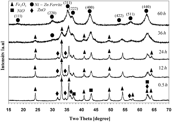
X-ray diffraction patterns of ball milled powders for various milling time: formation of Ni–Zn ferrite is observed after 60 h of milling
for milling time of 0·5 h, XRD pattern presents Fe2O3, NiO, ZnO compounds simultaneously. It can be observed from this figure that the diffraction line intensities of the Fe2O3 and ZnO compounds gradually decreased with increasing milling time. Fig. 2a shows that the most intense reflection of Fe2O3 (2θ = 33·28°) and the strongest isolated reflection of ZnO (2θ = 31·94°) are almost vanished after 60 and 36 h of milling time respectively. In addition, all diffraction lines of NiO compound broadened and shifted to the left with increasing milling time. Diffraction line shifting and broadening for the most intense reflection of NiO (2θ = 43·3°) is shown in Fig. 2b. At milling time of 60 h, all diffraction lines of the starting material are vanished or changed to the reflection angles of Ni–Zn ferrite in such a way the characteristic diffraction lines of Ni–Zn ferrite phase are appeared distinctly
the XRD results presented in Figs. 1 and 2 show that the ZnO diffraction lines are first vanished during ball milling in comparison to the characteristic diffraction lines of NiO and F2O3 compounds. This finding, which is consistent to the Bid et al. 10 and Jalaly et al. 11 reported data, indicates higher solid state diffusion rate of ZnO phase
diffraction line broadening of Ni–Zn ferrite phase indicates a decrease in the crystallite size and the introduction of lattice strain during the high energy ball milling. The Rietveld analysis of the diffraction pattern exhibits that the average crystallite size, the lattice strain and the lattice parameter values of Ni–Zn ferrite phase are 6·18±0·30 nm, 0·33±0·05% and 0·83977±0·00042 nm respectively. It is interesting to note that Bid and Pradhan10 obtained a crystallite size and lattice parameter of 4·6 and 0·8393 nm after 11 h of milling using a high energy planetary ball mill.
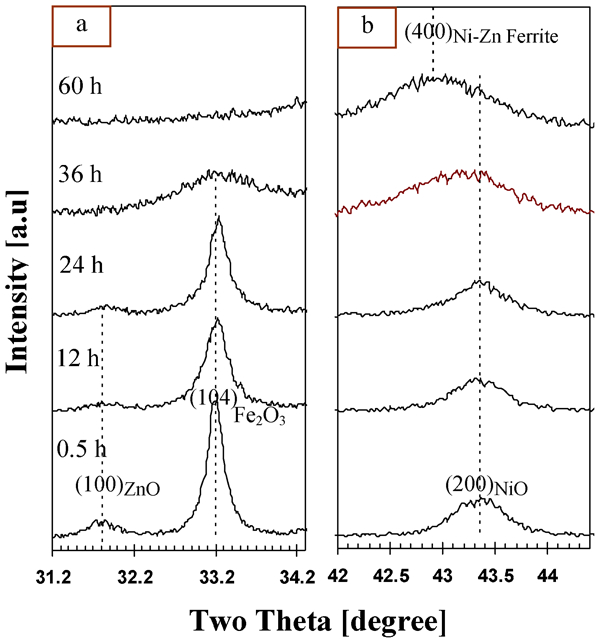
Magnified view of XRD patterns of ball milled powders extracted from Fig. 1
Figure 3 displays the XRD patterns of the as synthesised ferrite powders prepared by the ball milling and the solid state reaction method. It is evident from this figure that not only diffraction lines of the mechanically alloyed powder are broadened, but also its broadened diffraction lines are shifted to lower angles as compared to those of the calcined powders. Diffraction line broadening is attributed to the second order internal stress (non-uniform or microstress) while angular diffraction line shift is resulted from the first order internal stress (uniform or macrostress).16 Angular shift of diffraction lines of the mechanically alloyed powders to the lower angles indicates a tensile residual stress within the ferrite crystal lattice arising from high energy ball milling. The mean value of uniform lattice strain can be estimated roughly by the following equation:

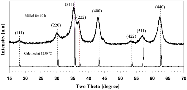
X-ray diffraction patterns of Ni Zn ferrite powders obtained after 60 h milling time and same powders prepared using solid state reaction method (calcined at 1250°C for 2 h)
The nanocrystalline structure of the as synthesised powder was also confirmed by high resolution transmission electron microscopy (HRTEM) micrographs. Figure 4 shows the HRTEM bright field image and the corresponding selected area electron diffraction (SAED) of the Ni–Zn ferrite powders formed within 60 h of ball milling. The corresponding diffraction pattern and the lattice fringes indicate an almost completely random orientation of the resultant nanocrystalline structure. The average grain size estimated from the HRTEM images is between 5 and 10 nm, which is relatively consistent with the value estimated by the analysis of the XRD data.
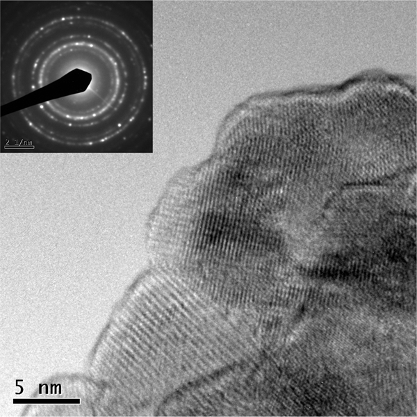
Image (HRTEM) and corresponding SAED pattern of Ni Zn ferrite powders obtained after 60 h milling time
Magnetic properties
Figure 5 shows the hysteresis curves (magnetization vs. external field) for 2 different samples that undergo mechanical alloying at different milling times (36 and 60 h). Magnetic properties such as saturation magnetization, remanent magnetisation, coercivity and susceptibility can be determined from these curves.
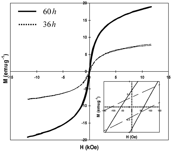
Hysteresis curves for samples milled at 30 and 60 h
The variation of saturation magnetisation and coercivity as a function of milling time are illustrated in Fig. 6. Referring to this figure, the saturation magnetisation increases and coercivity decreases with increasing the milling time. The increase in saturation magnetisation can be attributed to the Ni–Zn ferrite phase formation at the high milling time. The progress of mechanochemical reaction between initial oxide powders (i.e. ZnO, NiO and Fe2O3) during the ball milling can be monitored by means of saturation magnetization variations.
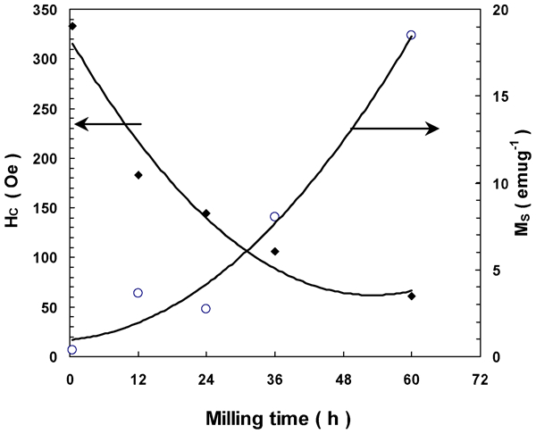
Saturation magnetisation and coercivity as function of milling time
The coercivity variations in the nanocrystalline mechanically alloyed soft magnetic powders are affected by several factors, which in some cases make it difficult to interpret coercivity as a function of milling variables. Zeng et al. 17 summarised various contributions to coercivity, including residual internal stress, crystallite size, contamination (oxidation, inclusions), various defects (dislocations), non-spherical shape, surface irregularities, surface anisotropy, interparticle interaction, etc. Nevertheless, it seems that the gradual increase in Ni–Zn ferrite with milling time is the dominant factor controlling coercivity variations during the ball milling. Figure 6 also shows that coercivity varies inversely with saturation magnetisation.
The variation of remanence ratio (Mr/Ms) versus milling time is shown in Fig. 7. The variation of the remanence ratio with milling time is very similar to that observed for the coercivity. The remanence ratio decreases from 0·106 at 0·5 h to 0·039 at 60 h. These values are much less than the theoretical value for an isotropic (0·5) or cubic Stoner–Wohlfarth particle (0·83).18 In the theoretical calculations, the effect of free poles formed on the most boundaries is ignored. These free poles set up demagnetising fields which can cause the actual value of Mr/Ms to be substantially lower than the calculated value.18
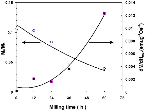
Remanence ratio and maximum susceptibility as function of milling time
The variation of magnetic susceptibility (dM/dH) with applied magnetic field (Fig. 8) shows peaks separated by magnetic field 2Hm, indicating the maximum susceptibility. The maximum susceptibility shows an upward trend with increasing milling time (Fig. 7), which is approximately similar to that observed for the magnetisation variations (Fig. 6). The overall trend of the milling process shows a progressive narrowing of the susceptibility peaks with increasing the milling time, which can be ascribe to the coercivity variation with milling time. As shown in Fig. 6, the coercivity also decreases with increasing the milling time. The narrowing of the susceptibility peaks has been also observed by Muthuselvam et al. 19 for Co2FeO4 ferrite prepared by mechanical alloying and subsequent annealing. They have found that a single phase structure of Co2FeO4 ferrite exhibits a narrow susceptibility peak due to the homogeneous structure of ferrimagnetic domains. The results of our mechanical alloyed samples are also consistent with their findings. As illustrated in Fig. 1, a single phase structure of Ni–Zn ferrite gradually develops with milling time, which in turn leads to the narrowing of the susceptibility peaks.
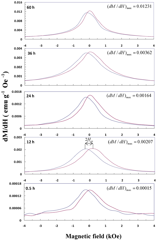
Field dependence of magnetic susceptibility of ball milled powders for various milling time: separation of peaks is represented by field 2Hm
In conclusion, all of the measured magnetic properties confirm that soft magnetic properties of the synthesised powders have been improved with the increase in milling time.
Conclusions
The structure and magnetic properties of ball mill prepared nanocrystalline Ni0·64Zn0·36Fe2O4 ferrite have been carefully studied as a function of milling time. The important observations can be summarised as follows.
Nanocrystalline Ni0·64Zn0·36Fe2O4 ferrite powders with the average crystallite size of 6·18 nm, non-uniform lattice strain of 0·33% and lattice parameter of 0·83977 nm has been formed after 60 h of ball milling. High resolution transmission electron microscopy micrographs also confirmed the nanocrystalline nature of the prepared powders.
In addition to the non-uniform lattice strain, a uniform lattice strain was also observed in the as milled powders. The mean value of uniform lattice strain for 60 h ball milled powders was found to be 0·40%.
Saturation magnetisation increased from 0·37 to 18·5 emu g−1 and coercivity decreased from 333·1 to 60·7 Oe when milling time increased from 0·5 to 60 h. Improvement of soft magnetic properties can be ascribed to the gradual increase in Ni–Zn ferrite phase with increasing milling time.
Footnotes
Acknowledgements
The authors would like to thank the Shiraz University for providing support to this research.