
Abstract
To better understand transient oxide formation on the surface of standalone NiCrAlY in a vacuum, plasma sprayed NiCrAlY samples were subjected to a series of heat treatments at temperatures ranging from 1000 to 1100°C and for two different holding times. The morphology, composition and type of transient oxide(s) formed after heat treatment were characterised using scanning electron microscopy, energy dispersive X-ray spectroscopy and X-ray diffraction (XRD). All samples exhibited the formation of a top layer of discrete or island like Ni rich oxide in cellular shaped alumina. The alumina layer, although difficult to be detected in cross-section, was very dense and covered most of the coating surface. The transisent oxide formation on the heat treated surfaces was further analysed using XRD and α-Al2O3 was detected on all heat treated samples in addition to NiO and possible spinel. Cr2O3 seemed to be present on the samples heat treated at 1000 and 1050°C but not on the sample heat treated at 1100°C. Increased Al surface content, in comparison to the as sprayed sample, was found on all heat treated samples. High Al content, corresponding to the extent of alumina formation on heat treated samples, was observed for samples heat treated for a longer time as more oxygen diffusion took place. The coating surface Al content increased with heat treatment from 1000 to 1050°C and reduced from 1050 to 1100°C.
Introduction
Gas turbine components are regularly subjected to service conditions that include exposure to a harsh combustion product atmosphere in temperatures that exceed 1000°C. A hot section component must not only withstand these conditions but meet expected service lifetimes that range between 10 000 h (for civil aeroengine applications) to 100 000 h (for industrial/power generation applications). 1 1,2 One of the elements in ensuring component durability is the application of MCrAlY type coatings to provide adequate oxidation and corrosion resistance to substrate superalloys and a ceramic thermal barrier coating to provide thermal insulations to the substrate components. Whether as a standalone environment resistant coating or as a bond coat in a thermal barrier coating system, MCrAlY has been designed to form an adherent, dense, stable and slow growing thermally grown oxide (TGO) layer upon exposure to oxidising atmosphere. The TGO layer formed must also resist coating spallation under cyclic thermal environment.3 In practice, MCrAlY materials that have shown the most promise are those that generate a slow growing α-aluminium oxide TGO layer. 4 4,5 However, depending upon coating composition, microstructure, coating application method and post-coating thermal treatment, a MCrAlY coating on first contact with the oxidising environment, can also form other types of oxide, such as transition metal oxides, spinels and other less stable aluminas. The coating composition must be formulated in such a way as to ensure the formation of a continuous layer of thermodynamically stable aluminium oxide, instead of transition metal oxides or spinels which are less protective in comparison to alumina.4
In general, high temperature iron-, nickel- and cobalt based alloys have been designed to form protective scales of either chromium oxide (Cr2O3), aluminium oxide (Al2O3), or silicon dioxide (SiO2) when exposed to oxidising conditions.6 However, at temperatures above 900°C, Cr2O3 has the tendency to react further with oxygen to form volatile CrO3, severely limiting the lifetime of Cr2O3-forming alloys. While both Al2O3 and SiO2 are able to withstand higher temperatures up to around 1200–1300°C, SiO2 formers are limited to environments with adequate oxygen activity due to the propensity to form volatile SiO at low oxygen activities.7 As such, the design of MCrAlY relies on the formation of stable alumina on the surface. In fact, MCrAlY coatings are commonly termed as secondary aluminium oxide formers that generally develop chromium oxide scales on the external surface of the coating. Subsequently an aluminium oxide layer is formed underneath the chromium oxide layer during exposure to an oxidising environment.8 For binary Ni–Al or Co–Al alloys, substantial amounts of aluminium are required in order to form and maintain a complete coverage of alumina on the surface. The addition of approximately 10 at-% of Cr, however, decreases the minimum amount of aluminium needed to form the surface scale to about 5 at-%.9 This helps to improve the ductility of the coating alloys.
While the thermodynamically stable hcp α-phase of aluminium oxide is desired as the primary TGO constituent,10 there are several meta-stable phases of aluminium oxide with different crystal structures that have also been shown to exist: γ-Al2O3 (cubic spinel structure), δ-Al2O3 (tetragonal structure), and θ-Al2O3 (monoclinic structure).11 – 13 These metastable aluminium oxide structures form more porous blade or whisker type surface morphologies and tend to grow at a significantly faster rate than α-Al2O3 structure. All meta-stable aluminium oxide phases can be converted to the α-phase through appropriate heat treatment.6 Since temperature and atmosphere and other service environments cannot be controlled, the formation of stable α-Al2O3 is best achieved through pre-oxidation heat treatment. Indeed, studies have suggested that significant gains in overall lifetime of the thermal barrier coating system can be achieved through pre-oxidation heat treatments at temperatures ranged between 1000 and 1100°C and in both vacuum and air furnace environments.14 – 18 In particular, it has been found that pre-oxidation heat treatments in a low pressure oxygen environment could favour the preferential formation of a largely continuous alumina on the MCrAlY bond coat, reducing or preventing the formations of chromium oxide, spinels [Ni(Cr,Al)2O4], and nickel oxide (NiO).
The objective of this study is to better understand the formation of transient oxide(s) on the surface of standalone NiCrAlY in vacuum pre-oxidation heat treatment without the influence of ceramic top coat. A new air plasma spray technique using a Mettech Axial-III, was employed to prepare the samples. Vacuum furnace heat treatments were conducted at various temperatures and durations. The morphology, composition and type of transient oxide(s) formed after heat treatment were characterised using scanning electron microscopy (SEM), a energy dispersive X-ray spectroscopy (EDS) and X-ray diffraction (XRD).
Materials and experimental procedures
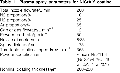
NiCrAlY coating was applied using Axial III Plasma Spray System (Northwest Mettech Corp., North Vancouver, Canada). The torch employed in this system injects powder axially, between three electrodes, ensuring that virtually all of the powder injected passes through the hottest part of the plasma plume. The high velocity plasma was generated using the processing parameters listed in Table 1, achieving a particle velocity in excess of 400 m s−1.
Plasma spray parameters for NiCrAlY coating
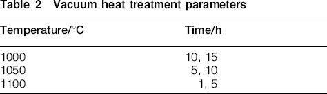
A Hastelloy X substrate was grit blasted with aluminium oxide (#24 mesh size) before coating. The coated samples were subsequently polished to 1200 grit finish with silicon carbide paper to level the surface and remove any surface oxide(s) formed during APS coating process. Heat treatments were carried out in vacuum (10−5 and 10−6 torr) furnace at temperatures and durations given in Table 2.
Vacuum heat treatment parameters
Microscopic examination was carried out using a scanning electron microscope (SEM). A Philips XL30S FEG SEM with a Phoenix EDS detector system and a Hitachi S-570 SEM with a Link Systems LX-5 model 5697 EDS detector were used to generate images and EDS data to measure the chemical compositions of surface oxides. Since the oxide films have thicknesses less than 1 μm, it is probable that the EDS spectra obtained from surface features contained information pertaining to the oxide layer as well as the underlying oxide layer and the NiCrAlY coating. X-ray diffraction analysis was used to identify the crystal structures of phases present on the coating surface. X-ray diffraction analysis was performed using a Bruker AXS D8 Advance diffractometer controlled with general area detector diffractometer software.
Results and discussion
Microstructure of as sprayed NiCrAlY
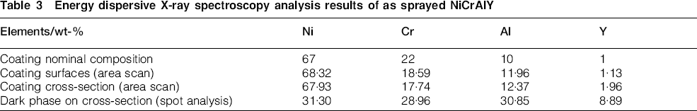
The surface morphology of the as sprayed NiCrAlY coating, as shown in Fig. 1, shows the typical constituents that are present on a thermally sprayed coating surface: coating splats, unmelted particles, porosity and glazed areas indicating melting and resolidification. Table 3 summarises the compositions measured from the coating surface (Fig. 1a) and cross-section (Fig. 1b and c). The results are in consistent with the powder material composition. The high Al and Cr contents detected from the dark phase on the cross-section suggest oxide formation during spraying operation. Figure 1b shows the image of polished coating surface prepared for subsequently vacuum heat treatment. The average coating thickness after polishing was ∼120 μm.
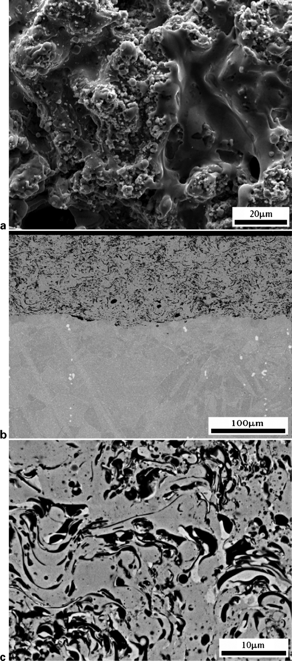
Secondary electron micrographs showing a as sprayed surface and b, c cross-sections of APS NiCrAlY
Energy dispersive X-ray spectroscopy analysis results of as sprayed NiCrAlY
Microstructure after vacuum heat treatment at 1000°C for 10 and 15 h
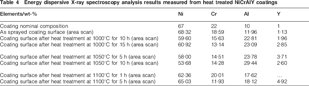
The NiCrAlY coating samples after vacuum furnace heat treatments of 1000°C for 10 and 15 h (Fig. 2a and c) show smoother surfaces compared to the as sprayed surface. This is due to surface polishing before the heat treatments and possibly a sintering effect from heat treatments. Some pores are observed from the cross-sections of the samples (Fig. 2b and d). The compositions, measured from the sample surfaces using area scan, revealed a significant increase in Al and Y concentrations and a decrease in both Ni and Cr concentrations (Table 4) compared to that measured from the as sprayed sample (Table 3). This Al enrichment suggests alumina formation during heat treatments. The cross-sections did not provide a clear indication of continuous surface scale layer (Fig. 2b and d). Increasing heat treatment time did not result in significantly changes to the microstructure other than slight increase in Al and Y measured from the heat treated surface.
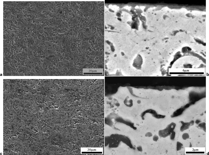
Secondary electron micrographs showing surface and cross-section features of APS NiCrAlY coating after vacuum furnace heat treatments at 1000°C for 10 and 15 h: a surface, 10 h; b cross-section, 10 h; c surface, 15 h; d cross-section, 15 h
Energy dispersive X-ray spectroscopy analysis results measured from heat treated NiCrAlY coatings
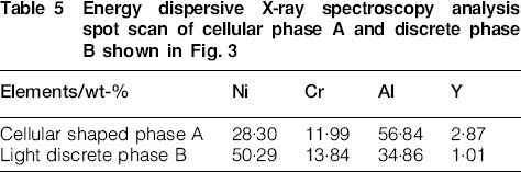
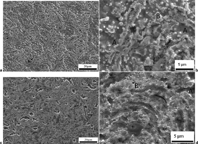
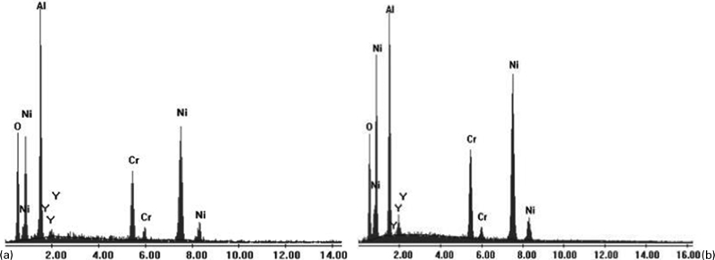
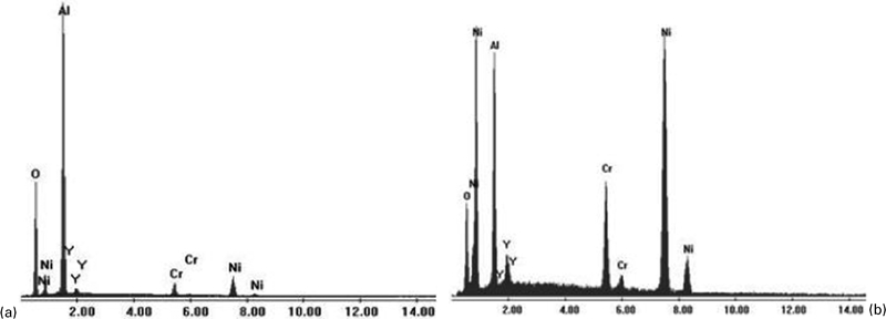
To identify phases formed on the surface, the NiCrAlY sample heat treated at 1000°C for 15 h was examined in detail at higher magnifications. Two distinct phases can be discerned from Fig. 3: bright discrete or island-like phase B and cellular type phase A. Energy dispersive X-ray spectrometry spot analysis was carried out on these two phases with their respective EDS spectra given in Fig. 4. The cellular type phase A was found to be rich in Al and Y (Table 5) in comparison to the as sprayed coating composition while the discrete phase B had less Al and more Ni than the cellular constituent. The presence of strong oxygen peak on the EDS spectra (Fig. 4) further confirms the formation of oxide(s). Although not considered accurate, EDS detected ∼40 at-% of oxygen in both phases, indicating the likelihood of the formation of Al2O3 and Ni rich oxide (NiO or aluminium spinel NiAl2O4) in the sample. The inclusion of Ni and Cr in the EDS analysis of alumina is believed to be a result of electron beam penetration into the substrate NiCrAlY layer. However, the large amount Al detected in the Ni rich oxide could be due either to the Al signal from underlying Al2O3 or the formation of aluminium spinel. No Cr rich oxide was found on the surfaces after vacuum heat treatments.
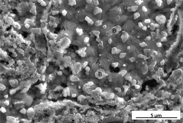
Two types of oxide constituent (A and B) present on NiCrAlY coating surface after vacuum furnace heat treatment at 1000°C for 15 h
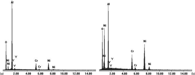
Energy dispersive X-ray spectra from a cellular type phase A (Al2O3) and b discrete Ni rich particle B
Energy dispersive X-ray spectroscopy analysis spot scan of cellular phase A and discrete phase B shown in Fig. 3
Microstructure after vacuum heat treatment at 1050°C for 5 and 10 h
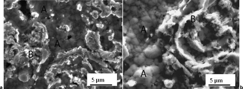
Vacuum furnace heat treatments at 1050°C for 5 and 10 h resulted in the formation of two types of surface oxides as shown in Fig. 5 with one of them being a lighter discrete and island-like oxide and the other a cellular shaped background phase. Less of the discrete phase was observed in the sample heat treated for 10 h and the surface of the sample was mostly covered with island-like phase (Fig. 5d). The compositions measured from the sample surfaces (Table 4) showed a significant increase in Al, from 23·78 to 29·44 wt-%, with the increase of heat treatment time from 5 to 10 h suggesting more alumina formation on the surface. Also noted in the table is the increased Al content, from 22·81 to 29·44 wt-%, when heat treatment temperature stepped up from 1000 to 1050°C (both for 10 h). Based on the EDS spot analysis (Fig. 6), the oxides formed on the heat treated surfaces (Fig. 5) were Al2O3 (A) and Ni rich oxide (B) (NiO or NiAl2O4). Again no Cr-rich oxide was found on the surface. Although not shown here, no visible surface scale was observed on the cross-sections of the heat treated samples.
Surface features typical of NiCrAlY coating surface after vacuum furnace heat treatments at 1050°C for a, b 5 h and c, d 10 h (A: Al2O3; B: Ni rich oxide)
Energy dispersive X-ray spectra from a cellular type phase A (Al2O3) and b discrete Ni rich particle B for samples heat treated at 1050°C
Microstructure after vacuum heat treatment at 1100°C for 1 and 5 h
The surface conditions of the NiCrAlY coating samples after vacuum heat treatments at 1100°C for 1 and 5 h (Figs. 7 and 8) show a similar surface feature (cellular Al2O3 phase with island-like Ni rich oxide) to those formed at 1000 and 1050°C. Very little discrete Ni rich oxide was observed and the island-like Ni rich oxide became more irregular and lifted from the alumina. The compositional analysis from the sample surfaces (Table 4) showed an increase in Al concentration and a decrease in both Ni and Cr concentrations as compared to the original coating composition and the Al concentration increased with the heat treatment time. However, the Al detected on samples heat treated at 1100°C for 1 and 5 h was lower than that from the samples heat treated at 1000 and 1050°C, indicating thinner alumina layer formed at this temperature.
Surface features typical of NiCrAlY coating surface after vacuum furnace heat treatment of 1100°C for a 1 h and b 5 h (A: Al2O3; B: Ni-rich particle B)
Energy dispersive X-ray spectra from a cellular type phase A (Al2O3) and b Ni rich particle B for samples heat treated at 1100°C
Comparing the Al surface content in samples heat treated at 1050°C for 5 h and 1100°C for 5 h, it became clearly that the Al content has reduced from 23·78 to 18·12 wt-% with the increase in temperature. High temperature would generally encourage more alumina formation and a higher weight percentage of Al would be detected from the surface. This was observed from samples heat treated at 1000°C for 10 h and 1050°C for 10 h. The opposing trend of alumina formation at 1100°C was possibly due to more protective Al2O3 layer formed at 1100°C and the lower residual oxygen content in the vacuum furnace at high temperatures. The cross-sections did not reveal any discernable surface scale after heat treatments at 1100°C for 1 and 5 h. Although the oxides have been observed to form on all samples after heat treatment, the thickness of these oxides could be too thin to be resolved under SEM observation.
XRD results
X-ray diffraction was carried out for samples in the as sprayed and heat treated conditions to examine the formation of surface oxide and also to determine the nature of the oxide. As shown in Fig. 9, the as sprayed NiCrAlY sample shows the diffraction peaks for γ-Ni, γ′-Ni3Al and β-NiAl, typical for NiCrAlY material. For the samples subjected to vacuum furnace heat treatment at various temperatures the XRD spectra show the presence of the γ-Ni and γ′-Ni3Al phase, while β -NiAl is only detected on samples heat treated at 1000 and 1050°C. With regard to surface oxide formation, α-Al2O3 was detected on all samples heat treated at various temperatures in vacuum. Al peaks for sample heat treated at 1100°C is much weaker comparing to other heat treated samples, corresponding to the lower Al content measured from EDS analysis. Similarly, NiO and possible spinel (overlaps with NiO and γ/γ′) were found on the samples analysed and the extent of these phases was lower in the sample heat treated at 1100°C. Cr2O3 seems to be present on the samples heat treated at 1000 and 1050°C but the peaks corresponding to Cr2O3 are not discernable for the sample heat treated at 1100°C.
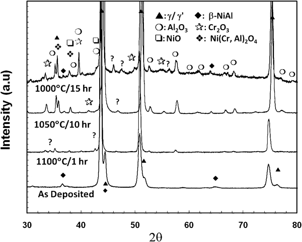
X-ray diffraction spectra for plasma sprayed NiCrAlY in following conditions: as deposited and vacuum furnace heat treatments at 1100°C for 1, 1050°C for 10 h and 1000°C for 15 h
Discussion
In this study, the transient oxide formation on APS NiCrAlY samples was evaluated after heat treating coating samples at various temperatures in vacuum. Before each heat treatment, the samples were polished to remove oxide scale which might have formed during the thermal spray process.19 This was done in order to examine the oxide formation without the influence of prior oxide formation. Upon reaching heat treatment temperature, base (Ni) and active elements (Al and Cr) in the NiCrAlY coating reacted with residual oxygen in the vacuum furnace to form their perspective oxides. As observed in this study, the pre-oxidation process resulted in the formation of Ni rich oxide (NiO or spinel) and alumina; Cr2O3, observed on the surface of the same NiCrAlY after heat treatment in air,19 was found on some of the vacuum heat treated samples.
In the isothermal oxidation process of MCrAlY coatings, the sequence and morphology of phase evolution in the transient stage (first 5–10 h) can have an impact on the stable oxide formation and long term coating system stability. Various types of oxides, such as NiO, Cr2O3 and spinels (NiCr2O4) 20 20,21 can form along with alumina in a NiCrAlY system. Spinels, though, are more often seen at a later stage of the oxidation process and form when either Al or Cr is being depleted causing NiO+Al2O3→NiAl2O4.22 For all six samples heat treated at 1000, 1050 and 1100°C, the EDS analysis only detected the presence of Ni rich oxide and alumina. The Ni rich oxide is likely to be NiO since it formed during the initial stage of the heat treatment when Al supply was abundant. Although both Al and Cr were present in the EDS analysis of the Ni rich oxide, the X-ray signals could have been emitted from the NiCrAlY coating and alumina formed under the Ni rich oxide. However, the presence of spinel (NiAl2O4) should not be ruled out without further analysis of the surface oxide with TEM.
The type of alumina formed (θ, γ or α-Al2O3) during the transient stage of oxidation has been reported to be dependent upon heat treatment temperature, time and alloy composition. Needle shaped θ-Al2O3 was found to form during air furnace heat treatment of NiCrAlY in the temperature range of 850–1100°C. 19 23 19,23,24 Other research on a high velocity oxy-fuel nano-NiCrAlY coating showed that after 24 h thermal exposure at 1000°C, α-Al2O3 was detected.25 Furthermore, on a magnetron sputtered Ni–30Cr–12Al–0·3Y coating, needle-like θ-Al2O3 was observed after 5 h at 900°C while complete α-Al2O3 (with wrinkled ridges) formed after 1 h oxidation heat treatment at 1100°C.26 When heat treatment was carried out at 1000°C, θ-Al2O3 initially formed and after 10 h of oxidation treatment α-Al2O3 began to form. Upon reaching 50 h α-Al2O3 became the dominant oxide with only traces of θ-Al2O3. In this study, under vacuum heat treatment conditions, no blade shaped γ-Al2O3 or needle shaped θ-Al2O3 was observed; instead, the alumina formed on the surface assumed cellular shape. X-ray diffraction analysis further revealed the presence of α-Al2O3. This observation suggests that the heat treatment in vacuum at a temperature range of 1000–1100°C encourages stable α-Al2O3 formation on NiCrAlY. Lower oxygen content reduces the growth rate of alumina and enhances the formation of a denser, cellular type of α-Al2O3.
Conclusion
In this study, air plasma sprayed NiCrAlY samples were subjected to vacuum heat treatment in the temperature range of 1000–1100°C with two different holding times at each temperature to examine the effect of temperature and time on the surface alumina formation. All samples exhibited the formation of two typical phases: a top layer of discrete or island-like Ni rich oxide and alumina in cellular shape. The cellular shaped alumina formed on the sample heat treated at 1100°C for 1 h was further analysed using XRD and identified to be α-Al2O3. The Ni rich oxide was confirmed to be NiO and likely to spinel as well. Increased Al surface content, in comparison to the as sprayed sample, was found on all heat treated samples. High Al surface content, corresponding to the extent of alumina formation, was observed for samples heat treated for a longer time as more oxygen diffusion through oxide layer took place. However, when the same heat treatment time was used, the Al content increased for a heat treatment temperature from 1000 to 1050°C and reduced when a temperature from 1050 to 1100°C was used. The increase in Al content with temperature is attributed to the faster oxygen diffusion and reaction with Al. The reduced Al content, hence reduced alumina formation, is possibly due to denser Al2O3 formation at 1100°C and the lower residual oxygen content in the vacuum furnace at a high heat treatment temperature.
Footnotes
Acknowledgements
The authors greatly thank NSERC for providing financial support for this research. A Research Assistantship for Philip Puetz was provided by the NSERC/CRD funding program.