
Abstract
Electrochemical method has been developed for the fabrication of alginic acid–haemoglobin and hyaluronic acid–haemoglobin films. The deposition yield was studied as a function of the haemoglobin concentration in the polymer solutions and deposition time. The results of deposition yield measurements and Fourier transform infrared spectroscopy showed the formation of composite films containing haemoglobin in alginic acid or hyaluronic acid matrix. The haemoglobin content in the composite films was varied by the variation of haemoglobin concentration in the solutions. Electron microscopy studies showed that the film thickness can be varied in the range of 0–50 μm by the variation of deposition time at a constant deposition voltage. The method enabled the formation of uniform films of controlled thickness. The films were deposited as monolayers or multilayers, containing individual alginic acid–haemoglobin and hyaluronic acid–haemoglobin layers. The proposed method can be used for the surface modification of biomedical implants and fabrication of thin films for biosensors.
Introduction
Alginate and hyaluronate are biocompatible, biodegradable and non-toxic polymers, which attracted significant interest for biomedical applications.1 Many investigations were focused on the deposition of alginate and hyaluronate films for the surface modification of biomedical implants and fabrication of biosensors.1 Alginic acid and hyaluronic acid are attractive building blocks for the fabrication of new biocompatible and biodegradable composite materials for tissue engineering.2 It is known that alginate solution can form gels of appreciable mechanical properties under the action of Ca2+ ions, which provide complexing of the carboxylate groups of alginate.3 It should be noted that hyaluronate is a natural biopolymer, which presents at high concentrations in skin, joints and cornea.4 The association of alginate with hyaluronate has been recently suggested to combine the gel forming and mechanical properties of the first polymer with the healing capacities of the second one.5 – 8 It was shown that alginic acid gel promotes hyaluronic acid retention in the composite materials.9
Alginate and hyaluronate provide mild conditions for the immobilisation of proteins, such as human haemoglobin.10 – 12 Haemoglobin is under investigation for the surface modification of biopolymer implants for the reduction of inflammatory response.13,14 It was shown that haemoglobin can modify the cellular interactions which occur at the tissue modified implant interface.13 Many investigations were focused on the fabrication of composite films, containing haemoglobin in alginate or hyaluronate matrix for application in biosensors.11,15 – 17
Significant interest has been generated in the development of new methods for the deposition of monolayer or multilayer composite films of alginate and hyaluronate16,18,19 containing haemoglobin and other functional materials. The interest in electrochemical methods stems from the high purity of the deposited materials and the possibility of deposition of uniform coatings on the substrates of complex shapes. Electrodeposition is a very simple, fast and low cost technique which can be used for the deposition of various materials and composites.20,21 In the previous investigations, electrochemical methods have been developed for the fabrication of alginic acid22 and hyaluronic acid4,23 films. These results pave the way for the deposition of composite films containing proteins, such as haemoglobin, in a polymer matrix.
The goal of this investigation was the development of electrochemical methods for the deposition of alginic acid–haemoglobin and hyaluronic acid–haemoglobin films. The films were obtained as monolayers or multilayers. The results presented below indicated that the film composition, microstructure and deposition yield can be controlled.
Experimental procedures
Sodium alginate, human haemoglobin, NaOH (Aldrich) and sodium hyaluronate (Alfa Aesar) were used as starting materials. Electrodeposition was performed from aqueous 0·5–2 g L−1 sodium alginate or 0·5–2 g L−1 sodium hyaluronate solutions containing 0·1–2 g L−1 haemoglobin. The pH of the solutions was adjusted to 8 by NaOH. The electrodeposition cell included a substrate centered between two Pt counterelectrodes. The distance between the substrate and counterelectrodes was 15 mm. The deposition voltage was 20 V and deposition time was varied in the range of 1–10 min. The films were obtained on Pt, 304 stainless steel and graphite substrates and dried in air for 48 h. Deposition yield was studied for the films deposited on stainless steel substrates. A minimum of three samples were prepared in each deposition experiment. All the deposits were obtained using fresh solutions. The deposition yield measurements were repeatable and the error was less than 5%.
The films were removed from Pt substrates for Fourier transform infrared spectroscopy (FTIR). FTIR investigations were performed on Bio-Rad FTS-40 instrument. The cross-sections (fractures) of the films deposited on graphite substrates were investigated using a JEOL JSM-7000F scanning electron microscope (SEM).
Results and discussion
The electrochemical method developed in this investigation for the deposition of composite films was based on the codeposition of haemoglobin and biopolymers, such as alginic acid and hyaluronic acid. Alginic acid (AlgCOOH) films can be deposited electrochemically from sodium alginate (AlgCOONa) solutions.22 The deposition involves the electrophoresis of anionic AlgCOO− macromolecules.22 The electrochemical decomposition of water in the anodic reaction


Chemical structures of a alginic acid and b hyaluronic acid
Electrodeposition of haemoglobin was investigated using aqueous 0–2 g L−1 haemoglobin solutions at pH 8. The haemoglobin molecules were negatively charged in such solutions. It was suggested that electrical field provided electrophoretic motion of anionic haemoglobin towards the anode and accumulation of the haemoglobin macromolecules at the anode surface. However, no deposition of haemoglobin films on the anodic substrates was observed from aqueous 0–2 g L−1 haemoglobin solutions.
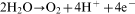
The codeposition of haemoglobin with alginate and hyaluronate resulted in the formation of composite films. Figure 2 shows the deposit mass as a function of the haemoglobin concentration in the 2 g L−1 sodium alginate solutions. The addition of haemoglobin to sodium alginate solutions at pH = 8 resulted in a significant increase in the anodic deposition rate. The deposition yield in the electrophoretic deposition process is usually described by Hamaker equation,26 which predicts a linear increase in the deposit mass M with increasing deposition time t and particle concentration C in the suspensions

Deposit mass as function of haemoglobin concentration in 2 g L−1 sodium alginate solutions at deposition voltage of 20 V and deposition time of 2 min
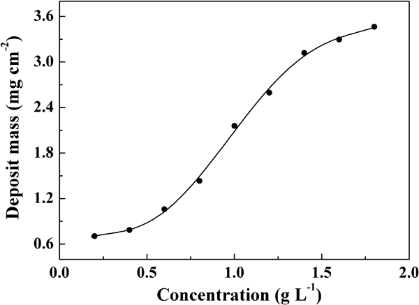
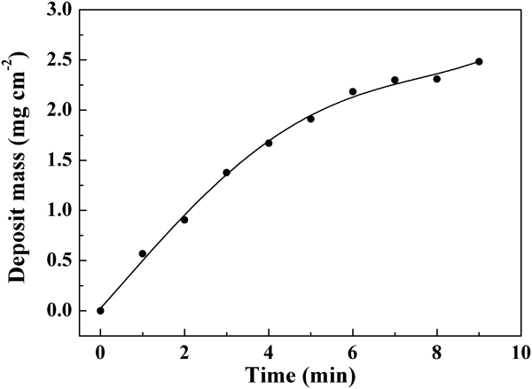
The Hamaker equation suggests that all the particles transported electrophoretically to the electrode surface are deposited. It should be noted that electrophoretic deposition requires the use of stable suspensions of charged particles or polymer macromolecules. However, the particles or polymer macromolecules, which are very stable in the bulk of the suspensions or solutions, must coagulate at the electrode surface to form a film. Recent studies27 highlighted the importance of pH variation and particle interactions at the electrode surface which promote deposit formation. The results of deposition yield measurements (Fig. 2) indicated that the amount of haemoglobin in the composite films can be varied. The deposit mass increased with increasing deposition time (Fig. 3), indicating the possibility of the formation of films of different thicknesses. However, the slope of the curve decreased with time owing to the decreased deposition rate. The decrease in the deposition rate with time can be attributed to the increasing resistance of the deposited layer.28
Deposit mass as function of deposition time at deposition voltage of 20 V for 0·6 g L−1 sodium alginate solution containing 0·4 g L−1 haemoglobin
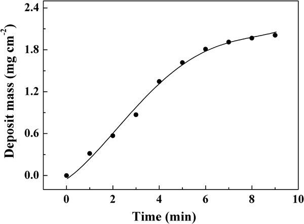
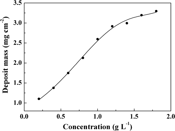
The addition of haemoglobin to the hyaluronate solutions resulted in an increasing deposition yield, as shown in Fig. 4. It is known that hyaluronate is negatively charged at pH>1·8.23 Therefore, haemoglobin and hyaluronate were negatively charged in the bulk solutions. The anodic codeposition of haemoglobin and hyaluronate can explain the significant increase in the deposition yield (Fig. 4) with increasing haemoglobin concentration in the hyaluronate solutions. The increase in the deposition yield can be attributed to the increase in the haemoglobin content in the composite films. The amount of the deposited material can be varied by the variation of the deposition time, as shown in Fig. 5.
Deposit mass as function of haemoglobin concentration in 2 g L−1 hyaluronate solutions at deposition voltage of 20 V and deposition time of 2 min
Deposit mass as function of deposition time at deposition voltage of 20 V for 0·6 g L−1 hyaluronate solutions containing 0·4 g L−1 haemoglobin
The deposits prepared by the anodic electrodeposition were studied by FTIR. Figure 6a–c compares the FTIR spectra of the alginic acid deposit, haemoglobin and composite alginate–haemoglobin deposit. The band at 1735 cm−1 in the spectrum of alginic acid (Fig. 6a) is related to the stretching of the protonated carboxylic groups of alginic acid.22 A similar band was observed in the spectrum of the composite haemoglobin–alginate film (Fig. 6c). The FTIR spectrum of haemoglobin (Fig. 6b) showed amide I adsorption at 1673 cm−1 and amide II adsorption at 1543 cm−1 in agreement with the literature data.29,30 The amide I (1670 cm−1) and amide II (1540 cm−1) absorptions of haemoglobin were observed in the FTIR spectrum of a composite film (Fig. 6c). Therefore, the FTIR data indicated the formation of composite haemoglobin–alginate films. The FTIR spectrum of the hyaluronic acid deposit showed a band at 1705 cm−1 attributed to the stretching of the protonated carboxylic groups. The peaks at 1659 and 1528 cm−1 can be attributed to the amide I and amide II groups of hyaluronic acid31 respectively. The FTIR spectrum of composite haemoglobin–hyaluronic acid deposit showed a broad band at 1720 cm−1 attributed to the stretching of the carboxylic groups of hyaluronic acid, enhanced amide I absorption at 1671 cm−1 and a broad peak around 1540 cm−1 attributed to amide II absorption. The shift of the amide I and amide II absorptions and broadening of the peaks was due to the overlap of the peaks related to the amide I and amide II bonds of haemoglobin and hyaluronic acid in the composite films.
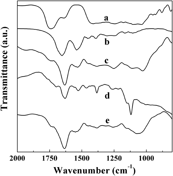
FTIR spectra for a alginic acid deposit prepared from 2 g L−1 sodium alginate solution, b as received haemoglobin, c alginic acid–haemoglobin deposit, prepared from 0·6 g L−1 sodium alginate solution containing 0·4 g L−1 haemoglobin, d hyaluronic acid deposit prepared from 2 g L−1 sodium hyaluronate solution and e hyaluronic acid–haemoglobin deposit prepared from 0·6 g L−1 sodium hyaluronate solution containing 0·4 g L−1 haemoglobin (deposition voltage 20 V)
Figure 7 shows SEM images of the cross-sections (fractures) of the films prepared from the alginate and hyaluronate solutions containing haemoglobin. The SEM images indicated the possibility of the fabrication of relatively thick films with thickness of 10–50 μm. The composite films can be obtained as monolayers or multilayers, containing alginate–haemoglobin and hyaluronate–haemoglobin layers, as shown in Fig. 8. The SEM images indicated the formation of relatively uniform layers (Fig. 8a). The thickness of the layers can be varied by the variation of the deposition time. The SEM images at a higher magnification (Fig. 8b) indicated that the individual layers were porous.
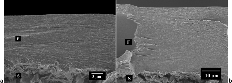
SEM images of a alginic acid–haemoglobin film prepared from 0·6 g L−1 sodium alginate solution containing 0·4 g L−1 haemoglobin and b hyaluronic acid–haemoglobin film prepared from 0·6 g L−1 sodium hyaluronate solution containing 0·4 g L−1 haemoglobin: deposition voltage 20 V; F = film; S = substrate
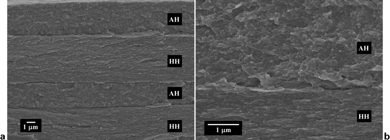
SEM images a and b at different magnifications of alginic acid–haemoglobin (AH)/hyaluronic acid–haemoglobin (HH) multilayer films: deposition voltage 20 V
As pointed out above, the combination of alginate and hyaluronate offers the possibility to combine the advanced properties of both materials, such as mechanical and gel forming properties of alginate with biocompatibility of hyaluronate and improve the hyaluronate retention in the composites. This investigation showed the feasibility of the fabrication of biopolymer composites, containing functional biomolecules by electrodeposition. However, there were no quantitative means for determining the relative composition of components. Compared to other film deposition methods,16 electrodeposition offers the advantage of high deposition rate and the possibility of film formation on the substrates of complex shape. The electrochemical method developed for the incorporation of haemoglobin into the biopolymer films can be used for the fabrication of composite coatings for implant materials with improved biocompatibility and thin films for application in biosensors. The haemoglobin can be used as a model protein for the development of electrochemical strategies for the incorporation of other proteins and enzymes with pH dependent charge into the biopolymer films. Further progress in electrochemical deposition is able to meet the needs for advanced methods for the industrial production of biosensors and other biomedical devices.
Conclusion
Electrodeposition method has been developed for the fabrication of composite alginic acid–haemoglobin and hyaluronic acid–haemoglobin films. The results of deposition yield measurements and FTIR data showed the formation of composite films, containing haemoglobin. Film thickness can be varied in the range of 0–50 μm by the variation of deposition time. The films can be obtained as monolayers or multilayers of controlled thickness. The method developed in this investigation can be used for the incorporation of other proteins and enzymes into the biopolymer matrix and the formation of composite films by electrodeposition. The proposed method can be used for the surface modification of biomedical implants and fabrication of thin films for biosensors.
Footnotes
Acknowledgements
The authors gratefully acknowledge the financial support of the Natural Sciences and Engineering Research Council of Canada.