
Abstract
This paper describes an amine free sol–gel method for silica coating of Au nanoparticles and shows an X-ray image of a colloid solution of the silica coated Au (Au–SiO2) particles. The Au nanoparticles that had a size of 16·9±1·2 nm were prepared through a conventional citrate reduction method. The silica coating was performed with a sol–gel reaction of tetraethylorthosilicate (TEOS) catalysed with NaOH in the presence of Au nanoparticles. The silica shell thickness was varied from 37 to 68 nm for TEOS concentrations of 1×10⁻³–20×10−3M at 4·3×10−5M of Au, 10·7M of H2O and 1·0×10−3M of NaOH. The optical properties of the Au–SiO2 particle colloid solution were related to the refractive index around the Au particles and the intensity of scattering from silica shells. The as prepared colloid solution could be concentrated up to an Au concentration of 4·3×10−2M with salting out. The concentrated colloid solution showed a high contrast X-ray image.
Keywords
Introduction
Recent development on imaging techniques using X-ray has enabled to simply examine the inside of living bodies.1 – 4 Contrast agents are usually used in imaging to obtain clear images. Since commercial X-ray contrast agents are solutions that dissolve chemicals with contrast properties at the molecular level, they flow fast in living bodies, or their residence time in living bodies is short. Therefore, it may be hard to take high contrast images for a long period using solution type commercial agents. The formation of nanoparticles of the contrast chemicals is a promising technique for increasing their residence time because viscous resistance of fluid acts on the particles.
Most commercial contrast agents, which are solutions of iodine compounds, are facing problems on their allergic reactions in patients. Coating of nanoparticles of contrast chemicals with inert shells is a candidate for the prevention of allergic reactions because the shell materials can keep the contrast agents from the living bodies. Coating can also prevent the nanoparticles from contacting other nanoparticles, so that aggregation of nanoparticles, which spoils blood flow, will be controlled.
Metallic Au nanoparticles are also expected to be used as contrast agents for imaging tissues in the living bodies at nanometre level5 – 12 because metallic Au is chemically stable and has high absorption property for X-ray. However, several kinds of metallic nanoparticles reveal toxicity in the living bodies,13 – 17 and nanoparticles tend to aggregate, which deteriorates properties characteristic of nanoparticles. Coating of the Au nanoparticles with silica that is chemically stable and inert for the living bodies may solve these problems.
Various methods for preparing silica coated metallic Au nanoparticles or Au–silica core–shell composite particles (Au–SiO2) have been reported.18 – 22 Most silica coating methods reported are based on a Stöber method using silicon alkoxide as silica source and amine as catalyst. Since amines are harmful to living bodies, methods without using amines are desired for the aim of using Au–SiO2 particles in living bodies. The present work proposes a method using NaOH instead of amine for preparing Au–SiO2 particles. X-ray imaging with the use of Au–SiO2 particles was also demonstrated.
Experimental
Chemicals
Hydrogen tetrachloroaurate (III) tetrahydrate (HAuCl4.4H2O, >99%) and trisodium citrate dihydrate (Na-cit, 99%) were used as a starting chemical for preparing Au nanoparticles and a reducing reagent respectively. Tetraethylorthosilicate (TEOS, 95%) and NaOH solution (5M) were used as a silica source for silica coating and a catalyst for hydrolysis of TEOS respectively. Ethanol (99·5%) was a solvent in silica coating. Sodium chloride (NaCl) (>99·5%) was used for concentrating particle colloid solution with salting out. All chemicals were purchased from Kanto Chemical and used as received. Water that was ion exchanged and distilled with Shimadzu SWAC-500 was used in all the preparations.
Preparation of materials
Preparation of Au nanoparticles
Au nanoparticles were prepared by reduction of gold salt with Na-cit.23 Freshly prepared 47 μL of 0·34M Na-cit in H2O was added to 9·95 mL of 4·83×10−4M HAuCl4 in H2O at a constant temperature of 80°C under vigorous stirring, which resulted in an Au concentration of 0·24×10−3M. The colour of the mixture turned wine red within a few minutes, which implied production of Au nanoparticles.24 An image of the Au nanoparticles taken with a transmission electron microscope (TEM) is shown in an inset in Fig. 1. Typically, spherical Au nanoparticles with an average diameter of 16·9±1·2 nm were observed, though occasionally some oblong particles were also present. Figure 1 shows the extinction spectrum of the Au colloid solution. A peak observed at 521·0 nm could be attributed to the surface plasmon resonance of Au nanoparticles, which confirmed that Au nanoparticles were produced.
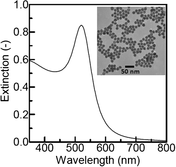
Ultraviolet–visible extinction spectrum of colloid solution of Au nanoparticles: inset shows TEM image of Au nanoparticles
Silica coating of Au nanoparticles
Silica coating of the Au nanoparticles, or fabrication of Au–SiO2 particles, was performed with a sol–gel method using TEOS and NaOH in the presence of Au nanoparticles. Tetraethylorthosilicate and the Au nanoparticle colloid solution were added to ethanol. After 15 min, NaOH aqueous solution was added to the Au–H2O–TEOS–ethanol solution. The reaction temperature and time were 35°C and 24 h respectively. The initial concentrations of Au, H2O, NaOH and TEOS were 4·3×10−5, 10·7, 1·0×10−3–2·0×10−3 and 0·5×10−3–20×10−3M respectively.
Characterisation
The obtained particles and colloid solutions were characterised by TEM, ultraviolet–visible spectroscopy and X-ray imaging using computed tomography. Transmission electron microscopy was performed with a JEOL JEM-2000FXII microscope operated at 200 kV. Samples for TEM were prepared by dropping and evaporating the particle suspensions onto a collodion coated copper grid. Volume average particle size was determined by counting more than 200 particles. Silica shell thickness was estimated as the difference between Au particle and composite particle sizes. Ultraviolet–visible extinction spectra were measured with a Shimadzu UV-3101PC spectrophotometer. X-ray images of the colloid solutions were taken with a Shimadzu SMX-100CT-SV3. Samples for X-ray imaging were prepared by concentrating the as prepared colloid solution using a salting-out technique. The salting-out was performed by adjusting the NaCl concentration in the colloid solution to 10 g L−1 with addition of saturated NaCl aqueous solution. After the salting out, the colloid solution was concentrated by removing the supernatant by decantation, adding the water or saline and shaking with a vortex mixer. The Au concentration was increased up to 4·3×10−2M with this concentrating process.
Results and discussion
Effect of NaOH concentration
In silica coating of metallic nanoparticles, silane coupling agents such as (3-aminopropyl) trimethoxysilane and 3-mercaptopropyltrimethoxysilane are often used for increasing the affinity between the metallic particle surface and the silica nuclei. Although no silane coupling agents were used in the present work, the Au nanoparticles were successfully silica coated. The citrates not only reduce the HAuCl4 but also colloidally stabilise the Au nanoparticles through adsorption of the citrates on the Au particle surface. Possibly, they also increased the affinity between the metallic Au particle surface and the silica nuclei. As a result, the successful silica coating was attained even with no silane coupling agents.
In the NaOH concentration range of 0·5×10−3–2×10⁻³M, clear colloid solutions with wine red colour were obtained. Over 2×10⁻³M, the solutions became unclear, and precipitates were produced. Figure 2 shows the TEM images of Au–SiO2 particles prepared at various NaOH concentrations, at which clear colloid solutions were obtained. High contrasts were observed between the core of the Au nanoparticles and the silica shell. At 0·5×10−3 and 1×10⁻³M, most particles contained a single Au core. For concentrations as high as 1·5×10−3 and 2×10⁻³M, many cores were composed of multiple Au nanoparticles. The increase in NaOH concentration leads to an increase in the ionic strength of the solution. Thus, the high NaOH concentrations used in this work should reduce the double layer repulsion between Au particles. At high NaOH concentrations, the Au cores probably aggregated before being coated with silica because of the increased ionic strength that would favour Au particle aggregation.
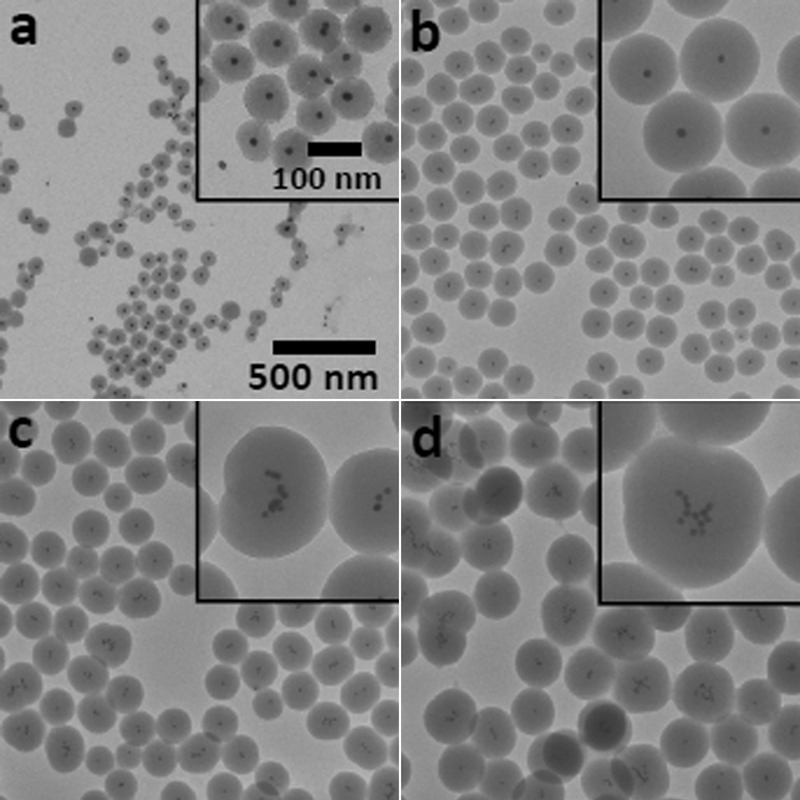
Images (TEM) of Au–SiO2 particles prepared at NaOH concentrations of a 0·5×10−3, b 1·0×10−3, c 1·5×10−3 and d 2·0×10−3M: all Au, H2O and TEOS concentrations were 4·3×10−5, 10·7 and 10×10−3M in series of experiments respectively
Silica shell thickness
Figure 3a–f shows TEM images of Au–SiO2 particles formed using various TEOS concentrations. At a TEOS concentration as low as 0·5×10−3M, aggregation of the Au nanoparticles took place, rather than formation of core–shell particles. This was probably related to a decrease in the ionic strength of the solution derived from the low TEOS concentration, which resulted in an increase in electrostatic repulsion between the Au particles and the silica nuclei. Consequently, the silica nuclei were not deposited onto the Au particle surface, and then the Au particles aggregated. In the range of 1×10⁻³–10×10−3M, quasi-perfect core–shell particles were produced, and the shell thickness increased with the increase in TEOS concentration. Figure 3g shows the shell thickness versus TEOS concentration. The shell thickness increased from 37 to 68 nm with the increase in TEOS concentration from 1×10⁻³ to 10×10−3M. For the concentration as high as 20×10−3M, the network structure of silica gel was observed between the Au–SiO2 particles, though the core–shell structure was formed. The ionic strength of the solution should be high at this TEOS concentration, compared to lower TEOS concentration, which reduces the double layer repulsion between silica nuclei. As a result, silica nuclei aggregated and formed a gel network structure. In conclusion for this section, the thickness was varied from 37 to 68 nm as the initial TEOS concentration increased from 1×10⁻³ to 10×10−3M. This means that the shell thickness can be controlled within a certain threshold.
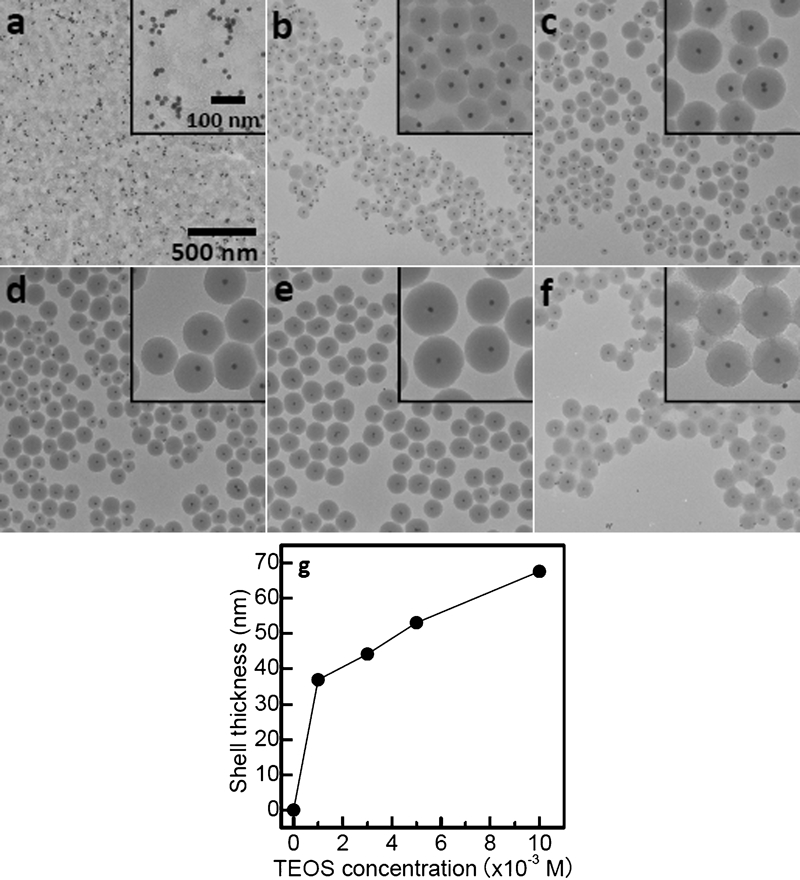
Images (TEM) of Au–SiO2 particles prepared at TEOS concentrations of a 0·5×10−3, b 1×10⁻³, c 3×10⁻³, d 5×10⁻³, e 10×10−3 and f 20×10−3M: all Au, H2O and NaOH concentrations were 4·3×10−5, 10·7 and 1·0×10−3M in series of experiments respectively; silica shell thickness as function of TEOS concentration is shown in g
Ultraviolet–visible spectroscopy
Figure 4 shows the extinction spectra of the colloid solutions of Au and Au–SiO2 particles with different silica shell thicknesses. Ethanol was added to the Au colloid solution to adjust the water concentration of the dispersion medium to 10·7M [1∶4 (v/v) water/ethanol], which was the same concentration as that in the silica coating. Surface plasmon absorption bands were observed at ∼525 nm for all the Au–SiO2 colloid solutions examined, and the plasmon band positions appeared to depend on TEOS concentration or shell thickness. The band position as a function of shell thickness is shown in an inset in Fig. 4. For the silica shell thickness of 36·9 nm, the plasmon band red shifted to 526·5 nm with respect to the uncoated Au nanoparticles (λmax = 522·0 nm). Above 36·9 nm, the plasmon band tended to blue shift back with an increase in the shell thickness. According to a study on the optical properties of core–shell particles,18 the red and blue shifts of surface plasmon absorption are due to a local increase in refractive index around the Au particles and due to an increase in intensity of scattering from silica shells with the increase in shell thickness respectively. A similar mechanism could be considered in the present work, which supported that the Au nanoparticles could be silica coated, and the silica shell thickness could be varied with the present method.
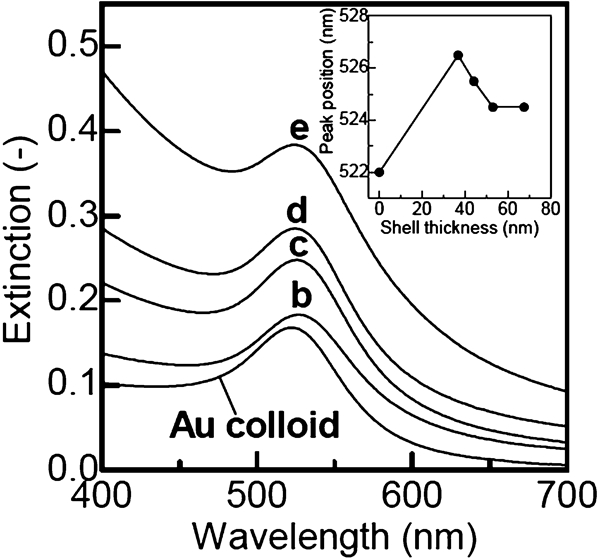
Ultraviolet–visible extinction spectra of Au colloid solution (shell free) and Au–SiO2 particle colloid solutions. Symbols b–e stand for symbols in Fig. 3. Inset shows surface plasmon peak position of Au–SiO2 particle colloid solution versus silica shell thickness. Peak position of shell free Au colloid solution is also shown as position at thickness of 0 nm
X-ray imaging
The Au–SiO2 particle colloid solution concentrated with salting-out, namely, the sample for X-ray imaging, was also colloidally stable. In a preliminary examination for X-ray imaging using the imaging set-up, saline could not be clearly imaged. In contrast, the concentrated colloid solution exhibited a high contrast image with a white image of the backyard, as shown in Fig. 5.
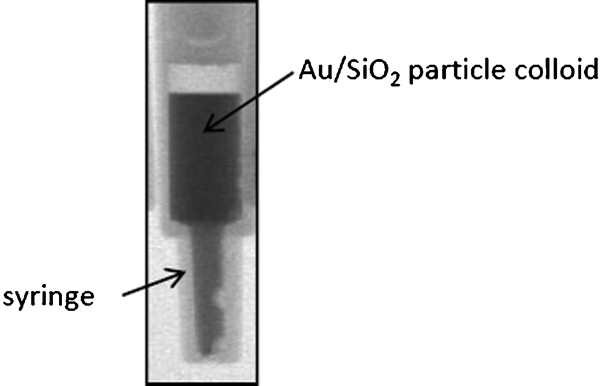
X-ray image of concentrated Au–SiO2 particle colloid solution: sample same as sample e in Fig. 3, which was concentrated for taking image
Conclusions
Amine free silica coating based on a sol–gel process was performed for Au nanoparticles. A quasi-perfect core–shell structure composed of Au core and silica shell was formed at 10·7M H2O and 1·0×10−3M NaOH in the presence of 4·3×10−5M Au nanoparticles with a size of 16·9±1·2 nm. Under these concentrations, the silica shell thickness increased from 37 to 68 nm with the increase in TEOS concentration from 1·0×10−3 to 20×10−3M. The red and blue shifts of surface plasmon absorption were qualitatively represented with a local increase in refractive index around the Au particles and an increase in intensity of scattering from silica shells with the increase in shell thickness respectively. The as prepared colloid solution of core–shell particles could be concentrated up to 4·3×10−2M Au with salting out. A high contrast X-ray image could be taken using the concentrated colloid solution. The results obtained in the present work indicated that the Au–SiO2 particle colloid solution would be used for studies on medical examination.
Footnotes
Acknowledgements
The authors express their thanks to Professor T. Noguchi of the College of Science, Ibaraki University, Japan, for his support in TEM observations.