
Abstract
The aim of this study is to design an artificial skin dress. A multilayer skin dressing included synthesised castor oil based polyurethane (PU) as the outer layer and two biopolymeric layers of heparin and chitosan as the inner layers were prepared. The surface of PU film was activated using two steps oxygen radio frequency plasma treatment. The surface of the modified PU films characterised by attenuated total reflection Fourier transform infrared spectroscopy, scanning electron microscopy and water drop contact angle measurements. Scanning electron microscopy observations confirmed the presence of grafted poly acrylic acid on the surface of PU films. Also, heparin and chitosan were immobilised on PU films. In vitro cell culture showed that the samples have an excellent biocompatibility with L929 fibroblast cells. Cell adhesion and proliferation of cells on the chitosan/heparin immobilised surfaces showed better behaviours compared with poly (acrylic acid) grafted PU film.
Introduction
Skin covers the entire external surface of the body. Fire, heat, chemicals, UV, electricity or diseases cause millions of dermal wounds every year. In the last decades, many efforts have been made to mimic the human skin and create skin substitutes.1 – 6 Regenerative medicine is one of the best approaches in this area with the potential of repairing the damaged tissues.
Among all the materials used in regenerative medicine, polyurethane (PU) is a synthetic material which is considered as the major class of applicable elastomers with good biocompatibility and high flexibility. These properties as well as excellent mechanical properties make it a suitable material for use in skin repair.7 Among different grades of PU, castor oil has a very low toxicity and also low cost, available as a renewable agriculture resource. Therefore, it can be prepared for use as a potential wound dressing material.8
Surface properties of the skin substitute are very important and have to satisfy the requirements of being a biomaterial. One of the promising approaches in this field is plasma surface modification. With this process, new functional groups are added to the membrane (PU thin film) surface and get cross-linked.9 Recently, various research groups have concentrated on modification of surface properties of PU membrane by plasma treatment and introduction of specific functional groups to the surface.10 On the other hand, there are some cases in recent researches focused on incorporating carboxylic groups in polymeric surfaces by graft copolymerisation of acrylic acid by immersing the polymer in a solution of poly-AAc.11
For the purpose of surface modification via plasma treatment, different biomolecules can be used to be grafted on surface. Heparin and chitosan, used in this work, are in a category which accelerates wound healing by painless wound surface protection.12 Heparin inhibits burn induced inflammation and restores blood flow in a shorter time. In addition, use of heparin and chitosan together had been reported to attenuate chitosan's protective effect.12 Therefore, immobilisation of heparin molecules is important in wound healing, which can be easily carried out by binding it to biodegradable chitosan carrier.13
Plasma graft copolymerisation techniques include plasma induced graft copolymerisation and simultaneous plasma treated graft copolymerisation. In the former, a polymeric substrate is treated by plasma before it undergoes subsequent graft copolymerisation in monomer solution. A mechanism of peroxide induction is proposed for this reaction. Whereas, in the latter, a polymeric substrate which preadsorbs a layer of reactive monomer, is treated by plasma. Therefore, the monomer with required functional groups is specifically introduced onto the surface of polymers.14, 15
In this work, the biocompatibility of PU was improved by grafting heparin and chitosan on its surface by two step plasma treatment. In brief, the castor oil based PU films were immersed in a solution of different percentages of poly-AAc, and then plasma treatment copolymerisation was carried out. It is worth mentioning that two step plasma treatment (TSPT) has some advantages related to conventional methods such as shorter polymerisation time and higher grafting degree. Meanwhile, castor oil has a very low toxicity available as a renewable agriculture resource that this source is an appropriate material for biomedical applications. Finally, the characterisation and properties of grafted films were evaluated by scanning electron microscopy (SEM), attenuated total reflection Fourier transform infrared spectroscopy (ATRFTIR), drop contact angle measurement, and in vitro cell behaviour investigation.
Materials and methods
Polyurethane synthesis
Castor oil (Sigma, 259853) as the soft segment and hexamethylene–diisocyanate (HDI-Merck, 822066) as the hard segment were used to synthesise PU with the ratio of 1∶1 to prevent the presence of free monomers and unreacted active groups.
The prepolymer with desired viscosity was prepared and a homogenous film was obtained by spin coating the prepared solution. Then, the films were placed in nitrogen atmosphere for 24 h to complete the polymerisation of films.
Two step plasma treatment
A custom made plasma apparatus was utilised for both plasma pretreatment and copolymerisation of silicone films. Details of this system were described in previous works.16 This system consists of a 34 MHz capacitively coupled radio frequency discharge.
First step
The PU films were placed on bottom of reaction chamber, and pretreated with 50 W of oxygen plasma for 30 s with the frequency of 34 MHz. Then the plasma pretreated films were immersed in aqueous monomer solution of poly-AAc (Merck, 800181) with the concentration of 30, 50, 70 and 90% for 10 min at room temperature, and finally dried at 40°C for 5 min.
Second step
In this step, the dried plasma pretreated films with a preadsorbed layer of reactive monomer on their surfaces were placed into the reaction chamber for plasma graft copolymerisation with the power of 60 W under the pressure of 30 mTorr for 3 min. The residual monomers and homopolymers were removed by soxhlet extraction in distilled water for 72 h.
All the samples were weighted after and before plasma treatment to obtain grafted weight and untreated weight respectively, with a microbalance, Scientech Zeta 210, to calculate grafting amount.
Chitosan and heparin grafting
Chitosan (Aldrich, 448877) solution in acetic acid (1M, pH 3·5) was prepared and homogenised by mechanical stirring at 4°C for 72 h. The 20 mM heparin (Caspian Tamin Co, Iran) solution in distilled water (pH = 4) was also prepared according to previous works.17 Different ratios of chitosan/heparin (0∶4, 1∶3, 1∶1, 3∶1 and 4∶0) were used for grafting the biopolymer films. The films were previously activated by immersing them in 10M water soluble carbohydrates for 30 min at 4°C, and then washed in 1mM hydrochloric acid. Treatment of activated films for 24 h with different solutions by various ratios of chitosan/heparin caused the grafting of biopolymers.
Scanning electron microscopy
The morphologies of all samples were studied using a Philips (XL30) scanning electron microscope. All the samples were coated by gold to improve the resolution.
Spectroscopy of ATRFTIR
A Brucker (equinox55) ATRFTIR spectrophotometer was used to observe the formation of graft copolymerisation on the surface of films.
Contact angle measurement
The static contact angles of all films were measured with the Kruss G10 contact angle measurement equipment. Five points of surface were investigated with sessile drop method.
In vitro cell behaviour investigation
L929 fibroblast cell line was cultured in DMEM (BioSeram, LM-D1111/500) containing 5% fetal bovine serum (Sigma, S1810-100) and 1% antibiotic penicillin/streptomycin (GIBCO, 15140).
The prepared PU films were cut with the dimensions of 1×1 cm2 and sterilised by physiologic serum for 24 h. Then 5×10⁴ cells were seeded on each film and incubated in a humidified atmosphere with 5%CO2 and the temperature of 37°C. Cell viability was investigated after 12 and 72 h. Cell attachment was evaluated after 12 h culture of cells in humidified atmosphere. Spreading of cells after 72 h was analysed by Motic Images Plus 2·0 software.
Results and discussion
Graft copolymerisation
The amount of poly-AAc grafted (μg cm−2) on surfaces was calculated according to the following equation
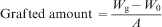
The results showed increase in grafted amount on PU surfaces with increasing concentration of acrylic acid monomers (Fig. 1).
Change in graft amount on surface of PU films as function of poly-AAc
In conventional plasma methods, activation of the surface of the film increases the grafting amount. As all the initiator peroxide groups were used in graft copolymerisation, the reaction takes place between monomers and viscosity is increased by formation of a homopolymer. While in TSPT, the sample is first placed in monomer solution and then, plasma polymerisation occurs. Therefore, lower amount of graft polymerisation is caused by lower inactive monomers on the surface of the film because of the hydrophobic nature of it. In TSPT method, polymerisation is carried out in plasma environment, therefore, not only free radicals are available during the process, but also, new free radicals are formed.
The thickness of the chitosan/heparin grafted PU films may be in the range of micrometre, which depends on the parameters of the plasma (gas, power, pressure, treatment time, flowrate, etc.). The surface properties of the mentioned films are measured only by the composition of the grafted layers.18
Spectroscopy of ATRFTIR
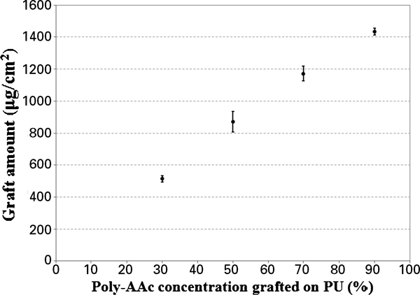
According to Fig. 2, the presence of grafted polymer in the surface of PU is confirmed, which PU IR spectra before and after oxygen plasma treatment is compared with modified samples. A wide band at 3337 cm−1 in plasma treated sample in comparison with non-modified PU film shows more formation of hydroxide groups after treatment. Appearance of a yoke-shape band in the range of 1741 and 1698 cm−1 in non-modified PU film is related to ester group in urethane polymeric chain bonding. This peak intensifies in modified samples around 1698 cm−1 because of the presence of carboxylic groups in poly-AAc.
Spectra (ATRFTIR) of modified and non-modified PU films with different concentrations of poly-AAc: a 0%; b 30%; c 50%; d 70%; e 90%
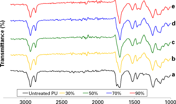
Figure 3 shows the result of ATRFTIR after grafting chitosan and heparin. The medium and wide bands around 3361 cm−1 are related to chitosan and represent N–H groups. The band at 1741 cm−1 indicates carbonyl group (C = O). The amino absorption (1580–1600 cm−1) became weak and the band appeared at 1233 cm−1. This phenomenon showed that chitosan immobilised on the PU film surface forms a complex with heparin by electrostatic interaction.19
Spectrum (ATRFTIR) of chitosan (Ch) and heparin (H) grafted PU films with different Ch/H ratios: a pure PU; b 1∶3; c 1∶1; d 3∶1
Contact angle measurement
The surface of PU film is almost smooth which becomes rough by grafting poly-AAc, because of the fact that increasing the amount of grafted polymer leads to a rougher surface. The result of contact angle measurement is shown in Fig. 4.
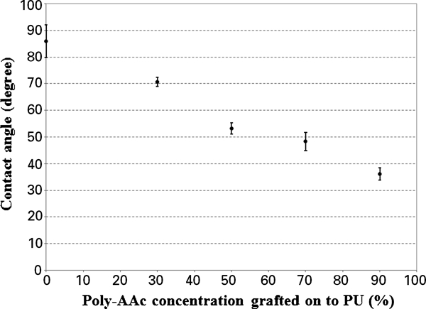
Contact angle of samples as function of poly-AAc concentration
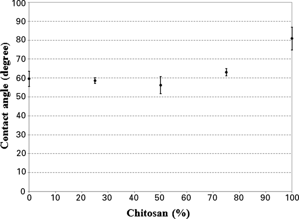
The values of contact angle as the amount of grafted chitosan and heparin are shown in Fig. 5. As it can be seen in this figure, this parameter did not significantly decrease with the addition of chitosan grafting. Since chitosan is a hydrophobic polymer, thus, with increasing amount of chitosan grafting (>50%), the contact angle increased.
Contact angle of samples as function of heparin and chitosan percentage
Peroxide groups or active sites are formed by plasma pretreatment and consequently, the value of contact angle decreases with increasing hydrophilic properties. Increase in hydrophilic properties is accompanied with increase in more inactive acrylic acid monomers on the surface of the films and as a result, higher grafting amount was obtained.
Scanning electron micrographs
In agreement with contact angle results, the SEM imaging shows an almost smooth surface in PU film and rougher surface in poly-AAc grafted ones (Fig. 6a and b respectively). In Fig. 6c, the presence of chitosan and heparin is obvious on the surface. The ratio of chitosan/heparin affects the surface charge of chitosan–heparin–polyelectrolyte mixture. In the ratio of 1∶1, the amount of NH3+ in chitosan equals to amount of COO− and SO3− in heparin. This electrostatic interaction makes the mixture of chitosan/heparin more compact and more homogenous.20
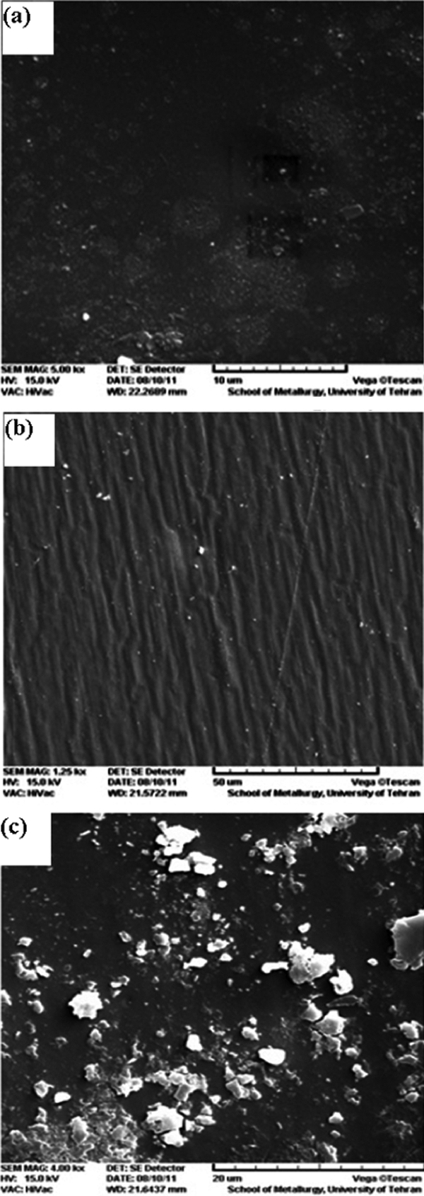
Images (SEM) of a pure PU, b 90% poly-AAc grafted PU and c Ch/H grafted PU with ratio of 1∶1
In vitro cell culture
Spreading of cells in heparin grafted PU is significantly higher than chitosan grafted ones (Fig. 7). This phenomenon is remarkably lower in poly-AAc grafted samples.21 Chitosan acts as a positive charge centre against the cell membrane with negative charge and makes the cells become round.22
Cell spreading is low in all samples compared with control
Cell morphology and cell attachment was investigated by confocal microscopy imaging. Contact angle value, active sites on the surface, appropriate charge, hydrophilic and hydrophobic properties are the most important factors in cell attachment.23 In chitosan grafted films the lower contact angles resulted in less hydrophilic property. On the other hand, chitosan as a positive charge centre decreases cell attachment and spreading. Better cell attachment is observed in heparin grafted samples with a lower contact angle and more hydrophilic characteristics.
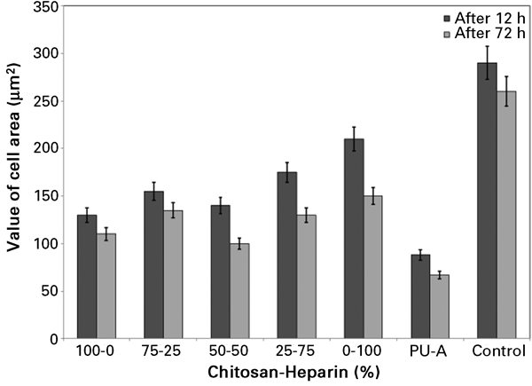
Cell density after 12 and 72 h of culture was evaluated (Fig. 8). The maximum density is observed in a chitosan/heparin (Ch/H) ratio of 1∶1. Poly-AAc grafted PU films contain carboxylic acid groups which decrease cell viability and attachment. Fibronectin and vitronectin of serum can react with heparin in all the samples containing heparin. This reaction improves and stimulates cell attachment on the surface.24
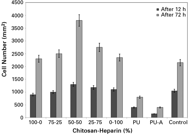
Cell density shows maximum in sample with equal amount of chitosan and heparin
Cell proliferation was assessed by comparing 12 h cell culturing with 72 h one. Grafted biopolymers show more proliferation, whereas in poly-AAc grafted PU films, cell density after 72 h is not as much as the biopolymer ones. The maximum cell density after 72 h was obtained in Ch/H ratio of 1∶1 which is in agreement with the other researches.25
Chitosan can react with integrin or other cell surface receptors and make them to form clusters. This clustering creates a DNA stimulating signal to enter the synthesis phase in cell growth cycle.
In this work, TSPT was used for PU surface modification and immobilisation of chitosan and heparin on the surface. This technique introduced some advantages compared with conventional plasma method as shorter time of polymerisation, more homogenous modified surface, higher grafting amount on the surface, and no need to control pH during the process or oxygen content before the process.
Conclusion
A TSPT was successfully employed to simultaneously graft copolymerised chitosan and heparin poly-AAc mixture onto the PU film. In this method, pretreatment with plasma and activation of PU film surface resulted in decreasing the contact angle (from 86 to 36°) and increasing the surface tension which led to better grafting of polymers. According to ATRFTIR, the presence of chitosan and heparin on the surface of the modified PU film was confirmed in comparison with non-modified PU ones.
Scanning electron cross-sectional micrographs showed a uniform grafting layer on the surface of the modified PU films. Since chitosan is a hydrophobic polymer, thus, with increasing amount of chitosan grafting (>50%), the contact angle increased. Appropriate biocompatibility was observed in modified samples; however, chitosan grafted films’ cell spreading decreased in some extent. The maximum cell density was obtained when the equal amount of chitosan and heparin was grafted on the surface.