
Abstract
Here we report for the first time to the best of our knowledge the processing techniques, nucleation kinetics and the nanoindentation behaviour of a 1·5 μm magnesium hydroxide thin film chemically deposited on a commercially available soda lime silica glass substrate at room temperature. The phase and microstructure of the films were analysed by X-ray diffraction, scanning electron microscopy, field emission scanning electron microscopy as well as transmission electron microscopy. An exponential nucleation kinetics was identified for the growth of the thin films. The nanomechanical properties, e.g. nanohardness and Young's modulus of the films were measured by the nanoindentation technique at ultralow loads of 50, 70 and 100 μN. Finally, the nature of deformation of the thin film was analysed in terms of the energetics of the nanoindentation process and the microstructure.
Introduction
Nowadays, thin films are considered as a highly promising material for biomedical, energy, optical, sensing related applications1 – 4 as well as for corrosion protection of magnesium alloys.5 – 8 In this regard, magnesium oxide is a very important protective material that can be in situ formed from the magnesium hydroxide [Mg(OH)2] thin film9 precursors. The most recently emerging application of magnesium hydroxide is as a buffer layer for the copper indium gallium selenium10 and dye sensitised solar cells.11 Most recently reported methods for preparation of magnesium hydroxide include hydrothermal synthesis,12 precipitation13 and surfactant mediated solution.14 The hydrothermal method12 requires autoclaving under stringent temperature and pressure control thereby making it comparatively more energy and cost intensive. The wet precipitation method,13 although simple to handle, requires the use of dispersant and does not provide the formation of a coating on a given substrate as it precipitates only the nanoparticles of Mg(OH)2. Similarly, the method of surfactant mediation14 requires stringent control of interface chemistry and yet provides only ordered array of Mg(OH)2 nanoparticles and not a coating on a given substrate.
However, the chemical deposition process10 has many distinct advantages over the aforementioned processes. First of all, it can be used for deposition over a relatively larger area. Second, the process can be fully automated by relatively simpler means. Third, the process involves simple chemical principles that can be handled with relative ease. In fact, the process can even be exploited for fabrication of low cost solar cells.10 Another great advantage of this process is that it is flexible. Thus, if needed, changes can be incorporated in the chemical bath composition without much hazard. Finally, a huge capital cost is usually not involved in the chemical deposition process. By controlling the pH of the corresponding solutions and the dipping time as well as the number of dipping, the microstructure, thickness and hence the physical/chemical properties of the thin films can be easily fine tuned. Furthermore, for solar cell applications, the films of the buffer layer will need to have a thickness, e.g. ⩽1–2 μm similar to those of the CIGS thin films themselves.10 This can be more easily achieved by chemical deposition in a manner that is less cost intensive. The thickness compatibility is required to ensure lesser recombination at the interface so that a beneficial effect occurs in terms of the efficiency of the solar cells.10, 11
On the other hand, the nanoindentation technique has recently15 – 17 emerged as a very important technique for characterisation of nanomechanical properties of thin films and coatings. In spite of the wealth of literature,9 – 14 there has not been any systematic study of the nucleation kinetics as well as the nanomechanical properties of chemically deposited magnesium hydroxide thin films. Therefore, the objective of the present work was to evaluate the nucleation kinetics and the nanomechanical properties of chemically deposited Mg(OH)2 thin films on soda lime silica (SLS) glass substrates.
Materials and methods
Magnesium hydroxide thin films were deposited onto precleaned and dried commercially available SLS glass slides (25×25×1·15 mm) by the conventional chemical deposition method from 0·1M aqueous solutions of magnesium nitrate, Mg(NO3)2 (reagent grade; Ranbaxy, Mumbai, India) and sodium hydroxide, NaOH (reagent grade; Qualigens Fine Chemicals, Kolkata, India) using the following chemical reaction at room temperature

The film thickness was measured by a profilometer (Form Talysurf 120; Taylor Hobson, UK). The phase purity was evaluated by X-ray diffraction (XRD; monochromatic Cu Kα 1 radiation, 35 mA, 40 kV; X'pert Pro MPD diffractometer; PANalytical, The Netherlands). Surface morphology of the coating was characterised by scanning electron microscopy (SEM; s430i; Leo, UK), field emission scanning electron microscopy (FE-SEM; Supra VP35; Carl Zeiss, Germany) and transmission electron microscopy (TEM; Tecnai G2 30, S-Twin, 300 kV; FEI, The Netherlands; LaB6 filament, line resolution 0·14 nm and point resolution 0·2 nm). To avoid charging, a 50–70 nm carbon coating was deposited on the Mg(OH)2 thin film by the arc deposition technique before insertion in the sample chamber for SEM. Before TEM observation, the sample was deposited onto Cu grids, supported with a carbon film. The samples were deposited onto the support grids by deposition from a dilute suspension in acetone.
The nanoindentation experiments18 – 20 were conducted with a commercial nanoindenter (TriboIndenter Ubi 700; Hysitron Inc., Minneapolis, USA). The details of the experimental procedures have been given elsewhere18 and hence, will be only briefly mentioned here. The nanohardness and Young's modulus data of the present Mg(OH)2 thin films were calculated from the load P versus depth of penetration h data of the aforesaid nanoindentation experiments by using Oliver and Pharr19 method. The machine gave the reduced Young's moduli data of the Mg(OH)2 thin films. These data were used to calculate the Young's moduli data of the present Mg(OH)2 thin films following the method given in Ref. 19 assuming a Poisson's ratio value of 0·25, typical of ceramics19 because no experimental data on Possion's ratio of Mg(OH)2 thin films were available in literature.
The machine had a load resolution of 1 nN along z axis while the depth resolution was 0·04 nm along z axis. Furthermore, the thermal drift was maintained at <0·05 nm s−1. All the nanoindentation experiments whose results are reported in the present work were conducted at room temperature (∼30°C) and ∼70% relative humidity. The machine provided a P–h plot in nanoindentation mode using a tetrahedral diamond Berkovich tip of ∼150 nm tip radius and semi-apex angle ∼65·3°. The diamond Berkovich tip had a Poisson's ratio of 0·07 and a Young's modulus of 1140 GPa.20 In all the experiments, a constant loading rate of 10 μN s−1 was used. The unloading rate was the same as the loading rate. The dwell time at peak load was 0 s. As soon as the peak load was reached, the sample was unloaded at the given unloading rate instantly and the loading and unloading P–h data were automatically recorded by the machine.
At least 16 nanoindents (i.e. 4×4 array) were made at each of the five randomly chosen different locations of the sample. Thus, each reported value of nanohardness was an average of at least 80 or more individual data points. In addition, the unloading part of the experimental P–h plot was analysed according to the Oliver–Pharr model19 following the equation

For conducting the nanoindentation experiments, the Mg(OH)2 thin films were mounted on a motorised table that allowed for a movement in the plane normal to the axial motion of the tip. The transducer that measured both load and depth consisted of a three-plate capacitor. It had the same tetrahedral diamond Berkovich tip of ∼150 nm tip radius as mentioned above, attached to its central plate. The instrument was calibrated before each and every experiment by performing indents of increasing depth in a standard fused quartz sample provided by the supplier of the machine. The standard fused quartz sample had a known18 nanohardness of 9·25±0·93 GPa and a reduced Young's modulus Er of 69·6±3·48 GPa. In the present experiments, three different peak loads, e.g. 50, 70 and 100 μN were applied.
Results and discussion
The thickness of the Mg(OH)2 film was measured as ∼1·5 μm. The phase purity of the synthesised thin films was confirmed by the typical XRD data (Fig. 1a; JCPDS file no: 7–239). The crystallite sizes estimated by the Scherer's method21, 22 were 1·75, 4·77, 3·70, 9·34, 5·99, 2·89, 5·57 and 4·26 nm corresponding to the broadened peaks (001), (101), (102), (110), (111), (103), (201) and (202) respectively. Corresponding to the strongest peak (101), the lattice parameters were calculated as a = b = 3·1449 Å and c = 4·78500 Å which matched well with reported data.21
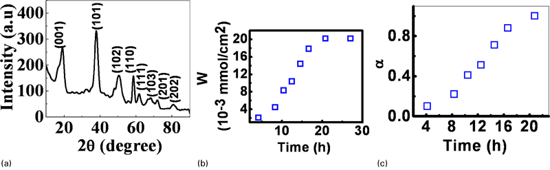
a XRD spectra of Mg(OH)2 thin film, b amount of Mg2+ ions and c α as function of time for deposition of Mg(OH)2 thin film
Figure 1b shows that the amount of Mg+2 ions W (mmol cm−2) increased slowly with time up to about 10 h and abruptly after ∼10·45 h before becoming nearly constant beyond ∼20·83 h; whereby with a sluggish growth rate, the final dried film thickness of 1·5 μm is reached after ∼27·08 h. This result suggested that the equilibrium between film formation and dissolution had happened during the present chemical deposition process.
Figure 1c shows the data on the extent of reduced reaction α for Mg+2 ions versus time where α (Ref. 23) represents the fraction of Mg+2 ions that can be obtained after a given time t divided by the equilibrium amount of Mg+2 ions in the film. The value of α varies from 0 at the beginning of the reaction to 1 at equilibrium reached after ∼21 h (Fig. 1b). The data are best fitted by the expression: α = 0·0105t1·5368 with a correlation coefficient R2 of ∼0·98. Following Ref. 23, simple algebraic manipulation leads to the nucleation kinetics equation: dα/dt≈0·38α0·4 that approximately resembles the Maple equation for exponential nucleation.
The cross-sectional SEM (Fig. 2a) confirmed that film had a relatively dense microstructure although there was apparently some residual porosity. The FE-SEM photomicrograph gave evidence that the film had a layered platelet like architecture comprising of the Mg(OH)2 nanocrystallites (Fig. 2b). The nanocrystallites appeared from the FE-SEM photomicrograph to have formed the platelets (Fig. 2c) in a given layer. Further, such layers appeared from the FE-SEM photomicrograph to be stacked upon one another with a typical inter layer distance of about 10-30 nm (Fig. 2d).
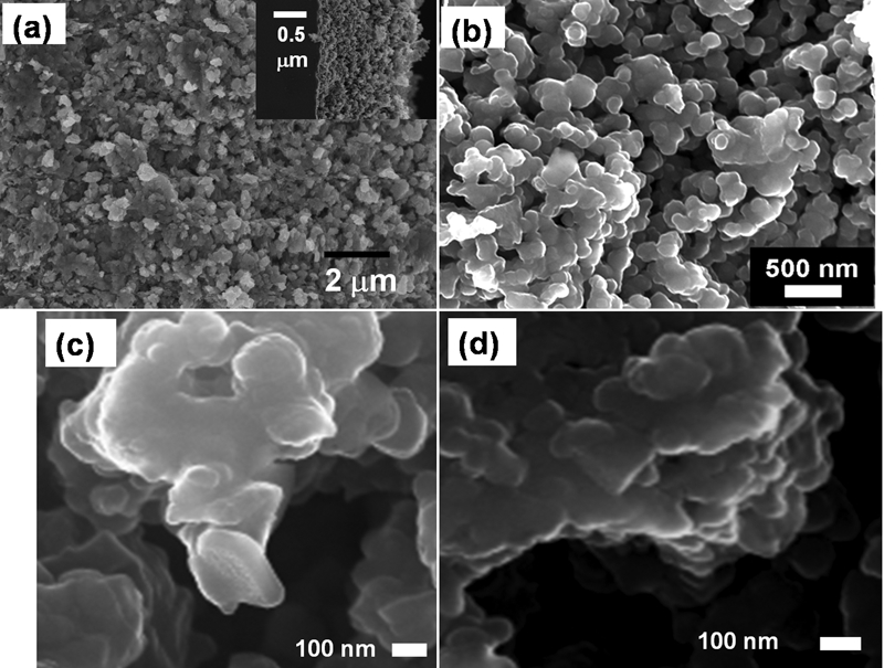
Microstructure of Mg(OH)2 thin film: a cross-sectional SEM showing realatively dense microstructure (inset shows film on glass substrate); FE-SEM images showing b a layered platelet type architecture, c nanocrystallites forming platelets and d stacking of layers
The TEM photograph confirmed that the platelets had a hexagonal shape (Fig. 3a). Their size had a Gaussian distribution with an average of about 108·04±23·53 nm (Fig. 3b) and a selected area electron diffraction pattern typical of thin films (inset, Fig. 3a). The average size of the platelets as obtained from the Gaussian distribution tallies with the typical size (107·17 nm) of a single platelet (Fig. 3c). The platelet composed of a large number of about 1–10 nm size nanocrystallites. The average size of such a range matched favourably with the average (4·79±2·30 nm) of those estimated from the XRD data (Fig. 1a). It had a thickness of ∼15·6 nm. In terms of order of magnitude, these data also tally with the average of the typical inter layer distance of about 10–30 nm as observed from FE-SEM image (Fig. 2d).
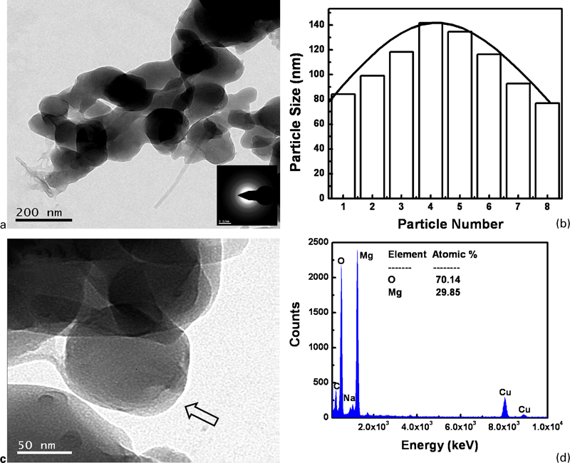
Images (TEM) of Mg(OH)2 thin film showing a platelet structure of nanocrytallites that formed layered structure (inset: typical selected area electron diffraction pattern), b Gaussian size distribution of platelets, c single platelet and d energy dispersive X-ray data of film
This picture also matches with the fact that the crystal structure of Mg(OH)2 is a layered CdI2 type arrangement. It comprises of successive hexagonal Mg2+ ion layers and OH− ion layers stacked one upon each other. The sixfold coordination of the magnesium cation by hydroxyl groups forms a
 octahedron.24,
25
octahedron.24,
25
Typical energy dispersive X-ray data showed ∼29·85at-%Mg and ∼70·14 at-%O (Fig. 3d), suggesting that the present films were somewhat rich in oxygen content and lean in magnesium content compared with those of an ideal defect free material.26 The peak for carbon came from the carbon coating and that of copper from the copper grid used to hold the sample.
For nanoindentation loads P of 50, 70 and 100 μN, the maximum depths of penetrations were ∼90, 100 and 140 nm which were well below 10% of film thickness (1·5 μm) (Fig. 4a). The average elastic recovery19 was ∼70%.
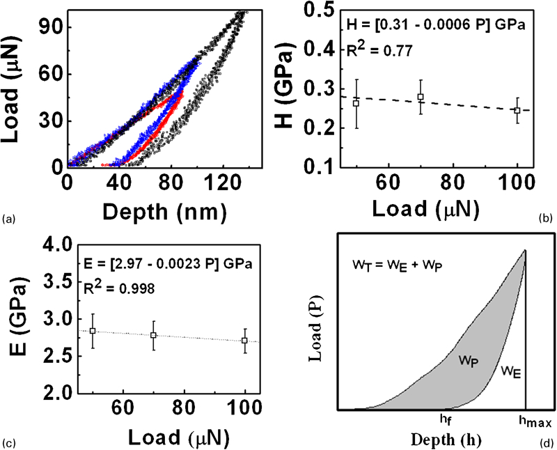
Nanoindentation data of Mg(OH)2 thin film a typical load depth plots, b nanohardness, c Young's moduli and d schematic of elastic and plastic energy spent in nanoindentation experiments
The nanohardness and Young's moduli of the Mg(OH)2 thin films were 0·26±0·06, 0·28±0·04 and 0·24±0·03 GPa (Fig. 4b) and 2·83±0·23, 2·77±0·19 and 2·70±0·16 GPa (Fig. 4c) respectively at loads of 50, 70 and 100 μN. The best fit equations given as insets of Fig. 4a and b respectively, predicted nanohardness of 0·31 GPa and Young's modulus of 2·97 GPa.
To the best of our knowledge, this is the first experimental data on nanohardness and Young's modulus of chemically deposited Mg(OH)2 thin films on SLS glass substrates. There is unfortunately no other published data on the nanomechanical properties of magenesium hydroxide either in thin film or in bulk form either. So, a direct comparison of the data from the present work with those from literature was not possible. However, from the reported data for exfoliated CoAl layered double hydroxide/polyamide 6 nanocomposites by in situ polymerisation,27 the nanohardness and Young's modulus of a CoAl layered double hydroxide were evaluated by the rule of mixtures to be ∼0·34 and 7·73 GPa which compared favourably with the present data of 0·31 and 3 GPa for nanohardness and Young's modulus respectively. There was no particular reason or bias for choosing this particular material's data except that to the best of our knowledge it was the only one data on nanomechanical properties of layered double hydroxide available in literature.
The amounts of energy spent in plastic, i.e. irrecoverable deformation, elastic, i.e. recoverable deformation and their sum total during the nanoindentation experiments are termed19 as WP, WE and WT respectively. This aspect is schematically indicated in Fig. 4d. The grey shaded part in Fig. 4d represents the energy spent in plastic deformation, i.e. WP. The white shaded part in Fig. 4d represents the energy spent in plastic deformation, i.e. WE. The total area under the load–displacement plot represents the total amount of energy spent in deformation, i.e. WT = WE+WP. In the present Mg(OH)2 thin films chemically deposited on SLS glass substrates, depending on the peak load, the ratio WP/WT varied in the range from ∼0·57 to 0·43 while the ratio WE/WT varied in a complimentary fashion in the range from ∼0·43 to 0·57 (Fig. 5), assuming that WT = WE+WP.19
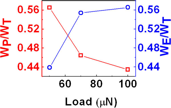
Energy ratio as function of load applied in nanoindentation experiments on Mg(OH)2 thin films
The present data (Fig. 5) matched well with reported experimental data28 on other materials. The decreasing trend of WP/WT (Fig. 5) with the increase in load happened because the ratio hf/hmax (Ref. 19) decreased from ∼31% at 50 μN load to ∼28% at 100 μN load (Fig. 4a). Here, hf has been already defined and hmax refers to the maximum depth of penetration for a given peak load attained during the nanoindentation experiments. These data implies that at 50 μN load, ∼69% of the maximum penetration depth was elastically recovered while at 100 μN load ∼72% of the same was elastically recovered. Thus, the increasing trend of WE/WT with the increase in nanoindentation load (Fig. 5) possibly reflected the fact that the current Mg(OH)2 thin films had a microstructure that was inherently capable of more elastic recovery the more it is deformed. It is suggested that the layered microstructure of the chemically deposited magnesium hydroxide thin films helped to accommodate the strain due to the applied nanoindentation load and thereby affect the elastic recovery through localised rearrangements on withdrawal of the nanoindenter during the unloading cycle.
Fitting the data from unloading part of the nanoindentation load versus depth plots (Fig. 4a) to the equation (1) gave ‘α’of about 0·017–0·024 (Fig. 6) which matched with experimental data reported for other materials19 and ‘m’ of about 1·79–1·92 with an average of 1·86±0·06 which suggested the conical punch geometry as the best approximation for the present nanoindenter following Sneddon's analysis.29
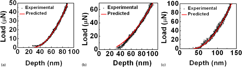
Fitting of unloading data from Fig. 4a to equation (1) at different peak loads (hollow symbols: experimental data; solid lines: predicted trend):
Finally, it should be mentioned that depending on load, the nanohardness of the Mg(OH)2 thin films was about 0·24–0·28 GPa. It is evident from Figs. 2 and 3 that the present film was not fully dense and it appeared to have some amount of residual porosity as mentioned earlier although an exact estimation of the same was beyond the scope of the present work. The presence of porosity in the film can degrade the nanohardness as it has been reported30 that the same of microplasma sprayed hydroxyapatite coatings could be much lower than those of their dense, bulk counterparts due to presence of about 10–20 vol.-% open porosity. A similar statement is true for the Young's moduli data of the present Mg(OH)2 thin films chemically deposited on SLS glass substrates because it has been demonstrated31 already that the presence of larger volume per cent porosity decreases the load bearing contact area and thereby the Young's modulus.
Conclusion
A simple, inexpensive chemical deposition technique produced a 1·5 μm nanostructred pure magnesium hydroxide film with an exponential nucleation kinetics on SLS glass substrates. By virtue of a strain tolerant layered microstructure the films showed nanohardness ∼0·3 GPa and Young's modulus ∼3 GPa. Thus, the nanostructured pure magnesium hydroxide film may be useful for protective as well as functional applications.
Footnotes
Acknowledgements
The kind permission of Director, CGCRI to publish this paper, the infrastructural supports received from the XRD and electron microscopy sections as well as the financial support of CSIR (project no. NWP 0027) are gratefully acknowledged.