
Abstract
The recovery of a metal workshop dated to the sixth century AD, superimposed to the Hadrian's Athenaeum in Piazza Madonna di Loreto, Rome, provided the opportunity to identify a series of metallurgical processes linked to the two chaînes opératoires related to copper, bronze, silver and lead productions. Some analysed fragments presented superficial treatment identified as silvering, the focus of this paper. The main aim is to investigate the signs of the surface treatment on the artefacts and discuss what methods would be best employed to analyse and interpret them in order to identify the process to which they relate. Different analytical techniques were used to obtain information on the superficial layer, and their pros and cons are presented so as to discuss how analytical limits can affect the interpretation of a process and the identification of an object. The conclusions reached here are necessarily limited because of the scarce number of samples analysed and the techniques used.
Introduction
A commercial excavation in Piazza Madonna di Loreto, Rome, uncovered the Adrian Athenaeum. The Athenaeum itself, almost completely obliterated by the latter uses of the space, was the extension of the Trajan Forum commissioned by the emperor Hadrian. Its initial use as a school room and its rich marble decorations were followed, during the sixth century, by a transformation of the space into a metallurgical workshop, where part of the materials present in the Athenaeum was reused for production purposes. Several furnaces with different shapes and different functions were set in place (Figs. 1 and 2) between the former rows of marble benches.
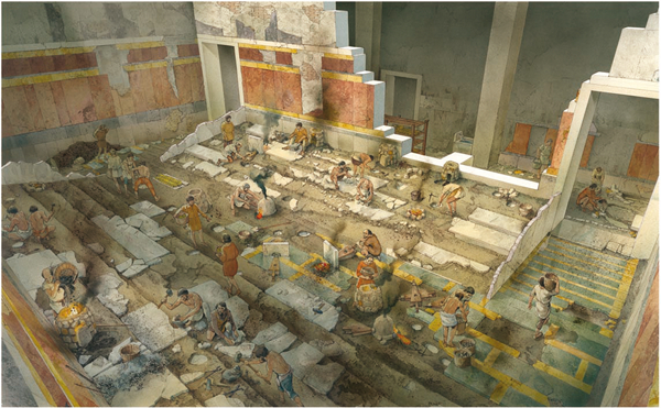
Reconstruction of transformation of Athenaeum into metallurgical workshop with systematic reuse of decorative material
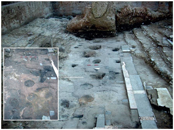
Image of excavated area of workshop showing evidence of furnaces (their density indicates intense level of activity at site)
A small portion of the material recovered at the site was selected for further archaeometric studies, and at least nine metallurgical processes were identified.1 This paper aims to investigate the silvering processes related to copper and copper alloy artefacts, how these processes are visible in the artefacts, and to understand which analytical method is more suitable for obtaining the appropriate information for interpreting the material. The level of corrosion of the artefacts and the discontinuous layer of silver makes it hard to gather information on their surface, and hence, a series of analytical techniques were used to investigate the problem and propose a technological approach that could be used when cutting a cross-section is impossible.
Experimental
Of the 39 samples analysed during the initial archaeometallurgical characterisation, five (Fig. 3) were identified as showing indications of silvering processes. These are two samples of metal debris (samples 13 and 16), one silvered metal fragment (sample 14) and two silvered metal objects.
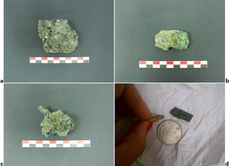
a macroscopic image of sample 13, b macroscopic image of sample 16, c macroscopic image of sample 14 and d macroscopic image of ingot and cross-section of planchet
The metal fragments (samples 13, 14 and 16) seem to be linked to the same process: the silvering of leaded bronze; on the other hand, the silvered metal objects (one planchet and one ingot) are related to the silvering of copper, and present different characteristics, possibly indicating two distinct production processes.2
The samples were analysed using a field emission gun scanning electron microscope (SEM) to investigate the corrosion layer and the distribution of silver and chlorine. The planchet was then chosen to investigate the potential use of time of flight secondary ion mass spectrometry (TOF-SIMS) and energy dispersive X-ray fluorescence (XRF) for routine analyses on silvered objects, as well as to determine the possible presence of mercury on the surface and the consequent identification of the use of mercury paste for the silvering process.
The samples could be cut and mounted in resin for the analyses with the SEM (Supra Zeiss). The mounted section was polished with carbide paper down to 8 μm and then further polished with diamond paste down to a quarter of a micrometre. The polished sections were then coated with a thin gold layer to increase their conductivity and improve the imaging and chemical analysis.
The TOF-SIMS is a very sensitive surface technique, whose range of action is limited to the first few nanometres of the surface. Considering the level of corrosion of the artefacts under consideration in this paper, this technique could only provide information on the top surface of the corrosion layer. In this case, the technique was used to investigate the cross-section and, in particular, the border between the corrosion layer and the actual surface of the artefacts, where the porosity observed with the SEM is located and where the ‘islands’ of silver were detected. The technique was used to inspect this boundary in order to verify the presence of a very thin continuous layer of silver that may have not been visible using the SEM, as well as the possible presence of mercury.
In order to perform the TOF-SIMS analysis, a 2 mm slice of the planchet was cut from the residual fraction after the mounting of the sample in resin; this section was freshly polished with carbon paste and then cleaned with acetone in order to reduce the level of contamination of the surface to be analysed. The instrument used was a Kore Technology Ltd time of flight secondary ion mass spectrometer, and the instrumental conditions used for the analyses were as follows: 25 keV indium primary ion source (FEI liquid metal ion gun) operating at 1 μA current. Secondary ions were analysed in a reflectron mass spectrometer and detected with a dual microchannel plate assembly. Flight times were recorded with a 0·5 ns time to digital converter. Spectra were taken from an area of approximately 150 μm2. Calibration of the mass spectra was established by defining a common series of CxHy peaks with a known mass using the mass calibration function in the instrument's software.
On the other hand, the XRF analyses were performed on the surface of the planchet. Different energies of the gun were used to investigate if the different degree of penetration of the X-ray could obtain information on the subsurface layer, the one that was observed with the SEM to contain the silver. The analyses were performed on the surface of the sample without further treatment, and a ZnTi pellet standard was used to cross-check the data. The ZnTi is a National Institute of Standards and Technology standard used to check the position of the peaks in the XRF; the standard is pure and does not contain any silver. The XRF instrument used (Oxford ED 2000) has a silver source. A thick aluminium filter is available in the instrument to avoid the detection of silver radiation coming from the tube. At high energy, the function of the filter is limited and part of the radiation reaches the detector, invalidating the detection of silver from the sample since part of the signal comes, in effect, from the tube itself. A series of analyses were performed on the ZnTi to investigate the energy at which only the signal from the sample is visible (no peaks of silver were detected from the standard).
Results
The results obtained from the investigation of the corrosion layer using SEM indicate that:
the corrosion layer is around 250 μm thick in every sample (Figs. 4–6); under a relatively compact crust of copper oxide, we observe an increasing porosity perpendicular to the surface
in the leaded bronze fragments, we observe that the porosity tends to be filled by coalescing lead metal, while it is formed of empty spaces in the two copper samples
in this area of porosity, silver ‘islands’ (around 10 μm in diameter) have been detected (Fig. 7); neither a continuous layer of silver nor the presence of silver in the body of the artefacts was detected in any of the artefacts analysed
high concentration of chlorine is detected in the corrosion layer and in the porosity area, in association with lead and silver in the leaded bronzes and in association with silver and copper in the copper artefacts.
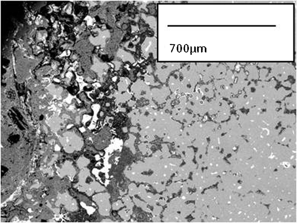
Polyhedral leaded bronze (sample 16) with areas of coalescing lead (on left close to surface in white) and deep corrosion layer (in darker grey on left)
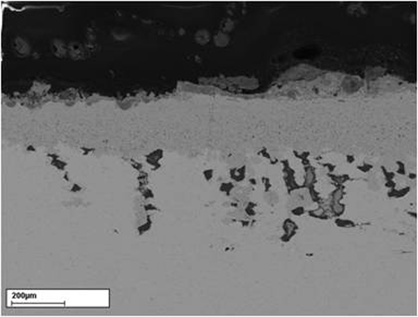
Image of surface of copper ingot showing high level of porosity and small inclusions attesting presence of silver (in white)
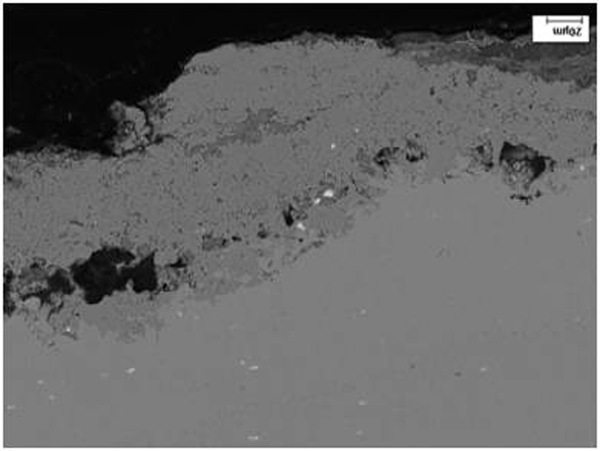
Image of surface of planchet showing clear-cut fracture parallel to surface indicating presence of chlorine and inclusions attesting presence of silver (in white)
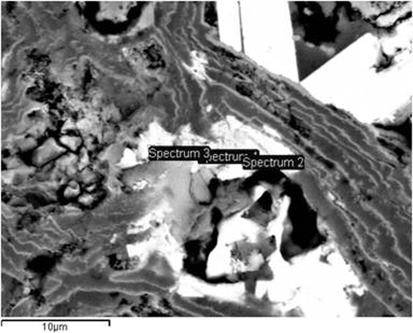
Detail of Fig. 4 showing area of association of silver and chlorine on surface
These observations suggested that if the silver was not looked for, it could ‘escape’ the analyses and mislead the interpretation to bronze or copper artefacts. To investigate this hypothesis, further analyses of the planchet were carried out using TOF-SIMS and XRF.
An initial mass spectrum was measured in the two areas analysed by TOF-SIMS, showing the main copper composition of the planchet (just below 63 and at 65; Figs. 8, 12 and 13), a small peak for silver (at 107; Fig. 9) and mainly the absence of mercury (the main peaks of which should be at 200, 198 and 202; Figs. 10 and 11).
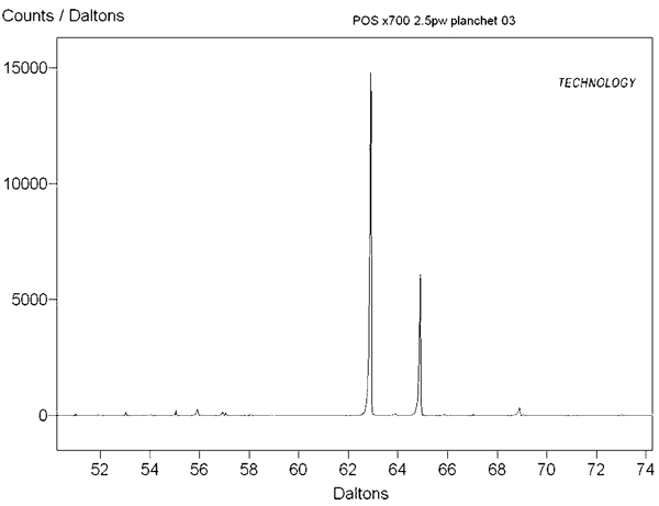
Portion of TOF-SIMS spectrum related to copper
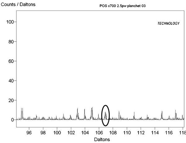
Portion of TOF-SIMS spectrum related to silver (at 107)
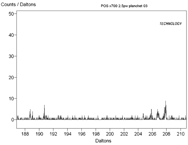
Portion of TOF-SIMS spectrum related to mercury (at 200, 198 and 202)
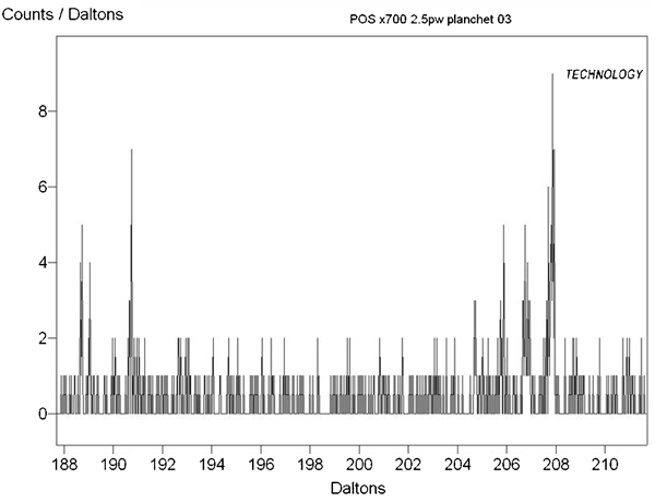
Detail of spectrum in Fig. 10 showing absence of peaks between 196 and 204, indicating absence of mercury in sample
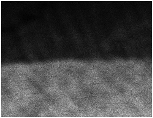
Total map showing corrosion products (upper dark grey layer) and body of object (copper: grey layer)
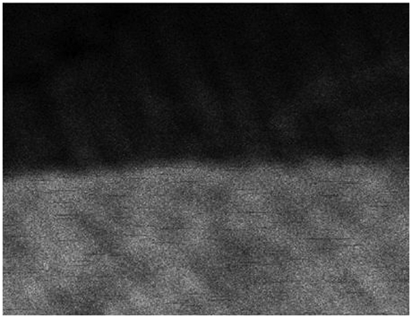
Map of copper showing strongest intensity in body of artefact, as expected
Subsequently, chemical maps of the two areas were measured in order to attempt a description of the distribution of silver in the sample. The map (Fig. 14) shows that silver is detected from under the layer of corrosion towards the body of the sample.
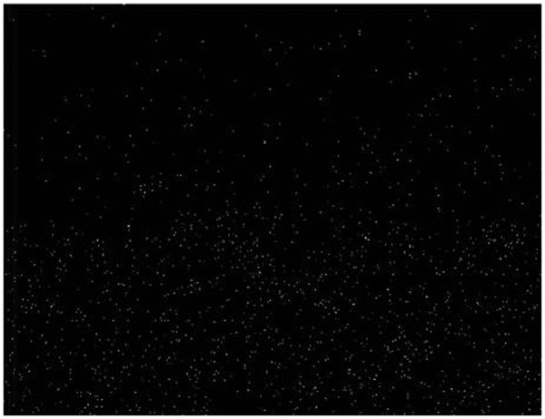
Map of silver showing barely visible line at border between corrosion products and body of object
No clear silver layer is detected on the surface of the sample; instead, it looks as if the silver diffused towards the body of the sample (Fig. 14). This observation, in correlation with the observations made using the SEM, seems to indicate that the silver is concentrated on the surface of the object (under the corrosion layer). Larger concentrations can be observed in the aggregates detected by the SEM. The diffusion towards the centre of the object seems to be related to the smearing of the silver through polishing on the most superficial layer (the one visible with TOF-SIMS).
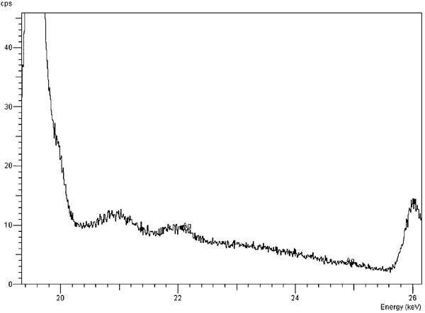
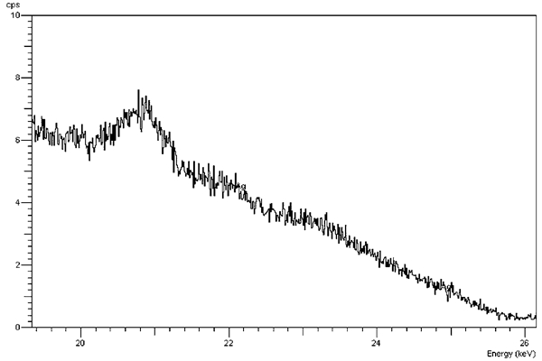
The measurements with XRF were carried out at 50, 45, 40, 35, 30 and 27 kV, using a thick aluminium filter on the zinc/titanium standard. At 50 kV, the intensity of the silver peak in the standard is ∼15 cps subtracting the background (Fig. 15); decreasing the energy of the beam, the intensity of the peak of silver decreases down to 2 cps at 27 kV (Fig. 16). The planchet was analysed through the corrosion layer at 50 and 27 kV (Figs. 17 and 18). At 50 kV, an intensity of 4 cps was detected for the silver Kα line (the same line observed in the zinc/titanium standard and described above). At 27 kV, there was no visible silver Kα line and the signal was completely lost. The data deriving from the zinc/titanium standard, however, lead to the conclusion that the silver visible at 50 kV is coming from the X-ray source, and the presence of silver in the sample could not be detected.
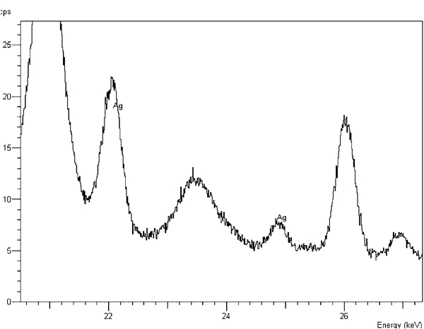
Kα and Kβ lines of silver measured in zinc/titanium standard using 50 kV beam and thick aluminium filter
Portion of spectrum of zinc/titanium measured at 27 kV showing small Kα peak of silver
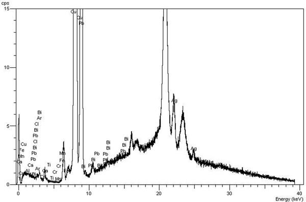
Complete XRF spectrum of planchet measured at 50 kV with thick aluminium filter (Kα and Kβ lines of silver are visible at 22 and 25 keV)
Detail of spectrum of planchet collected at 27 kV that shows no sign of silver
The area around 10 keV of the spectrum obtained at 50 kV was investigated to ascertain the presence of mercury. No peaks were visible in the area (Fig. 19), confirming the data deriving from the TOF-SIMS and the SEM regarding the absence of this element.
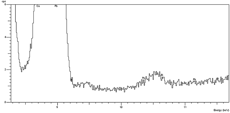
Detail of XRF spectrum of planchet collected at 50 kV showing no signs of mercury (at 10 keV)
Discussion and conclusion
The exploration of the materials and the investigation of the techniques reported above allow us to conclude the following.
The corrosion layer in all the samples is around 250 μm thick.
Silver is present under the corrosion layer. It is concentrated in small aggregates of around 10 μm in diameter. A very thin layer barely perceivable using TOF-SIMS is also present at the boundary between the corrosion layer and the body of the artefact.
Silver is not present in the body of the fragments or the artefacts.
The energy dispersive XRF with silver source used cannot be diagnostic of the presence of silver under the corrosion layer.
Mercury could not be detected using either XRF or TOF-SIMS.
As mentioned in the section on ‘Introduction’, the fragments presenting surface silvering are of two types: leaded bronze and copper. In all the objects, chlorine was present on the surface in high concentrations, indicating that this element was involved in the process of silvering. Vlachou et al.3 advance the hypothesis of the use of mercury paste in association with chlorides to treat the surface. The analyses of the planchet indicate that this artefact was treated with an alternative process (since mercury was not detected), suggesting the hypothesis of a treatment with hot cerargyrite baths.2, 4. It can be hypothesised that all the artefacts at the site received a similar treatment, but only further analyses of the leaded bronze fragments similar to the ones carried out for the planchet could provide conclusive answers. The bronze alloy presenting silvering treatment analysed here matches very well with the alloy presented by Vlachou et al.3 as the ‘best’ alloy for silvering with mercury paste. Nevertheless, the observations of the surface of the leaded bronze fragments do not present a continuous layer of silver, but a scattered rare presence of silver aggregates of small dimensions, even though Vlachou et al.3 show a different scenario in the case study they present. In any case, as already stated above, the data on the leaded bronze fragments are not yet conclusive, and further work is needed to reach final conclusions.
The main aim of this paper is anyway not to highlight the processes of silvering performed at Piazza Madonna di Loreto but to investigate the possibility of loss of data regarding the presence of silver in case of surface analyses through routine laboratory techniques. The possibility that silver is not present in a continuous layer, as in this case, and that it is not present in the body of the metal, but only in discrete particles scattered under the corrosion layer, decreases, first of all, its overall concentration in the object and makes its detection harder. In these conditions, already in cross-section, only a thorough investigation of the boundary between the corrosion layer and the body of the artefacts allowed the detection of a couple of silver aggregates per sample using SEM. A much more sensitive technique like the TOF-SIMS detects a more constant layer of silver (admittedly not even in a convincing layer shape), but again, this observation can only be made on a cross-section and would be impossible as a surface analysis since the TOF-SIMS examines only the first couple of nanometres of the surface.
The analysis of the surface carried out by XRF demonstrates the possibility of ‘missing’ the silver altogether. XRF (in its portable or laboratory form) is the principal technique that would allow routine non-destructive in situ or laboratory analysis. In this case, if the Sopraintendenza ai Beni Culturali di Roma would not have given permission for the sectioning of the planchet, no signs of the silvering process could have been observed and an important part of the archaeometallurgical information would have been lost.
The question that the author wants to conclude with is the following: how many of these types of data have been lost? How many copper and bronze fragments were actually silvered?
Careful planning and rethinking of the methodological approach to archaeological materials are a necessity that has to be continuously mediated with the availability of techniques, the possibility of invasive analysis and the possibility to move the materials from their environments. More importantly, the limitations of the data obtained by our instruments need always to be carefully taken into consideration and cross-checked.
Footnotes
Acknowledgements
The author thanks all the researchers from the Universita’ G. D'Annunzio in Chieti involved in the first screening and cataloguing of the materials, namely, S. Prosperi, A. Iacone, M. Tornese and S. Antonelli; without their contribution, it would have been impossible to obtain so much information from such a small amount of material selected for the analyses. In particular, thanks go to Dr La Salvia for the constant support and discussion of the materials. The author also thanks Professor T. Anson and L. Tammita for their support during the analyses and the experiments and Brunel University. Dr Serlorenzi and Mr Ricci are also acknowledged for allowing the analysis of the material.
This paper is part of a special issue on Arts and Surfaces