
Abstract
In the present work, the microstructure and structure of La0·25Ce0·52Nd0·17Pr0·06(OH)3 and La0·25Ce0·52Nd0·17Pr0·06O2 is obtained from transmission electron microscopy and X-ray diffraction measurements. Space group P63/m is assigned to the structure of La0·25Ce0·52Nd0·17Pr0·06(OH)3. Lanthanides are assigned to Wyckoff positions 2c. Cell parameters are a = 6·375(5) Å and c = 3·753(5) Å. The thermal decomposition of this compound was studied by differential scanning calorimetry. The process is exothermal with an enthalpy change ΔH° value of −254±10 kJ mol−1. The decomposition kinetics is complex and two global processes with Ea values of 98±4 and 61±2 kJ mol−1 were observed. The product is a lanthanide dioxide. Space group Fm3m is assigned to the La0·25Ce0·52Nd0·17Pr0·06O2. Lanthanides are distributed in Wyckoff positions 4a. The cell parameter is a = 5·479(5) Å. Nanopores in the oxide surface are obtained using this method and characterised by STEM measurements.
Keywords
Introduction
Hydrogen purification is a unitary operation consisting of the removal of contaminants from the hydrogen stream. This operation includes the removal of organic volatile compounds and gases such as O2, CO2 and CO. This task can be carried out by using multisubstituted lanthanide dioxides as a starting sieving material. The process is highly specific and the selected oxides should be thermally and chemically stable. Surface with pores is also desirable. Dioxides are usually obtained by heating the lanthanide salts1 by chemical coprecipitation1 or mechanical alloying.2 Thermal decomposition of the hydroxide is an alternative way of synthesis. Although it is successfully used to obtain the sesquioxides of the pure lanthanide metals,3–5 little is known about its use on the synthesis of lanthanide based dioxides. It is probably due to the lack of information on the structure and thermal stability of lanthanide based dioxides and hydroxides.
Although pure CeO2 or PrO2 might be used for this application, substitution of Ce4+ and Pr4+ ions with La3+ and Nd3+ ones produces a cubic dioxide structure with larger crystalline parameters due to the larger sizes of La3+ and Nd3+ ions leading to a more open structure allowing larger surfaces of contact with the gas stream. Unlike pure stoichiometric CeO2, multisubstituted lanthanide dioxides with mixed 3+/4+ cations produce secondary structures which are unstable on heating. These structures leave the surface of the main structure with pores without losing structural integrity. These are two desirable features used in hydrogen purification. In addition, La0·25Ce0·52Nd0·17Pr0·06 metal composition is cheaper than pure Ce or Pr metals used to synthesise the respective dioxides.
Then, the aims of the present study are the determination of the structure of a lanthanide based hydroxide, the analysis of the decomposition process and the synthesis of the dioxide. The results are used in the design of materials based on these dioxides applied to hydrogen purification.
Experimental
La0·25Ce0·52Nd0·17Pr0·06(OH)3 was synthesised from pulverised pieces of La0·25Ce0·52Nd0·17Pr0·06 (99·7%, Alpha Aesar) treated with nanopure water. Drying of samples was performed under Ar (99·999%) flow at room temperature in a sealed reactor. The lanthanides nominal composition was verified by neutron activation analysis using La2O3 (Reacton, 99·99%) and CeO2 (Reacton, 99·99%) as reference patterns. Room temperature X-ray diffraction (XRD) was measured on a Philips PW 1710/01 Instrument with Cu Kα radiation (graphite monocromator). Diffraction patterns were analysed by the Rietveld method using DBWS software.6 Particle sizes and morphology were observed by scanning electron microscopy (SEM 515 microscope). Characterisation by TEM was carried out using a Tecnai F20 G2, field emission microscope and a FEI CM20, operated at 200 keV. The diffraction patterns were indexed using jEMS software. Calorimetric measurements were carried out in a differential scanning calorimeter (TA Instruments, DSC 2970) under stagnant air between room temperature and 500°C. Heating rates selected were between 2 and 25°C min−1.
Results and discussion
Characterisation of La0·25Ce0·52Nd0·17Pr0·06(OH)3
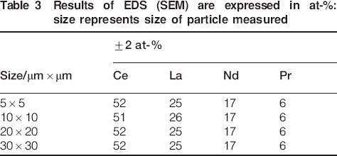
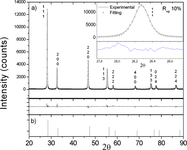
The X-ray diffractogram of La0·25Ce0·52Nd0·17Pr0·06(OH)3 is shown in black line in Fig. 1a. For comparison, the reference pattern of La(OH)3 (PDF file 036-1481) is presented in Fig. 1b. No preferred crystallographic orientation and no hkl extinctions are observed. The experimental data is refined using the Rietveld method. Refinement is shown in hollow dots in Fig. 1a. In this figure, Rwp stands for the goodness of the fit. The difference between the observed intensities and the calculated ones is shown in Fig. 1. Refinement results are summarised in Table 1. Lanthanides were randomly distributed in Wyckoff positions 2c. The microstructural parameters were obtained from the X-ray profiles. Instrument profile was obtained using a crystalline Si pattern. To analyse the peaks, a semiempirical ratio between the Lorentzian and Gaussian contributions was selected.7 The results are summarised in Table 2. Low values of strain ϵ and small crystallite sizes d are observed. No dependence of size to crystallographic orientation is seen. Measurements using EDS were conducted to the hydroxide particles. A sample is shown in the upper inset of Fig. 1. Results are summarised in Table 3. The compositional values are similar to the ones obtained by neutron activation analysis.
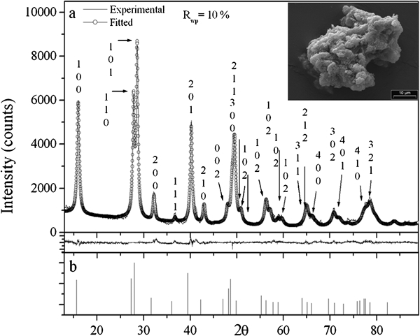
a experimental diffractogram of starting La0·25Ce0·52Nd0·17Pr0·06(OH)3 (black line) and Rietveld refinement (hollow dots) and b reference pattern of La(OH)3: upper right inset displays SEM image of starting particles
Structural parameters*
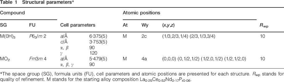
*The space group (SG), formula units (FU), cell parameters and atomic positions are presented for each structure. Rwp stands for quality of refinement. M stands for the starting alloy composition La0·25Ce0·52Nd0·17Pr0·06.
Microstructural parameters*
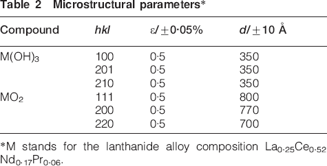
*M stands for the lanthanide alloy composition La0·25Ce0·52Nd0·17Pr0·06.
Results of EDS (SEM) are expressed in at-%: size represents size of particle measured
The nanostructure of the sample was analysed by TEM measurements. Two selective area diffraction patterns are shown in Fig. 2. The rings of the Fig. 2a are indexed according to a hexagonal structure. Cell parameters are a = 6·4±0·5 Å and c = 3·7±0·5 Å respectively. These results are in agreement with the XRD analysis presented in Fig. 1a. The diffraction rings shown in Fig. 2b are indexed according to a face centred cubic structure a cell parameter of 5·5±0·5 Å, not observed in Fig. 1. The nature of this minor phase is discussed later.

Selective area diffraction patterns of starting sample: a ring diffraction pattern indexed according to La0·25Ce0·52Nd0·17Pr0·06(OH)3 hexagonal structure; b minor structure indexed according to cubic structure
Bright field and dark field TEM images of the starting particles presented in Fig. 2b are shown in Fig. 3a and b respectively. A 300 nm size particle with sharp edges is observed in the bright field image. The dark field image was obtained by selecting with the objective aperture the ring indexed as 111 in the cubic structure presented in Fig. 2b and which does not overlap with a ring in Fig. 2a. The sizes of the grains that produce this ring were estimated to be between 2 and 20 nm. The hkl diffraction planes of this structure are not observed in Fig. 1.
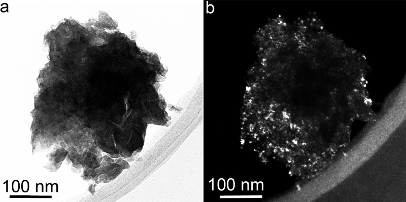
Bright field and dark field TEM images of same particle with structure indexed as cubic one showed in Fig. 2b
Thermal decomposition of La0·25Ce0·52Nd0·17Pr0·06(OH)3
The thermal decomposition of the hydroxide is shown in Fig. 4. An exothermal peak with shoulders is observed. The peak has the onset of 100°C and the offset of 346°C. The enthalpy change of thermal decomposition of La0·25Ce0·52Nd0·17Pr0·06(OH)3 is calculated from the differential scanning calorimetry (DSC) curve. The value found is ΔH = −254±10 kJ mol−1. The shape of the DSC curve also shows a shoulder that suggests that the decomposition consists of more than one step. The exothermal evolution is not reported for pure lanthanide hydroxides such as La(OH)3,8 Pr(OH)3,3,8 and Nd(OH)3.9 As an example, the thermal decomposition of La(OH)3 shows two endothermal peaks.8 This process is reversible according to two successive dehydration steps8


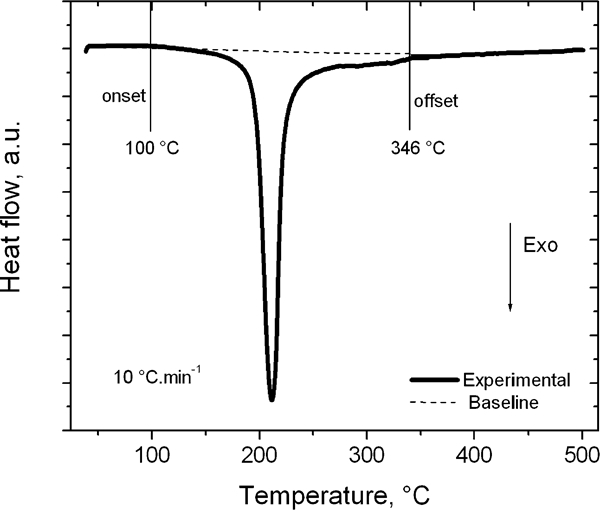
Measurement of thermal decomposition of La0·25Ce0·52Nd0·17Pr0·06(OH)3 using DSC
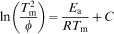
The activation energy Ea of the thermal decomposition is evaluated by DSC using the Kissinger formalism10 according to
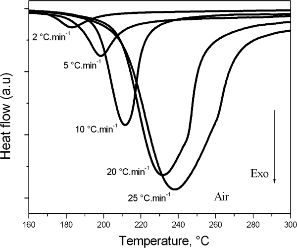
Curves of thermal decomposition of La0·25Ce0·52Nd0·17Pr0·06(OH)3 at different heating rates obtained by DSC
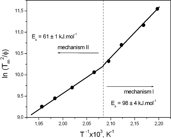
Kissinger plot of thermal decomposition of La0·25Ce0·52Nd0·17Pr0·06(OH)3
Since Ea values decreases as the temperature rises, the controlling global rate changes between two in-series processes.12 A change between parallel processes would have led to an increase in the Ea values as temperature increases, instead.12 Then, the changes in Ea should be related to changes in the controlling regime of the reaction. The mechanism I with a higher Ea value (98±4 kJ mol−1) is related to a heterogeneous reaction controlled by either mixed or chemical control since Ea values are larger than 40 kJ mol−1.13 In this sentence, mixed control means a reaction controlled by both diffusion in the pores of the sample and intrinsic chemical reaction. In the same way, chemical means intrinsic chemical reaction. As the temperature rises, in-series mechanism II appears and the Ea value decreases to 61±1 kJ mol−1. Then, the regime of the reaction is controlled by the previous regime and influenced by gas diffusion. In both cases, the final product is the same. Global mechanism I, with a higher Ea value, should be affected by the formation of intermediaries of reaction occurring in the lower temperature range. Unstable oxide hydroxides are the most likely candidates. These intermediaries are not independently observed by DSC. As temperature rises, the formation rate of intermediaries increases, reaction is accelerated and global Ea decreases pushing the reaction to global mechanism II with the lower Ea value.
Characterisation of La0·25Ce0·52Nd0·17Pr0·06O2
The XRD of the reaction products annealed at 1000°C during 24 h is shown in black line in Fig. 7a. For comparison, the reference pattern of CeO2 (PDF file 81-0792) is shown in Fig. 7b. No preferred orientation is observed in the sample. The refinement data is presented in black line with hollow dots in Fig. 7a. The difference between observed and calculated data is presented in the same figure. A zone of the diffractogram with the refinement for the 111 peak is presented in the upper right inset. Refinement details are summarised in Table 1. The cell parameters are larger than those of CeO2 (Fm3m, a = 5·411 Å, PDF file 81-0792). Then, the structure holds the ions of La and Nd which are larger than the ones of Ce. The microstructural data are presented in Table 2. No preferred orientation is observed and crystallite sizes are larger than the ones of the starting hydroxide. It indicates grain growth favoured by annealing the structure at 1000°C. The annealing also produces low strain values as observed in Table 2.
a experimental diffractogram of obtained La0·25Ce0·52Nd0·17Pr0·06O2 (black line) and Rietveld refinement (hollow dots, inset shows detail of refinement) and b reference pattern of CeO2 (JCPDF 81-0792)
The homogeneity of the product is analysed by TEM measurements. Using the refinement data obtained from the XRD experiment, the rings of the TEM diffraction pattern showed in Fig. 8a were indexed according to a cubic F structure with a cell parameter a = 5·4±0·5 Å. The indexation shows a very good agreement with the XRD results within the experimental errors. This kind of diffraction pattern shows high repeatability exhibiting a low preferred orientation and the absence of amorphous material. These assessments are in complete agreement with the shape of the peaks observed in Fig. 7 and the microstructural values presented in Table 2. The dark field image of the particles that exhibit this diffraction pattern shows the characteristics presented in Fig. 9a. The crystalline domains range from 2 to 50 nm. Also, the STEM image exhibits very well facetted nanoparticles with an average size of 50 nm as observed in Fig. 9b. These well defined boundaries define a shape coherent with the one of a cubic structure annealed at high temperatures (1000°C). This type of agglomeration of smaller particles also shows a ring diffraction pattern characteristic of a cubic structure.
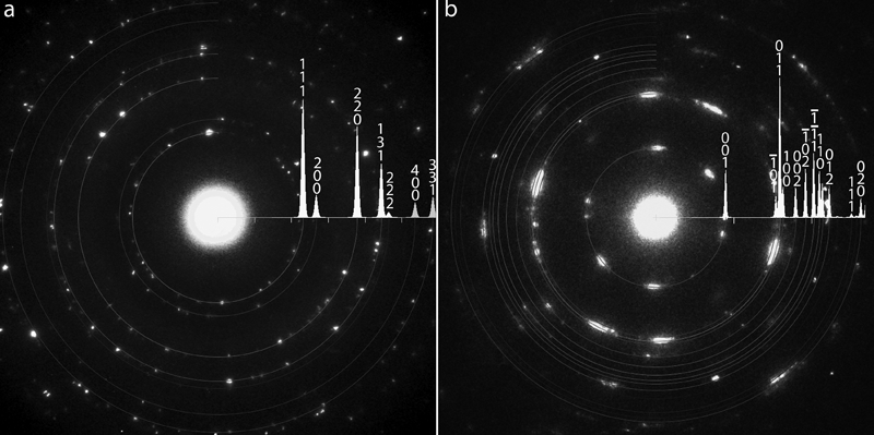
a diffraction ring pattern indexed La0·25Ce0·52Nd0·17Pr0·06O2 cubic structure and b diffraction ring pattern indexed according to monoclinic La*OOH (lanthanum rich oxide hydroxide) structure
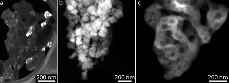
a dark field image of La0·25Ce0·52Nd0·17Pr0·06O2, and b and c STEM images of matrix of La0·25Ce0·52Nd0·17Pr0·06O2
These results are in agreement with those of Fig. 7a. Despite this agreement between micro- and nanostructure, the sample is not homogeneous. The diffraction rings of another structure found are observed in Fig. 8b. The cell was indexed as a monoclinic one with cell parameters a = 3·3 Å, b = 3·8 Å, c = 6·0 Å and β = 112°. This structure is not detected by XRD as observed in Fig. 7a. The structure might be an oxide hydroxide of an unstable lanthanum rich (La*) sesquioxide (La*2O3). This compound should be formed during the thermal decomposition of La0·25Ce0·52Nd0·17Pr0·06(OH)3 according to a reversible scheme similar to reaction presented in equations (1) and (2). The interplanar values of the diffraction rings are closer to the values of a LaOOH (lanthanum oxide hydroxide).8 This rich lanthanide oxide hydroxide (La*OOH) should be unstable on cooling/heating.8 The reaction occurs with loss of water and the monoclinic structure should not be coherent with the cubic La0·25Ce0·52Nd0·17Pr0·06O2 matrix. As a result, the final La0·25Ce0·52Nd0·17Pr0·06O2 structure should be porous. This feature is indeed seen in the STEM image of Fig. 9c as cavities. These voids were not present in the entire sample. Measurements by TEM indicate that voids were observed on nearly 10% of the sample surface. This is in agreement with the fact that the unstable La*OOH is the minor phase in the sample.
Structure stability and scheme of decomposition
At room temperature, the starting La0·25Ce0·52Nd0·17Pr0·06(OH)3 is isostructural to La(OH)3 as analysed by XRD and TEM. The structure is not pure and a secondary phase is observed. This second structure is detected only by TEM. The crystallite sizes of this structure are two orders of magnitude smaller than the main hexagonal La0·25Ce0·52Nd0·17Pr0·06(OH)3. The structure found is stoichiometric La0·25Ce0·52Nd0·17Pr0·06O2.
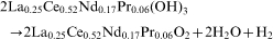
On heating, the thermal evolution consists on a single exothermal step. Unlike the case of La(OH)3,8 the presence of reaction intermediaries such as an oxide hydroxide is not observed as a single thermal evolution.8 The mechanism is not simple. It is deduced by the shape of the DSC curves of Fig. 5 and by the presence of a break in the Kissinger plot presented in Fig. 6. The thermal decomposition of La0·25Ce0·52Nd0·17Pr0·06(OH)3 is not reversible. The following global stoichiometry is proposed
The next step in this development is the controlled synthesis of La0·25Ce0·52Nd0·17Pr0·06O2 or lanthanide contents closer to these, at temperatures between 100 and 240°C to favour the formation of unstable intermediaries that leave the dioxide structure with a higher amount of pores on the surface. Larger amounts of unstable intermediaries should not destabilise the dioxide matrix and the acquired knowledge on the crystalline structures of the La0·25Ce0·52Nd0·17Pr0·06(OH)3 and La0·25Ce0·52Nd0·17Pr0·06O2 would take them apart them from the crystalline structures of these intermediaries favoured at low temperatures as suggested in mechanism II.
Conclusion
New findings are reported in the present work.
The crystalline structure of La0·25Ce0·52Nd0·17Pr0·06(OH)3. The structure is associated to space group P63/m with lanthanides randomly located in Wyckoff position 2c. The cell parameters are smaller than those of pure La(OH)3 and pure Nd(OH)3 indicating that the structure holds the ions of Ce and Pr. The cell parameters were also found as presented in Table 1.
The thermal decomposition process of this hydroxide. This finding includes the global stoichiometry, the calculation of the enthalpy change ΔH and activation energies of the process showing unique characteristics not observed in pure lanthanide hydroxides such as the exothermal formation of a dioxide.
The crystalline structure of La0·25Ce0·52Nd0·17Pr0·06O2. The structure is associated to space group Fm3m with lanthanides randomly distributed in Wyckoff positions 4a. The cell parameters are larger than those of pure CeO2 or PrO2 indicating that the dioxide structure holds the larger ions of La and Nd. The cell parameters were also found as presented in Table 1.
The formation of nanopores in the oxide surface due to the presence of an unstable secondary phase.
These findings are used in the development of advanced materials for hydrogen purification.
Footnotes
Acknowledgements
The authors wish to thank Dr D. Schryvers for helpful discussion of TEM results. M. R. Esquivel wishes to thank the Agencia Nacional de Promoción Científica y Tecnológica of Argentina (project no. PICT N° 33473), Universidad Nacional de Cuyo of Argentina (project no. 06/C310) and Comisión Nacional of Energía Atómica of Argentina for partial financial support. E. Zelaya thanks the support of a Bijzonder Onderzoeksfonds project of the University of Antwerp.