
Abstract
In this research, we characterise a new porous calcium phosphate composite composed of hydroxyapatite (HAp) and dicalcium phosphate dihydrate (DCPD) prepared by foam casting method using polyurethane foam, designed for osteoconductive and osteoinductive scaffolds. According to the obtained results, the values of elastic modulus, compressive strength and density of the samples reduced with increasing percentage of DCPD phase. The mechanical properties of the prepared samples were near the natural compact bone. After in vitro examination and confirming the bone-like apatite ability of the prepared composites, in vivo examination was employed, and for this purpose, a rabbit model was developed. New bones formed on the outside surface and on the macropore walls of the samples, as osteoids and osteoclasts, were evident 2 months post-operatively. Considering these properties, the prepared composite scaffolds made from HAp and DCPD could be considered as highly bioactive and potential bone tissue engineering implants.
Keywords
Introduction
Bone tissue engineering and regenerative medicine are new research areas with clinical applications in bone replacement on orthopaedic defects, bone neoplasia and tumours, pseudoarthrosis treatment, stabilisation of spinal segments as well as in maxillofacial, craniofacial, orthopaedic, reconstructive, trauma and neck and head surgery.1–6 It may provide solutions for generating a new bone tissue with good functional and mechanical qualities, reducing the risks and expenses of using autografts, allografts and metals.7–10
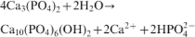
During the last decades, different biomaterials of biological or synthetic origin have been designed with the aim of acting as extracellular matrix composite scaffolds for new bone formation. Meanwhile, clinical uses require a series of biomaterial properties, such as bioactivity, osteoconduction, osteoinduction, biocompatibility and biodegradation.11–14 Biomaterials play a key role in several biomedical applications, and it is imperative that both the materials and the biological aspects are clearly understood for attaining successful biological outcome. Among biomaterials, calcium phosphates (CaP) are the most ubiquitous family of bioceramics well known for their use in biological applications.15–17 The chemical composition of CaP bioceramics is roughly equivalent to that of the inorganic matrix of human bone and is found to be the most suitable implant material. The major phase found in the bone is hydroxyapatite [HAp, Ca10(PO4)6(OH)2], and the other commonly known phases are octacalcium phosphate, tricalcium phosphate (TCP), dicalcium phosphate dihydrate (DCPD, CaHPO4.2H2O) and dicalcium phosphate bioceramics.
Hydroxyapatite is one of the frequently used bioceramics for bone and dental tissue reconstitution, which has excellent biocompatibility with hard tissues18–20 and high osteoconductivity and bioactivity despite its low degradation rate,21 mechanical strength and osteoinductive potential.22,23 It has also neither antigenicity nor cytotoxicity.24,25 In addition, it seems that using DCPD in bone tissue engineering is interesting, and a large number of studies have reported the advantages of using DCPD bioceramic in bone tissue engineering applications.26 For instance, a report indicated that the mechanisms of DCPD growth and dissolution were of interest because of the importance of this CaP in dental and bone regeneration applications.27 The properties of anhydrous and hydrated salt dicalcium phosphate, i.e. CaHPO4 and CaHPO4.2H2O, have been studied for many years as their mineral forms, i.e. monetite and brushite. As members of the series of CaP, they have considerable biological importance with respect to the biomineralisation processes in bones and teeth and find practical uses in dental cements and restorative materials. It is also important to point out that the degradability of different CaP varies with the ratio of Ca/P, with the highest being that of DCPD, which usually results in the most extensive bone remodelling around the scaffold.28–32
Previous studies have used different forms of HAp, but concerns have been raised regarding the limited degradation properties of this material.33,34 Therefore, biphasic composites of HAp and biodegradable material, like HAp/TCP, have been studied as an alternative to HAp ceramics. Porous biphasic calcium phosphate (BCP) composite bioceramics are expected to accommodate bone grow, and this characteristic provides osteoconductive properties.
Among the CaP ceramics, the BCPs, which are composed of different concentrations of the stable phase (HAp), and the more soluble phase (DCPD/TCP) have presented significant advantages over other CaP ceramics due to their controlled dissolution rate, different mechanical properties and balance between resorption/solubilisation, which guarantees the stability of the biomaterial while promoting bone ingrowth.35–37
The porosity and bioactive surface facilitate cell attachment, proliferation and differentiation and, consequently, provide a more biocompatible, osteoconductive and in some cases osteoinductive ceramic, which can favour increased bone formation.35–39 In fact, these chemical and physical properties, produced by variations of temperature, pH and duration of the sintering process, make each biomaterial unique and lead to different tissue responses.40–44 Currently, the BCP represents the most promising and best alternative for bone reconstructions as they can overcome the shortcomings of autografts and allografts, such as expense, limited supply, additional trauma in the case of autografts and risk of disease transmission in the case of allografts.
Porous bioceramics of HAp/DCPD generally biodegrade faster than bioceramics made of HAp and HAp/TCP due to the increased resorption rate of DCPD relative to HAp and TCP implants of similar structure.45 The bioresorbability of CaP ceramics appears to be dependent on their chemical/crystal composition and on the environment of the implantation site.46 Jarcho47 has proposed the existence of two different biological resorption pathways: one involving solution mediated processes and the second involving cell mediated processes. Contrary to expectations, the material more closely resembling the body's own hard tissue component (HAp) was found to dissolve much more slowly than many CaP not naturally occurring in bone when similar ceramic structures and degrees of purity were used.48 It can be stated that implants of crystalline HAp have a lower tendency to bioresorb because of their chemistry and small surface area.
Replication or slurry dipping is a simple method of producing porous and interconnected foams using ceramic slurry and has been used to produce CaP bone graft.49,50 The mentioned method produces scaffolds/bone grafts with poor mechanical properties due to the presence of the central void in the strut and the presence of micropores and microcracks due to non-uniform coating of the ceramic slurry.51 Electrospraying is another form of replication method that can produce interconnected pore structures, and it has been possible to obtain solid struts with no central hole,52 therefore leading to improved mechanical properties.53 However, foam preparation by electrospraying only requires well dispersed ceramic suspensions of high concentrations of 60–65 wt-%.52,53 The most conventional and popular manufacturing route for ceramic/bioceramic foams with open cells is using polyurethane template.54 The resulting material has a well defined cell size that is appropriate for bone tissue engineering applications.
In this study, we prepared porous composite structures of HAp/DCPD bioceramics containing different percentages of each component to evaluate the effect of DCPD addition on the chemical and mechanical properties of the final composites. Finally, in vitro and in vivo examinations were performed to evaluate the biological responses of the prepared samples.
Materials and methods
Preparation of composites
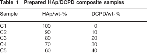
For the synthesis of composite samples, commercially purified HAp and DCPD powders (Merck Company, Germany) were used. Powders of HAp/DCPD, with different weight ratios, were prepared in a planetary ball mil (Retch PMA, Brinkman, USA) for 30 min to ensure homogeneity. The specific surface area was determined by 15-point BET measurement (Micromeritics Gemini 2360). Then, the powders P were suspended in distilled water W at a ratio of P/W = 55% (w/v), and different additives (e.g. colloidal silica, three poly phosphate and carboxy methyl cellulose) were mixed to the HAp/DCPD solution for 24 h to obtain a suitable bioceramic slurry. The porous scaffolds with different amounts of HAp and DCPD (see Table 1) were fabricated by polyurethane foam reticulate method. Polyurethane foam templates were purchased from Safoam (80 ppi, Iran). Typically, the polyurethane foam templates were replicated using the slurry by a repeated dipping and drying process. The samples were then air dried, and after drying, the samples were heated to 1050°C on a strict schedule, which minimised disruption during pyrolysis and allowed the bioceramic to achieve a high density. This heating schedule consisted of a heating rate of 0·5°C min−1 up to 800°C, held at this temperature for 1 h, then rapid heating of 5°C min−1 from 800 to 1050°C, held at 1050°C for 3 h, and then cooled in the furnace. Figure 1 shows the prepared composite samples using polyurethane foam template.
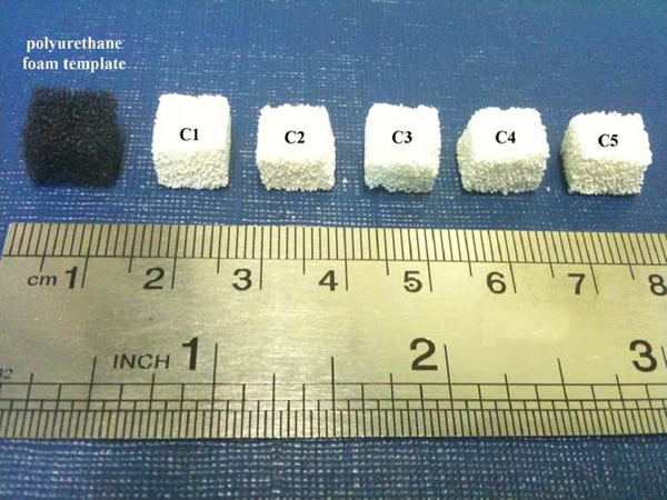
Prepared composite samples using polyurethane foam template
Prepared HAp/DCPD composite samples
Preparation of simulated body fluid (SBF)
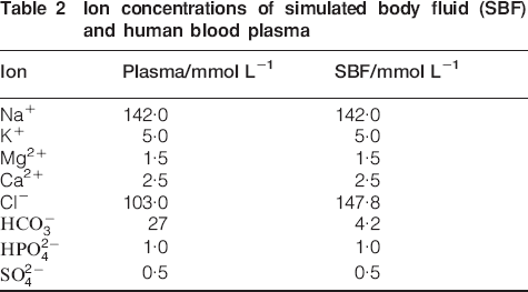
Reagent grade chemicals NaCl, NaHCO3, KCl, K2HPO4.3H2O, MgCl2.6H2O, CaCl2, trishydroxymethyl aminomethane [Tris buffer, (CH2OH)3CNH2] and 1 N HCl as required materials for the preparation of SBF were purchased from Merck Inc. The SBF solution was prepared by dissolving reagent grade NaCl, KCl, NaHCO3, MgCl2.6H2O, CaCl2 and KH2PO4 into distilled water and buffered at pH 7·25 with Tris buffer and 1 N HCl at 37°C. Its composition is given in Table 2 and is compared with the human blood plasma.
Ion concentrations of simulated body fluid (SBF) and human blood plasma
Sample characterisation
X-ray diffraction (XRD) analysis
The samples surfaces were analysed by XRD with a Siemens-Brucker D5000 diffractometer. This instrument works with voltage and current settings of 40 kV and 40 mA respectively and uses Cu Kα radiation (1·540600 Å). For qualitative analysis, XRD diagrams were recorded in the interval of 10°⩽2θ⩽50°at scan speed of 2° min−1.
Fourier transform infrared spectroscopy (FTIR) analysis
The samples were examined by FTIR with a Bomem MB100 spectrometer. For IR analysis, 1 mg of the scraped samples was carefully mixed with 300 mg of KBr (infrared grade) and pelletised under vacuum. Then, the pellets were analysed in the range of 400–4000 cm−1 with 4 cm−1 resolution averaging 120 scans.
Scanning electron microscope analysis
The morphology and microstructure of the synthesised samples were evaluated using SEM. The samples were coated with a thin layer of gold by sputtering (EMITECH K450X, UK), and then their morphology was observed on a scanning electron microscope (SEM-Philips XL30) that operated at the acceleration voltage of 15 kV.
Density, porosity and pore size measurement
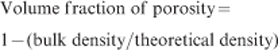
For density and porosity measurements, the sintered specimens were initially subjected to ultrasonic washing in distilled water for a few minutes. After drying the samples in a stagnant air oven at 90°C, their dry weights were recorded. The porous specimens were then boiled in distilled water for ∼3 h and allowed to cool in water for 24 h. Wet weight in air and wet weight suspended in water were determined using an analytical balance (Precisa, 300S, Switzerland). Bulk density, apparent porosity and volume fraction of porosity were calculated in our samples using the formula below


Mechanical behaviour
For analysing the mechanical behaviour of the composite samples, they were supplied in the form of cylinders with a mean diameter of 3·4±0·5 mm, which were filled down to a length of 6·3±0·7 mm. All the mechanical testing was performed using a Zwick/Roell 2005 with a crosshead speed of 0·01 mm s−1. The compressive strength and the modulus of elasticity were determined from mechanical test recordings. The modulus of elasticity was determined from the slope of the linear portion of the stress–strain curve.
Biological evaluation
In vitro study in SBF
We performed in vitro studies by immersion of the samples in SBF at a concentration of 1 mg sample/mL of the fluid at 37°C for different time periods. All the reacted solutions were saved for inductively coupled plasma atomic emission spectroscopy (ICP–AES) (Varian Co., USA) analysis of Ca to measure the ionic concentration in the SBF solutions. In addition, the pH of the SBF solutions was measured by a calibrated pH meter every step and using a Corning pH meter 320.
In vivo implantation procedure, histological testing
For in vivo implantation, all the specimens were supplied in the form of cylinders with a mean diameter of 3·4±0·5 mm, which were filled down to a length of 6·3±0·7 mm. The specimens were sterilised with ethylene oxide gas before implantation. Thirty-eight adult New Zealand white rabbits weighting 3·0–4·0 kg were used. The rabbits were assigned to one of four groups (1, 2, 3 and 6 months; n = 6 in each group) with respect to implantation time. Then, the specimens were inserted into defects made using a saline cooled, diamond tipped 3·5 mm trephine in the medial femoral condyle of rabbits. The implants were immersed in the solution for 5 min before implantation. Each rabbit was used for a bone graft. A cavity 3·5 mm in diameter and 7 mm in depth was drilled manually in the femoral condyles under general anaesthesia with antibiotic protection. After carefully washing with a physiological saline solution, the cavities were filled with bone graft. The cavities were randomly filled with one of the implants. After 1, 2, 3 and 6 months, the rabbits were sacrificed by an overdose of thiopental sodium, and the femoral condyles were removed for the assessment of the biomechanical properties of the implanted specimens and the observation of bone ingrowth by light microscopy. Experiments were performed according to the European Guidelines for Care and Use of Laboratory Animals (European Directive 86/609/CEE).
For histological examination, the samples were decalcified in 10% formalin solution and sectioned into pieces 5–6 mm thick and then stained with haematoxylin–eosin and masons trichrome stain. The sections were examined with a light microscope. The amount of fibrous tissue and new bone formation and the presence of remodelling were qualitatively assessed. The presence of new lamellar bone and osteoclasts along the trabeculae of the newly formed woven bone was considered a sign of remodelling.
Statistical analysis
All the experiments were performed in fifth replicate. Tukey's honestly significant difference multiple comparison testing was used to determine the significance of the deviations in the strength and modulus of each sample. For all the statistical tests, a p<0·05 was considered to be significant. All the statistical analyses were performed with the software program SPSS for Windows, version 9 (SPSS Inc., Chicago, IL, USA).
Results and discussion
X-ray diffraction analysis
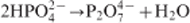
Figure 2 shows the XRD patterns of the prepared composite samples. As it can be seen in this figure, the first sample (C1) shows the main characteristic peaks of HAp [Ca10(PO4)6(OH)2, Joint Committee on Powder Diffraction Standards (JCPDS) no. 09-0432] as the major phase, with a minor phase of β-tricalcium phosphate [Ca3(PO4)2, β-TCP, JCPDS no. 09-0169]. It is also obvious that by further addition of the brushite phase to the composite samples, the main characteristic peaks of β-TCP became more apparent, and finally, for the C5 sample, β-TCP was the major phase. As the main product of pyrolysis of brushite at higher than 600°C is calcium pyrophosphate, calcium pyrophosphate at higher than 1000°C react with HAp to form β-TCP, as shown in the following reactions

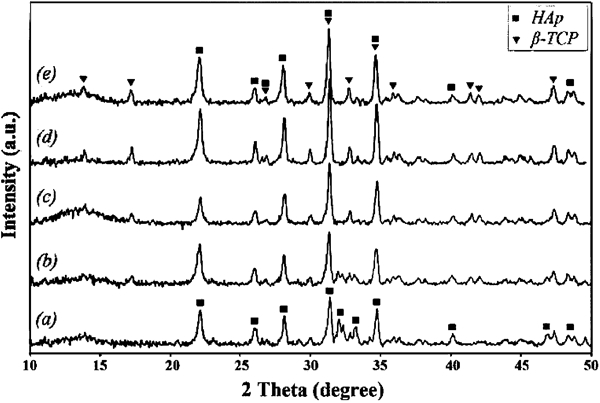
X-ray diffraction patterns of composite samples: a C1, b C2, c C3, d C4 and e C5
Fourier transform infrared spectroscopy analysis
Figure 3 shows the FTIR spectra, in the 400–4000 cm−1 spectral range, of the prepared composite samples. The C1 sample exhibited five important infrared bands of HAp located at 560, 605, 710, 1040, 1640 and 3479 cm−1. The characteristic
 and OH− absorption bands of HAp were observed in the as dried sample along with the additional broad bands at 1640 and 3479 cm−1 from the adsorbed H2O. In addition, the band at 1040 cm−1 arises from υ3 PO4, and the bands at 560 and 605 cm−1 arise from υ4 PO4. The FTIR analysis showed all the typical absorption characteristics of the HAp powder, and according to these explanations, it is obvious that the synthesised powder is certainly HAp. The weak absorption peak at 880 cm−1 was assigned to the P–O–H vibration in the
 group,56 which exists in non-stoichiometric HAp. It is worth mentioning that by further addition of brushite powder to the composite samples, the OH− absorption band disappeared, and the spectrum obtained was characteristic of β-TCP. In addition, the obtained results from the FTIR spectrum of composite samples became similar to that of the obtained results from XRD patterns.57
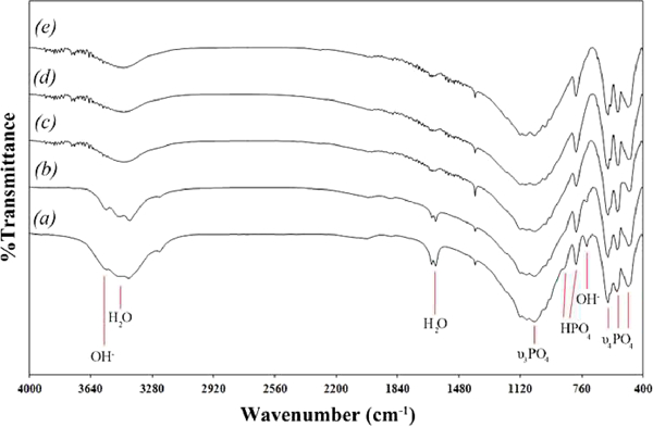
Fourier transform infrared spectroscopy patterns of composite samples: a C1, b C2, c C3, d C4 and e C5
Scanning electron microscope observations
In this study, SEM was used to observe the morphology of the scaffold microstructure. The low and high magnifications of SEM images captured from the top view of the C4 composite sample are shown in Fig. 4. The observations indicate a network of interconnected pores. In addition, the composites were porous non-parallel aligned, and the pores were close to spherical shapes with size ranging from 100 to 200 μm, which can be desirable for bone cell growth.58
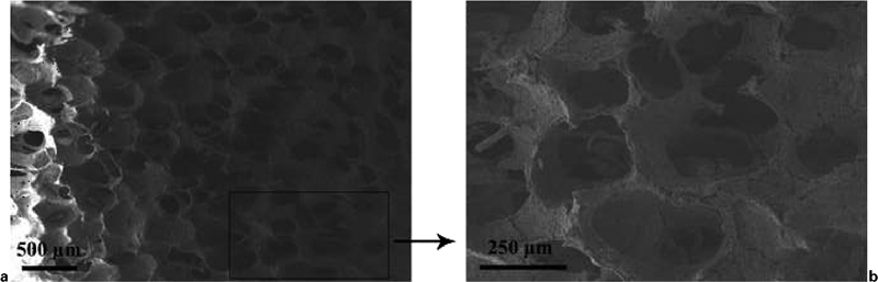
Images (SEM) s captured from top view of C4 composite sample
Mechanical properties
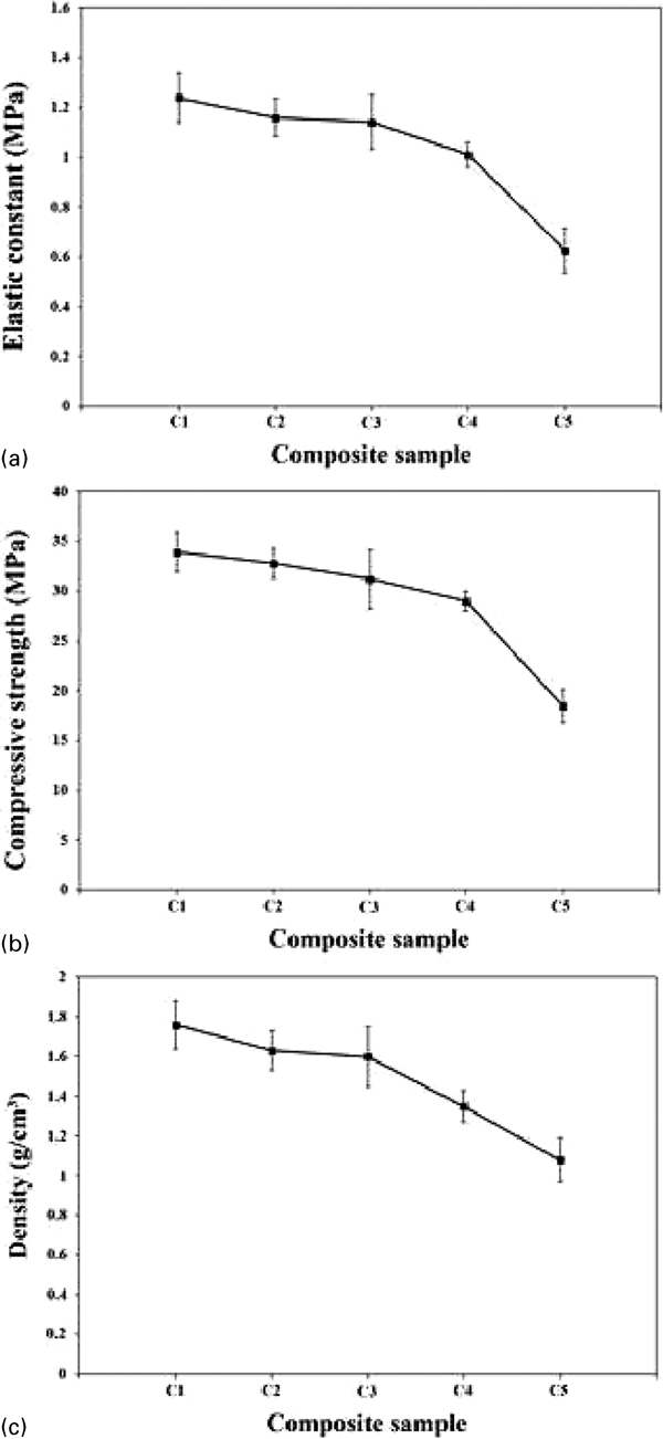
Generally, an ideal tissue engineering implant should be biocompatible and highly porous with adequate mechanical properties. For this purpose, the prepared nanocomposite samples were tested to determine the effects of using the DCPD as the second phase on the mechanical properties of the samples. Table 3 gives the data obtained from mechanical compressive tests of natural cancellous and cortical bone. 59 59,60 In addition, Fig. 5 shows the elastic constant E, compressive yield strength σyield and density ρ of the prepared samples. As it can be seen in this figure, the values of elastic modulus, compressive strength and density of the samples reduced with increasing percentage of DCPD phase.
Mechanical properties of prepared samples
Mechanical indices of natural compact and spongy bone
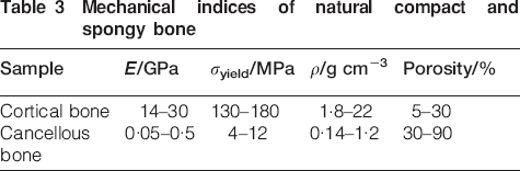
In addition, the elastic modulus values varied from 0·6 to 1·2 GPa, the σyield values varied from 15 to 35 MPa and the density values varied from 1 to 1·8 g cm−3. In addition, according to Fig. 6, which shows the percentage of porosity of the samples, the porosity values are from 34 to 39%. It is worth mentioning that the mechanical properties of the prepared samples were near the natural compact bone.
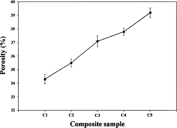
Porosity percentages of different prepared composite samples
In vitro characterisation
Changes in SBF composition
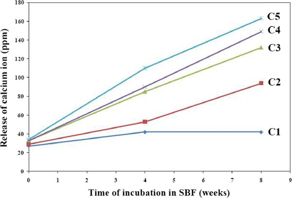
Figure 7 shows the variations of Ca concentration in the SBF solution for various periods, measured by ICP–AES, versus immersion time. When the prepared composite samples react with SBF, both chemical and structural changes occur as a function of time within the surface of samples,47 and accumulation of dissolution products causes both the chemical composition and the pH of the solution to change. As it can be seen in all the cases, the Ca concentration in the solution increased continuously during the first days of immersion. In addition, the pH variation with time increased from 7·4 to 8 during the 4 weeks of immersion, and then the pH increased slowly up to 8·3 until week 8.
Variations of Ca concentration for different samples in SBF solution for various periods measured by ICP–AES
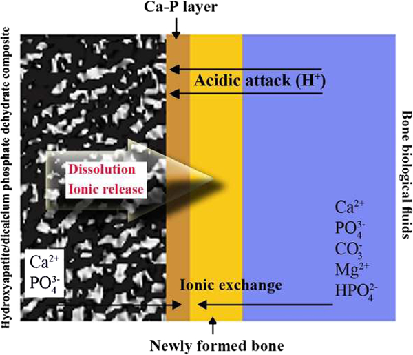
Mechanism of bioactivity process
Bioactive composite scaffolds from CaP ceramics elaborated under pressure and under temperature treatment of Ca(NO3)2, H3PO4, NH4OH and H2O.61 As it can be seen in the SEM images in this study, the structure of samples is clearly porous, but we can distinguish the microporosities (<10 μm) that permit diffusion of ions and fluids from macroporosities (100–600 μm) that permit cellular colonisation. The bioactivity process of different bioactive materials was studied in vitro and in vivo.62 In the case of this study, the bioactivity process occurs under an acidic attack with H+ at the material surface, as shown in Fig. 8. This leads the dissolution of CaP crystals and a high release of Ca2+,
 .63 The concentrations of calcium and phosphorus increase in the surrounding fluids, and this supersaturation induces reprecipitation of apatite crystals at the surface of porous scaffolds.64 These apatite crystals may incorporate Ca2+, Mg2+, CO3−,
 and organic molecules present in the surrounding fluids.65 This dissolution–reprecipitation process leads to the formation of a carbonated apatite layer at the material surface and permits a chemical bond with newly formed bone. The solubility of CaP base bioactive materials varies with different factors: porosity, grain size, crystallinity, sintering temperature and so on.
66
66,67 For instance, an increase in the sintering temperature leads to an increase in the HAp crystal size and finally reduces its solubility. On the other hand, the solubility increases with porosity and pore size.
and organic molecules present in the surrounding fluids.65 This dissolution–reprecipitation process leads to the formation of a carbonated apatite layer at the material surface and permits a chemical bond with newly formed bone. The solubility of CaP base bioactive materials varies with different factors: porosity, grain size, crystallinity, sintering temperature and so on.
66
66,67 For instance, an increase in the sintering temperature leads to an increase in the HAp crystal size and finally reduces its solubility. On the other hand, the solubility increases with porosity and pore size.
Bioactivity process of prepared samples
Scanning electron microscopy observations after immersion in SBF solution
Herein, apatite was incorporated onto the surface of the scaffolds in situ via the SBF technique. Figure 9 shows the SEM images of the scaffolds after immersion for 2 weeks. According to the observations, scattered and small particles were covered on the surface of the scaffold pore walls after immersion, as shown in Fig. 9a. A substantial amount of apatite microparticles formed on the surfaces of the pore walls throughout the scaffold. After that, the whole inner pore wall surfaces of the scaffold were covered by a layer of apatite, and the underlying surfaces were not clearly observable. In addition, it is predictable that a longer immersion time of the scaffolds led to more apatite formation.
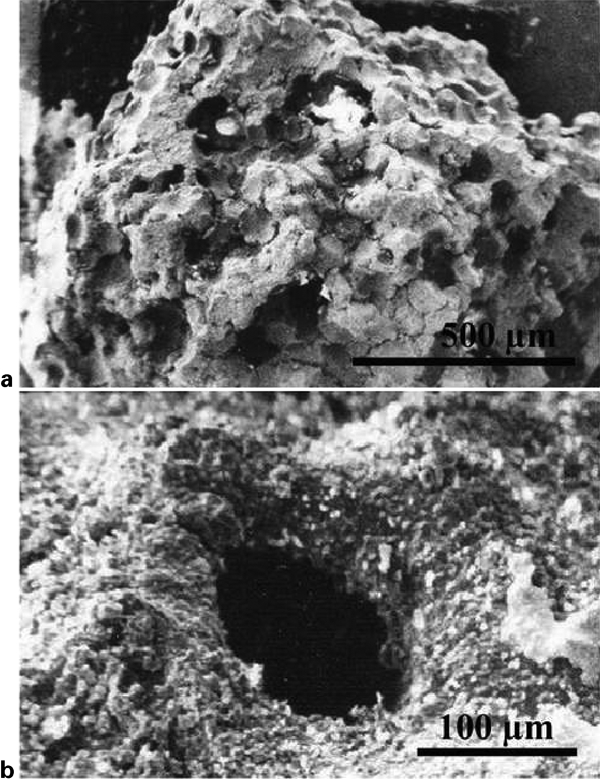
Images (SEM) of scaffolds after immersion in SBF
In vivo evaluations
Apatite mineralisation of bioactive materials is thought to be an important phenomenon in the chemical interactions between implant materials and bone tissue, which ultimately affects the in vivo osteogenesis of the bone grafting materials. Figure 10 shows the optical micrograph of in vivo examination of sample C4 after 2 months. According to the obtained results, no inflammation, tissue necroses or tissue rejection was observed after implantation. Generally, during the in vivo process at 1 month post-operation, fibrous connective tissue and blood vessels grow into the macropores, contributing to the early fixation of the specimens. However, there is no new bone formation in the macropores, and few macrophages are detected on the macropore surface. Herein, new bone formation occurred on the surface and macropore walls of specimens as osteoids and osteoclasts were evident at 2 months post-operatively. The fusion of the specimen and the host bone was realised through the bonding of new bone 3 months post-operatively. The new bone occupied most of the macropore spaces, and a new bone tissue layer developed on the inside surface of the specimen and replaced the previously mentioned connective tissue membrane 2 months post-operatively. In general, it is expected that in such implantation, osteoblasts develop 1 month post-operatively, bone marrow starts to develop in new bone tissues at 2 months post-operatively and bone tissue tends to mature with the development of osteocytes and bone marrow >2 months post-operatively.
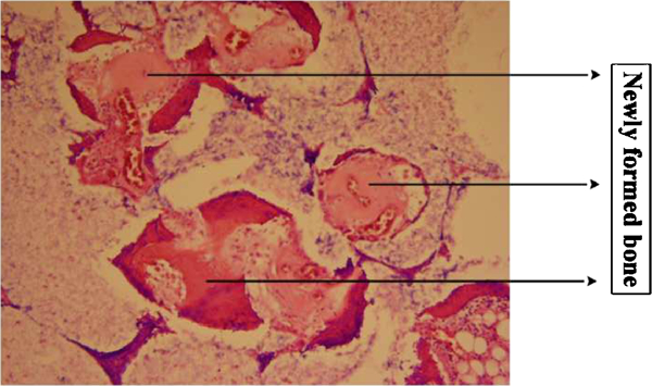
Optical micrographs of C5 composite sample in implant site 2 months post-operatively (H&E magnification, ×100)
The HAp, DCPD and biphasic CaP compositions are osteoconductive, as they support bone apposition and growth, but are slowly degraded in the body.68–71 Dicalcium phosphate dihydrates are more soluble in body fluids than HAp.72 The resorption rate of biphasic CaP is between that of pure HAp and pure DCPD. The biphasic CaP concept is based on an optimum balance between the more stable phase (HAp) and the more soluble phase (DCPD). Biphasic CaP ceramics are soluble and gradually dissolve in vivo, seeding new bone formation as they release calcium and phosphate ions into the biological medium.73 The formation of the dynamic interface between bioactive ceramics and host bone is believed to result from a sequence of events involving interaction with cells and the formation of a carbonate HAp by a dissolution/precipitation process.73,74 Bioceramics support bone formation while they partially dissolve and degrade in the body. It is noticeable that these properties are related to their physical, chemical and microstructural characteristics, which depend on their sintering temperature. During the CaP biomaterial synthesis process, the sintering temperature is a crucial step for obtaining bone substitutes with reproducible biological properties. It is worth mentioning that the sintering temperature effectively modifies the microstructure, specific surface area and dissolution characteristics of CaP ceramics. High microporosity and small crystal size are essential for the adhesion, proliferation and differentiation of osteogenic cells that will produce the bone extracellular matrix. On the other hand, high sintering temperature decreases the specific surface area and dissolution properties.66
It is important to point out that the histomorphometry of the amount of bone coverage in the C4 sample at 1, 2, 3 and 6 months after implantation is shown in Fig. 11. As it can be seen in this figure, with increasing implantation time, the amount of host bone increased.
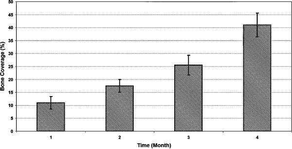
Histomorphometry of amount of bone coverage in C4 sample at 1, 2, 3 and 6 months after implantation
Conclusions
In conclusion, the experiments provide data to support the use of composite scaffolds in bone repair applications. Biomineralisation studies showed the deposition apatite phase on the surface of the scaffolds ascertaining the bioactive nature of the scaffolds. In addition, the in vitro and in vivo results showed that the scaffolds were biocompatible, and no inflammation, tissue necroses or tissue rejection was observed after implantation. The success of applying biomaterials composed of HAp and DCPD as scaffolds for generating a new bone tissue is related to the fact that this combination is biocompatible and forms a favourable three-dimensional matrix for human osteoblast cells to adhere and spread, associating the advantage of DCPD osteoinduction to the superior bioactivity and osteoconduction of HAp.