
Abstract
Co–CeO2 nanocomposite coatings were obtained by electrocodeposition of nanoparticulate cerium oxide bioceramic (25 nm mean diameter) into cobalt matrix on stainless steel support and the resulting properties were evaluated comparatively with pure cobalt layers. The surface and anticorrosion properties in the simulating body fluid (SBF) solution of the obtained Co–CeO2 nanocomposite layers are characterised using scanning electron microscopy with energy dispersive spectroscopy system, ultrahigh microtopography, Vickers surface microhardness, linear polarisation and electrochemical impedance spectroscopy techniques. The results indicate that CeO2 nanoparticles are uniformly dispersed into cobalt matrix and influence the surface morphology, microtopography, and microhardness and corrosion behaviour of nanocomposite coatings obtained. Coating thickness increases with increased current density as well as the surface roughness and microhardness. At the same time, the results obtained by corrosion tests show that when compared with the traditional polycrystalline cobalt films, the obtained Co–CeO2 nanocomposite layers exhibit the enhanced corrosion resistance in the SBF solution.
Introduction
In recent years much attention has been focused on the research and development of metal matrix composite coatings because such materials offer outstanding mechanical and multifunctional properties. These composite coatings possess enhanced properties such as wear, corrosion and oxidation resistance, dispersion hardening or self-lubrication relative to pure metal, so that they can protect the metal substrates more effectively against severe environmental conditions during operation,1 and provide improved electrocatalytic activity.2,3–10 The metal matrix composite can be produced through a number of routes including metal processing, powder metallurgy, electrodeposition techniques, etc.11 By electrodeposition technique the composite coatings are obtained during electrocodeposition of fine ceramic or polymer particles dispersed within a metallic matrix from metal electrolytic baths. There are reports on the incorporation of nanosized SiC, ZrO2, Al2O3, TiO2, La2O3 and CeO2 in the nickel matrix forming composites.3,5,12–19 The incorporated inert particles have played an important role in enhancing either the corrosion resistance or the wear resistance, many works being carried out on the Ni matrix. There are reports on the synthesis and properties of Ni–Al2O3 and Ni–ZrO2 composite coatings. 5 6 5,6,13 However, to the best of our knowledge, there are no reports on the synthesis and properties of Co composite coatings containing an oxide powder such as CeO2 and the performances characterisation of composite coatings obtained. Among the oxide ceramics, the silicon carbide, alumina and mixed alumina–zirconia systems are by far the most useful ceramics to prepare composite coatings. Because of their excellent mechanical properties, including strength, toughness and wear resistance, as well as the thermal and chemical stability, the alumina and zirconia composite coatings have been widely used in the engineering and biomedical fields for materials such as structural ceramics, thermal barrier coatings and total hip replacement.20 When organised well, the benefits of the individual components play a synergistic role in the final properties of the composite coatings obtained.21,22–28 Metals are generally chosen for their inert qualities whereas ceramics may offer bioactivity, resorption as biomaterials or hardness, wear corrosion resistance as hard facing materials for industrial use. Combining cobalt as the metal matrix and cerium oxide as the dispersion phase, the resulting composite coating could give some interesting properties with possibilities to use them as biomaterials or wear corrosion resistant coatings. Moreover, there are very few references in the literature regarding the electrodeposition of ceramic particles in the cobalt matrix.29, 30 Many of studies are carried out on electrodeposited nickel matrix.13–19,22–28
In the present work, efforts have been made to synthesise by electrocodeposition and to analyse the performance characteristics of the surface modified cobalt with CeO2 nanosized bioceramic particles (mean diameter 25 nm), in order to obtain a coating as biocompatible material or industrial use presenting at the same time good adhesion to stainless steel support, microhardness and corrosion performances. In the literature very limited studies could be found in the dispersion of nanoinert particles in the cobalt matrix and its influence on the overall properties including corrosion resistance in synovial solutions.
Material and methods
Electrochemical synthesis of Co–CeO2 nanocomposite layers
The composite materials were obtained using dispersed nanosized CeO2 particles (25 nm mean diameter) and cobalt plating electrolyte based on cobalt chloride and cobalt sulphate, using as support for coatings deposition stainless steel (304L). The compositions of the solution and operating parameters for electrodeposition are shown in Table 1. A bath solution without additives was used to avoid any reactions with the CeO2 particles and ensure that no impurities are present. Analytical reagents and double distilled water were used to prepare the plating solutions. Before deposition, the electrolyte was stirred for 24 h to ensure a good dispersion avoiding the agglomeration of particles suspended in the electrolyte.
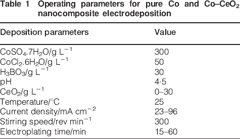
Operating parameters for pure Co and Co–CeO2 nanocomposite electrodeposition
The boric acid was used as a buffer to prevent pH changes of the electrolyte during the electrodeposition process. Particles were kept suspended with a magnetic stirrer and codeposited with cobalt resulting nanocomposite coatings of cobalt matrix with nanodispersed bioceramic of CeO2 noted in this paper as Co–CeO2.
A double electrode cell was employed to carry out electroplating experiments. The cathode, made of 304L stainless steel foils with an exposed area of 5 cm2, was positioned in the vertical plane with anode. A similar dimension of pure cobalt foil was used as the anode. The distance between cathode and anode was fixed at 4 cm. Schematic illustration of the electrochemical deposition process is shown in Fig. 1.
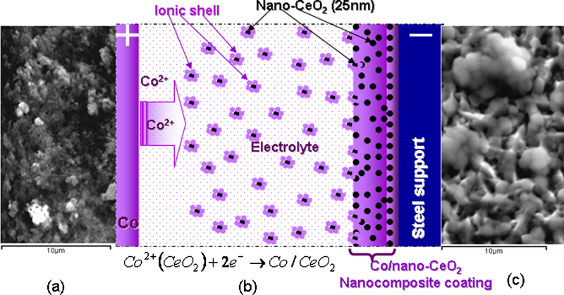
Schematic electrochemical codeposition of nano-CeO2 (25 nm) with cobalt to obtain Co–CeO2 nanocomposite coatings:
Pure dispersed cerium oxide (CeO2) was suspended in the electrolysis bath. The particles have had different shapes with an average particle size of 25 nm (Fig. 1a). The particles were kept in suspension by magnetic stirring at a rotation speed of 300 rev min−1. The cobalt foil was used as anode to make sure that the cobalt ions concentration remains constant during the electrodeposition.
Before plating, the stainless steel cathode was polished with emery papers, rinsed in distilled water and ethylic alcohol. For comparison, cobalt films were also deposited in the same electroplating conditions but without addition of CeO2 bioceramic particles.
Characterisation of Co–CeO2 nanocomposite
The surface morphologies and the components of the coatings were studied, using scanning electron microscopy (SEM) (JEOL JSM-T220, Oxford Instrument) with energy dispersive X-ray (EDS) analysis attachment.
Thickness of pure cobalt and composite deposits were verified by measuring the weight before and after deposition and also by examining the cross-section of the electrodeposited coatings using a light microscopy (MeF3 – Visilog F5).
On every electroplated surface the Vickers microhardness was measured on a Metkon type microscope with an applied force value of 0·025 kg (chosen to have a good readable tip print and avoid the influence on steel support). The average of five microhardness measurements was reported for minimising error percentage. Coating roughness was measured with a high resolution optical sensor (three-dimensional measuring station with a high resolution optical sensor, STIL).
Electrochemical studies to investigate the corrosion behaviour of Co–CeO2 nanocomposite layers and pure cobalt coatings were performed in three electrode cells using Hank simulating body fluid (SBF) as the solution having the composition present in Table 2.
Composition of SBF solution (Hank) used for corrosion resistance investigation of Co–CeO2 composite coatings
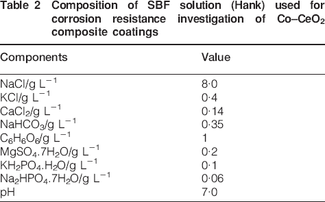
In order to minimise the influence of the coating thickness on the electrochemical measurement parameters, the sample thickness was maintained at 23±2 μm for both types Co–CeO2 and pure cobalt layers. The CeO2 dispersed phase in the Co–CeO2 composite layer was estimated at 15 wt-% after analysis. The sample was loaded in the Teflon sample holder and the exposed surface area to the corrosive medium was 1 cm2. Platinum grid of 1 cm2 area was used as the counter electrode and Ag–AgCl (saturated KCl) was used as the reference electrode. The corrosion resistance of the obtained Co–CeO2 nanocomposite films and the pure Co layer was evaluated by the electrochemical impedance spectroscopy (EIS) and polarisation measurement, using the Solartron electrochemical workstation (1286 potentiostat coupled with 1255 frequency response analyser). The sample was kept in SBF solution for 12 h in order to establish the open circuit potential EOCP or the steady state potential. The EIS measurements were conducted in a frequency range from 100 kHz to 0·01 Hz with 10 mV sinusoidal signal amplitude versus open circuit potential EOCP. The spectrum was recorded with a data density of 5 points per decade. After each experiment the impedance data were displayed as Nyquist plot. The Nyquist plot is a plot of real impedance (real Z) versus imaginary impedance (imaginary Z). Each experiment was repeated at least three times to verify the reproducibility of the results. The acquired data were curve fitted and analysed using ZView programme. After EIS measurements the system was allowed to attain the open circuit potential and a potentiodynamic curve was acquired.
Results and discussion
Preparation of Co–CeO2 composite layers
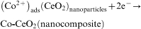
After immersing the nano-CeO2 dispersed phase (Fig. 1a) into cobalt electrolyte, it is supposed that cobalt ions are adsorbed on cerium oxide particles (Fig. 1b) being further reduced to cathode surface resulting in entrapped particles into electrodeposited cobalt matrix (Fig. 1c). The cobalt electrocrystallises and forms the metal matrix which includes the CeO2 particles to form Co–CeO2 nanocomposite coating as it is shown schematically in Fig. 1 and noted in this paper as Co–CeO2. In aqueous solutions, cobalt is present in the form of divalent cobalt ions with octahedral coordination, having four equatorial and two axial coordination sites, each occupied by water molecules. By adding CeO2 particles in the electrolyte, some ions could be adsorbed on their surfaces, transported in the electrolyte and further entrapped into cobalt growing electrodeposited coating (Fig. 1b), following the equation (1)
Influence of CeO2 nanometric particles on coating thicknesses
The coating thicknesses obtained at different current densities, determined by weighing the cathode before and after deposition or codeposition, are presented in Fig. 2. From Fig. 2 it could be noticed that by increasing the current density and maintaining the deposition time constant, the thickness of the deposited layers also increases. The Co–CeO2 nanocomposite coatings show a larger increase in thickness as compared with the pure cobalt layer at the same current density. This proves a higher current efficiency by adding the nanometric cerium oxide in the cobalt electrolyte to obtain Co–CeO2 nanocomposite coatings. By calculating the cathode current efficiency, it was found a mean value of 92% for pure cobalt electrodeposition and a higher mean value of 96% for Co–CeO2 composite codeposition.
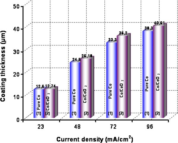
Thicknesses of layers versus current density obtained by: 1: pure cobalt electrodeposition; 2: Co–CeO2 codeposition of nanocomposite layers at 20 g L−1 of cerium oxide in cobalt electrolyte
SEM–EDS analysis of nanocomposite coatings
Figure 3 Figure 4 Figures 3–5 compare pure cobalt layers and Co–CeO2 nanocomposite coatings, carried out with 20 g L−1 of CeO2 in the plating bath at different current densities. The cobalt electroreduction is perturbed by nanometric CeO2 bioceramic particles embedded in the coating during the electroplating process as could be seen on SEM surface morphologies at different current densities.
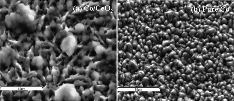
Images (SEM) of surface morphologies of electrodeposited layers obtained at 23 mA cm−2:
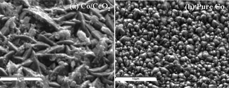
Images (SEM) of surface morphologies of electrodeposited layers obtained at 48 mA cm−2:
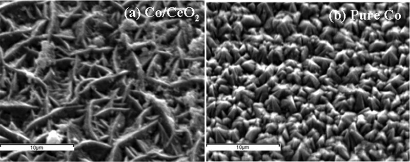
Images (SEM) of surface morphologies of electrodeposited layers obtained at 72 mA cm−2:
From Figure 3 Figure 4 Figs. 3–5, it could be observed that the pure Co surface remains regular with the increase in the current density (Figs. 3b, 4b and 5b), whereas the composite surface changes slightly from a slightly agglomerated surface to a more non-agglomerated surface with a spindle-like structure (Figs. 3a, 4a and 5a). Changes in cobalt matrix surface morphology compared with those of pure cobalt electrodeposits provide information on the effect of CeO2 dispersed phase in the electrocodeposition process of composite layers. Scanning electron microscopy–EDS analysis gave the information on the cerium oxide particles content embedded in the cobalt matrix and the mapping mode on the cerium element shows good uniformity of the dispersed CeO2 phase in the composite coatings as it is shown in Fig. 6.
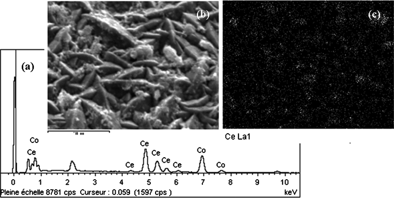
Scanning electron microscopy–EDS analysis of Co–CeO2 composite layer showing presence and uniformity of nanometric cerium oxide in cobalt matrix:
From Fig. 6a, the X-ray spectrum analysis of nanocomposite layer with cerium could be observed indicating the presence of nano-CeO2 on the SEM image performed on Co–CeO2 nanocomposite layer (Fig. 6b). Good uniformity of nano-CeO2 dispersion is shown in Fig. 6c by X-ray elemental mapping on the SEM image.
Microhardness of Co–CeO2 nanocomposite and pure Co layers
The microhardness of Co–CeO2 nanocomposite and pure cobalt layers has been determined by optical microscopy, using the Vickers method on a Metkon type microscope on the sample surface. The average of five microhardness measurements was reported for minimising error percentage. As expected there was an increase in the microhardness of the Co composite coatings after the addition of CeO2 particles (Fig. 7). The highest microhardness was exhibited by the composite electrodeposited at the highest current density of 96 mA cm−2.
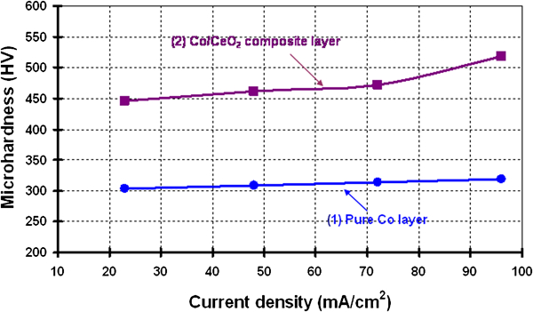
Microhardness of electrodeposited layers versus current density: 1: pure Co surfaces; 2: Co–CeO2 nanocomposite layers
Nanocomposite coatings, Co–CeO2 (25 nm) revealed a microhardness between 446 and 518 HV0·025 compared with pure cobalt layers which allow the values between 304 and 319 HV0·025. As it is known from classical metallurgy, hindering the dislocation movement could increase the hardness of a material by lattice distortions (the grain boundary density or the small grain size). The benefit of oxide (chromium oxide) in the microstructure of cobalt chromium alloys was observed by some authors in the spark plasma sintering of cobalt and chromium fine powders,31 as a new alternative method to improve the performances of alloy implants. This chromium oxide in the microstructure of the alloy can be more beneficial than carbides and the hardness of the alloy becomes closer to that of ceramics. Also its lower density enables the alloy to be lighter. The chemical stability of the oxide ensures that it remains intact and due to its insolubility in water, no carcinogenic or toxic reactions will occur.31 In our research results the cerium oxide nanoparticles embedded into cobalt matrix during the electroplating process can act as dispersion hardeners that limit dislocation movement. This assessment could confirm the differences in surface morphologies of the two types of coatings and good effect in improving microhardness of added CeO2 nanoparticles to cobalt matrix.
Surface microtopography of Co–CeO2 nanocomposite coatings
Surface microtopography of composite coatings could be an important factor for further engineering and applications of such materials, especially in implant applications for cells adhesion. The surface topography of artificial materials is known to play a significant role in interactions with biological systems such as proteins and cells.32, 33 Furthermore in a new technique using electrohydrodynamic print patterning that generates ordered topographies using proven biomaterials and composites,34 the authors notice that the topography, which includes the surface features and the features themselves, is a crucial physical cue for cells, influencing cell adhesion, proliferation and differentiation and should be considered when designing biomedical architectures.
Therefore, roughness plays an important role in determining how a real object will interact with its environment. Three-dimensional surface microtopography of pure cobalt and Co–CeO2 composite coatings were measured with an ultrahigh microtopography (STIL) with a high resolution optical sensor and further treated with an image surface analyser (Surface Map) software. The results presented in Fig. 8 show a slow increase in surface roughness with the current density for pure cobalt electrodeposited layers as well as for Co–CeO2 electrodeposited composite layers.
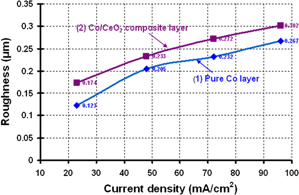
Variation of three-dimensional surface microtopography (Sa) with current density applied in electroplating process of: 1: pure Co layer; 2: Co–CeO2 (20 g L−1) nanocomposite coating
Surface roughness of Co–CeO2 nanocomposite layers is increasing from 0·17 to 0·30 μm, being higher as compared with the pure cobalt layer at the same current density (Fig. 8).
Corrosion behaviour of Co–CeO2 nanocomposite layers in SBF solution
The EIS and potentiodynamic measurements for the corrosion behaviour were performed in a three electrode cell in the SBF solution having the composition presented in the Table 2. The results are presented in Fig. 9 for Co–CeO2 nanocomposite coating and Fig. 10 for pure cobalt layer, where the points represent the experimental data and the line represents the fitting curve. The acquired data were curve fitted and analysed using ZView programme.
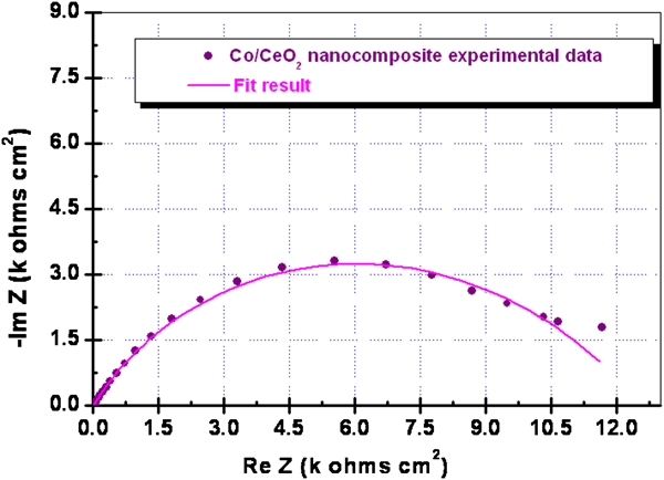
Electrochemical impedance spectroscopy Nyquist plot of experimental measurements and fitting impedance of Co–CeO2 nanocomposite coatings after 12 h of immersion in SBF solution
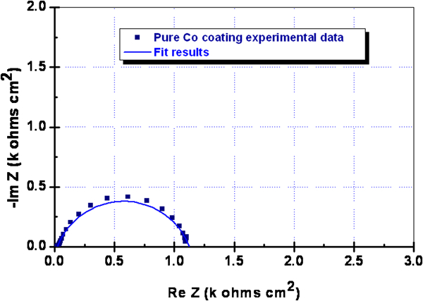
Electrochemical impedance spectroscopy Nyquist plot of experimental measurements and fitting impedance of pure cobalt coatings after 12 h of immersion in SBF solution
As shown in Figs. 9 and 10, the EIS measurements evidenced the higher corrosion resistance of the Co–CeO2 nanocomposite layers. Indeed the codeposition of the CeO2 nanoparticles into cobalt matrix improves the anticorrosion properties in the SBF solution.
The Nyquist plots of Co and Co–CeO2 coatings appeared as single semicircles. The EIS experimental data of pure cobalt layer could be fitted with a simple equivalent circuit model (Randles circuit) where instead that charge transfer resistance is the polarisation resistance, R1 of pure cobalt coating (Fig. 11a). Based on the above analysis, the EIS plot shown in Fig. 10 is analysed using the elements of the electrochemical equivalent circuit, resulting from fitting line which calculates for polarisation resistance of pure Co layer, a value of R1 = 1·11 kΩ cm2.
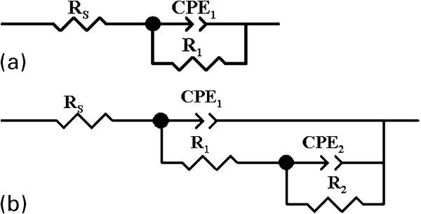
Equivalent circuit used to fit EIS experimental results for corrosion behaviour in SBF solution:
More complex equivalent circuit was necessary and proposed to fit the experimental impedance data of the Co–CeO2 nanocomposite layer (Fig. 11b).
The proposed equivalent circuit elements are as follows: Re is the electrolyte resistance between the reference electrode and the working electrode (the tested sample with pure cobalt or Co–CeO2 nanocomposite coatings), CPE1 is the double layer capacitance depending on frequency of cobalt, CPE2 is the double layer capacitance depending on frequency of insulating CeO2 bioceramic particles included into cobalt matrix or possible formed complex passive layer, R1 is the polarisation resistance of cobalt in pure cobalt layer or Co–CeO2 nanocomposite coating, R2 is the resistance of insulating CeO2 bioceramic particles included into cobalt matrix in the case of nanocomposite coating or possible formed complex passive layer.
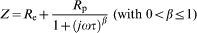
In this case the electrochemical impedance could be described by the following equation
Similar equivalent circuits were proposed to interpret the corrosion behaviour through EIS experimental data for composite coatings in nickel matrix having silicon carbide as reinforcing phase,2, 4 or copper matrix with the silicon carbide dispersed phase.35
After EIS measurements the system was allowed to attain open circuit potential and a polarisation potentiodynamic measurement was performed with a sweep rate of potential scan at 5 mV s−1. The diagrams are presented in Fig. 12 for both Co–CeO2 and pure cobalt layers.
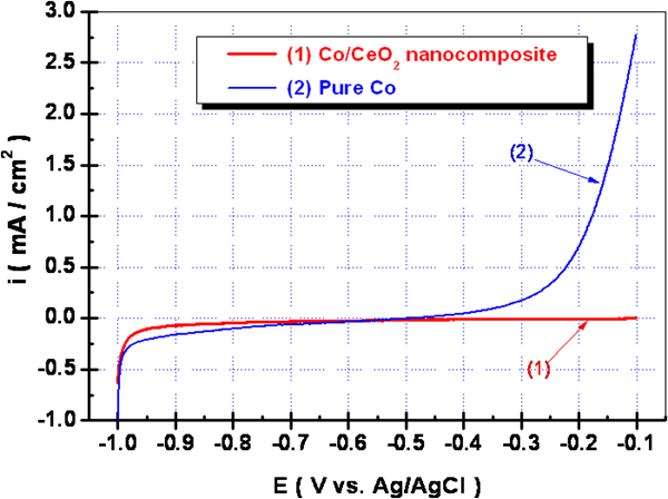
Potntiodyamic measurements in SBF solution performed with scan rate of 5 mV s−1 of potential from −1 to −0·1 V versus Ag–AgCl for: 1: Co–CeO2 nanocomposite layer; 2: pure cobalt layer
As shown in Fig. 12 the Co–CeO2 nanocomposite layers confirm the better corrosion resistance compared with pure cobalt coating having a larger passive domain and a lower passivation current density. No results regarding the corrosion behaviour of Co–CeO2 nanocomposite coatings in SBF solution were found in the literature.
Conclusion
Electrocodeposition of bioceramic CeO2 nanometric particles in the cobalt matrix was investigated with the main aim to evaluate the effect of nanoparticles in the electrolyte during the electoreduction of cobalt and performance investigation of Co–CeO2 nanocomposite layers obtained.
The morphology of Co–CeO2 nanocomposite layers is changed as compared with pure Co layers obtained at the same current densities. Good uniformity of nanometric cerium oxide dispersed particles into cobalt matrix is obtained and is proved by SEM–EDS cerium mapping on Co–CeO2 nanocomposite surfaces.
The Co–CeO2 composite coatings are uniform and well bonded to the steel substrate, the thicknesses increase with increasing current density and the microparticulates affect the electrodepositing process of the Co coating, by a slow increase in the Co–CeO2 nanocomposite coating thickness compared with pure Co obtained at the same current density, meaning probably a slow increase in current efficiency.
The Co–CeO2 nanocomposite coatings exhibit better corrosion resistance compared to plain Co, observed from higher polarisation resistance (∼10 times) on electrochemical spectroscopy and potentiodynamic diagrams plotted in the SBF solution.
The present study has shown that by adding nano-CeO2 bioceramic dispersed particles to cobalt by electrocodeposition the synergistic combination of corrosion resistance along with improved microhardness was imparted to the cobalt matrix. Because of the combined corrosion resistance and higher microhardness, Co–CeO2 nanocomposite layers may find a lot of engineering applications.
Overall, our work demonstrates the possibility to obtain Co–CeO2 nanocomposite coatings by the electrocodeposition process of CeO2 nanoparticles with cobalt. The benefits of nanocerium oxide addition in the cobalt films are: good uniformity of dispersed phase into cobalt matrix, higher microhardness of nanocomposite layers obtained and improved corrosion resistance in the SBF solution.
Footnotes
Acknowledgements
The Romanian National Research Authority (ANCS) is gratefully acknowledged for the support of this work as a Bilateral Project Romania– France, Programme Humbert Curien (PHC) – Brancusi: ‘Nano-structured coatings for functionalised surfaces’, no. 19603 PC (contract no. ANCS 214/2009-2010). Furthermore, the work is sustained by the bilateral research agreement between Research (Competences) Centre Interfaces – Tribocorrosion and Electrochemical Systems (CC-ITES) from Dunarea de Jos University of Galati (RO) and Chemical and Material Engineering Laboratory (LGPM) from Ecole Centrale Paris (F).