
Abstract
Albizia falcataria is a tropical hardwood tree extensively used in South East Asia, which has been reported as a causal agent of upper respiratory health problems through occupational exposure to sawdust. Limited data exist on its phytochemical profile, sawdust properties and toxicological effects. In this study, alkaloids, anthraquinones, flavonoids, saponins, steroids, tannins and triterpenoids were detected in A. falcataria sawdust. These compounds are not volatile, suggesting the route of exposure to be direct ingestion or physical contact with the sawdust. Toxicological screening with the bioluminescent bacterium, Vibrio fischeri exposed to aqueous (3·5% w/v NaCl) extracts of A. falcataria sawdust showed mean EC50 greater than 9800 mg L−1 for 5–30 min exposures. Microscopic analysis shows irregular sawdust morphology that can lead to irritation, and exacerbate health effects due to wood cell bound chemical exposure. It is recommended that A. falcataria be handled as a toxic wood species.
Introduction
The recent growth of tropical hardwood processing industries has been associated with increasing reports of acute respiratory health effects related to the occupational exposure to sawdust (Liswidowati et al. 2001). Sawdust exposure of many commercially valuable hardwood and softwood species has been reported in the literature as being a causal agent of various types of respiratory symptoms, including mucous and eye irritation, and induced occupational asthma (Baran and Teul 2007; Demers 2011; Demers et al. 1997; Douwes et al. 2006; Lampe and McCann 1985; Woods and Calnan, 1976). Of the current fast growing species of tropical hardwoods, the Albizia genus has seen significant growth in use for furniture, moldings, boxes, pallets, crating, fiberboard, particleboard and pulp feedstocks (Office of International Affairs, 1983; Krisnawati et al. 2011). As a result, the first reports of occupational exposure to sawdust from A. falcataria have recently surfaced in Japan, as causal agents of occupational asthma (Tomioka et al. 2006). No chemical causative agents have been identified for negative health effects in the literature.
Albizia is a genus of hardwood trees included in the taxonomic family of Fabaceae (Leguminosae), sub- family Mimosoideae. A. falcataria, a species native to the Molucca Islands of Indonesia, Papua New Guinea and the Solomon Islands is among the fastest growing tropical trees in the world, growing 15 m in 3 years under favorable conditions (Office of International Affairs 1983; Chudnoff 1984). It is known by other scientific names, Paraserianthes falcataria and Falcataria moluccana and under the trade names Moluccan sau, Albizzia, Molucca Albizzia, Batai, Mara or Pauh (Chudnoff 1984). Owing to its nitrogen fixing potential, A. falcataria can thrive in nutrient poor soils and has been widely used as both a fodder tree for grazing cattle and as a shade tree with agricultural plantation species such as coffee, tea and cacao, as well as a windbreak for bananas (Office of International Affairs 1983).
The growth in demand from the Asian wood products markets has resulted in increased acreage and production of Albizia, as well as other fast growing hardwoods, within several Southeast Asian nations. While board foot production has increased, efforts in improving the quality of these woods for improved durability, growth rate and insect resistance through amplified fragment length polymorphism based strategies (Aparajita and Rout 2009), optimisation of propagation techniques (Ghosh et al. 2010), and investigations to identify physical and chemical properties and quantify their variability within and between Albizia species is actively occurring (El-Harwary et al. 2011). These international research programs will likely further promote the inclusion of Albizia sp. and other tropical fast growing timber into value added products, increasing occupational exposure to sawdust and the reported health effects to the workers.
To date, investigations of the properties of whole extracts or pure chemicals isolated from Albizia wood species have focused on the isolation of compounds with agricultural, industrial, and health care applications. Amino acids, vitamins, and minerals have been quantified for the commonly grown fodder tree, A. lebbeck (El–Hawary et al. 2011). Cytotoxicity for the A2780 ovarian cancer line showed IC50 values (concentration producing 50% inhibition) of 0·8, 1·5 and 0·6 μg mL−1 respectively for alkaloids and triterpenoid saponins extracted from A. gummifera, gummiferaosides A–C (1–3) (Cao et al. 2007). Similarly, Julibroside J21 isolated from A. julibrissin was cytotoxic against the human Bel-7402 liver cancer cell line with an IC50 of 10 μg mL−1 (Zou et al. 2006). These studies indicate that Albizia species can possess bioactive chemical constituents that may contribute to negative health effects reported during occupational exposure to sawdust of A. falcataria. However, neither the chemistry nor the toxicity of A. falcataria has been thoroughly investigated or previously reported.
The aim of this study was to generate a baseline phytochemical characterisation of A. falcataria wood dust, including the analysis of volatile and semivolatile species, identification of sawdust particle geometric features that could contribute to human tissue irritation, and assessment of the cytotoxicity of crude aqueous extracts of A. falcataria wood dust. The association of these results with plausible mechanisms for explaining the reported health effects associated with Albizia falcataria sawdust exposure is discussed.
Materials and methods
Plant material
A. falcataria wood sawdust samples were collected from an active wood processing plant and shipped from Java, Indonesia by the PT Albasia Bhumiphala Persada company, a partner of Pacific Ring Incorporated (Seattle, WA, USA). Albasia Bhumiphala Persada produces butt jointed and finger jointed lumber core panel products and has reported cases of upper respiratory irritation to workers exposed to A. falcataria sawdust. For the current investigation, the same type of wood sawdust as present in the worker's typical environment was provided. This resulted in the analysis of sawdust generated from kiln dried A. falcataria wood panels with varying particle sizes.
Extraction and fractionation of compounds from A. falcataria
Evaluation of the possible bioactive compounds from A. falcataria required the production of extractives, or so called secondary metabolites employing a multiple, stepwise extraction process, generating polar and non-polar fractions that contained the compounds of interest in solution. A schematic of the stepwise extraction method used in this preparative process is shown in Fig. 1.
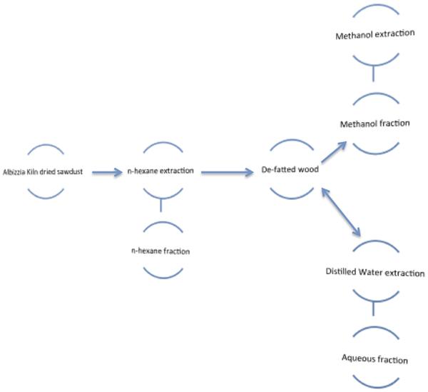
Schematic diagram of A. falcataria sawdust chemical extraction and fractionation
n-hexane extraction
The A. falcataria sawdust samples were dried at 50°C overnight in an oven and homogenised through a Wiley Mini Mill (Thomas Scientific, Swedesboro, NJ, USA) to pass through a 20 mesh sieve (0·853 mm opening). A total of 16 g of sawdust were processed as four, 4 g samples, carefully weighed into a Soxhlet extractor glass thimble. A flat bottom flask containing 150 mL of n-hexane (GC/MS grade; Burdick & Jackson, Morristown, NJ, USA) was installed at the base of the Soxhlet apparatus, and the mixture was heated, allowing it to reflux for 3 h. The defatted sawdust in the thimble was removed from the Soxhlet apparatus and set aside to air dry inside an extraction hood. The n-hexane liquid fraction was evaporated to dryness under reduced pressure using a rotary evaporator. The weight of the precipitated film formed after rotary evaporation was recorded and the film was stored at 4°C for volatile and semivolatile species analysis by GC/MS.
Methanol extraction
One half of the air dried, defatted sawdust produced after n-hexane extraction was exhaustively re-extracted with 150 mL methanol (HPLC grade; J.T. Baker, Phillipsburg, NJ, USA) in a Soxhlet apparatus. Exhaustive extraction occurred over a period of 12 h, replacing the methanol after 3 h of reflux. The collected extracts from each consecutive methanol extraction step were combined and concentrated under reduced pressure using a rotary evaporator. The weight of the precipitated film formed after rotary evaporation was recorded and the film was stored at 4°C for phytochemical, volatile and semivolatile species analysis by GC/MS.
Water extraction
The remaining half of the defatted sawdust produced after the n-hexane extraction were macerated with distilled water (2·5 times the wood volume), under agitation on a hotplate/stirrer set at 40°C and 600 rev min−1 for 3 h. This macerated mixture was filtered and exhaustively re-extracted with distilled water over a period of 12 h, replacing the distilled water after 3 h of maceration. The collected extracts from each consecutive extraction steps were combined and evaporated to dryness at 60°C. The weight of the resulting precipitate was recorded and the solids were stored at 4°C for phytochemical screening analysis and GC/MS analysis.
Phytochemical screening
Sample preparation
The individual films created by each extraction process were redissolved with suitable solvents prior to testing for the presence of bioactive phytochemical compounds. The n-hexane soluble extractive film was dissolved with 5 mL of n-hexane (GC/MS grade; Burdick & Jackson), the methanol soluble extractive film was dissolved with 5 mL of methanol (HPLC grade; J.T. Baker) and the water-soluble solids were dissolved in 5 mL of distilled water. Owing to the number of phytochemical screening tests performed, the original extraction steps were repeated three additional times to produce sufficient material. The number of screening tests was necessary to validate the presence of the phytochemical species in the samples using multiple approaches. This was needed, as the phytochemical profile of A. falcataria is not described in the literature.
Test for alkaloids, Dragendorff test
To a 1 mL aliquot of each of the redissolved extracts, 5 drops of 1M HCl (ACS grade; J.T. Baker) was added followed by 3 drops of prepared Dragendorff's reagent. This reaction immediately formed a precipitate that, based on the colour, indicated the presence of alkaloids with either an orange or orange red precipitate (Malec and Pomilio 2003).
Test for anthraquinones
To a 1 mL aliquot of each of the redissolved extracts, three drops of 1M dilute sulfuric acid (reagent grade; J.T. Baker) were added. This was partitioned with 3 mL of anhydrous benzene (99·8%; Sigma-Aldrich, St Louis, MO, USA), to form a visible layer. Three drops of 5 N ammonia (ACS grade; J.T. Baker) was added. A rose pink colouration indicated the presence of anthraquinones (Kumar et al. 2009).
Bornträger's test for anthraquinones
To a 1 mL aliquot of each of the re-dissolved extracts, 2 mL of 10% aqueous solution of ferric chloride (ACS grade; Fisher Scientific, Fair lawn, NJ, USA) and 1 mL of 12M concentrated HCl (ACS grade; J.T. Baker) were added and heated over a water bath. The extract was cooled, filtered and centrifuged; the resulting filtrate was thoroughly agitated with 2 mL diethyl ether (ACS grade; EMD Chemicals, Gibbstown, NJ, USA). The ether extract was further partitioned with 2 mL of concentrated 15M ammonia (ACS grade; J.T. Baker). A pink or deep red colouration of the aqueous layer was a positive indication for anthraquinones (Kumar et al. 2007).
Test for flavonoids
To a 1 mL aliquot of each of the redissolved extracts, three drops of 1M sodium hydroxide (ACS grade; J.T. Baker) was added until an intense yellow colour was produced. The dropwise addition of 1M dilute hydrochloric acid (ACS grade; J.T. Baker) to this solution will produce a colourless solution if flavonoids are present (Onwukaeme et al. 2007).
Shinoda test for flavonoids
A 1 mL aliquot of each of the redissolved extracts was evaporated and rediluted with 1mL of water, to which 1 mL of methanol (HPLC grade; J.T. Baker) and 4 mg of magnesium ribbon (99% pure; Fisher Scientific, Waltham, MA, USA) was added, followed by the addition of four drops of 12M concentrated HCl (ACS grade; J.T. Baker). The formation of a pink to red coloured solution indicated the presence of flavonoids (Maridass et al. 2008).
Frothing test for saponins
A 1 mL aliquot of each of the redissolved extracts was diluted with 5 mL distilled water and thoroughly agitated to induce the formation of a frothy foam. The presence of a persistent froth, which remained standing for 15 min or more, was a positive result for saponins (Onwukaeme et al. 2007; Mojab et al. 2003).
Emulsion test for saponins
The froth from the positive frothing test of saponins described above, was separated from the liquid phase and placed inside a glass test tube. To this froth, three drops of olive oil were added and observed for the formation of an emulsion. The presence of an emulsion indicated further evidence of saponins (Edeoga et al. 2005).
Liebermann–Burchard test for pentacyclicsteroids and terpenoids
To a 1 mL aliquot of each of the redissolved extracts, 1 mL of chloroform (ACS grade; Fisher Scientific), 3 mL of acetic anhydride (Reagent grade, Acros) and two drops of 18M concentrated sulfuric acid (Reagent grade; J.T. Baker) were added. A dark green colouration of the liquid is a positive result for the presence of steroids; a pink colour indicates the presence of terpenoids (Maridass et al. 2008).
Salkowski test for steroids
To a 1 mL aliquot of each of the redissolved extracts, 0·4 mL of chloroform (Reagent grade; Fisher Scientific) and 0·60 mL of 18M sulfuric acid (Reagent grade; J.T. Baker) were carefully added with a Pasteur pipette, to produce a visible interphase between liquid layers. A reddish brown colouration on the interphase indicated a positive result for the presence of steroids (Edeoga et al. 2005).
Test for tannins
To a 5 mL aliquot of each of the extracts, three drops of 1% lead acetate (ACS grade; Fisher Scientific) were added. The formation of a yellow precipitate indicated the presence of tannins (Ramaan et al. 2006; Santhi et al. 2011).
Braemer's test for tannins
To a 2 mL aliquot of each of the re-dissolved extracts, 2 mL of 10% aqueous ferric chloride solution (Laboratory grade, Fisher Scientific, Waltham, MA) was added. The formation of a dark blue, dark green or dark green/grey colouration of the solution indicated the presence of tannins (Maridass et al., 2008).
Gelatin test for tannins
A 2 mL aliquot of each of the redissolved extracts was mixed with 2 mL of a 2% aqueous gelatin solution. A white curdy precipitate was a positive result for tannins (Ramaan et al. 2006; Strumeyer and Malin 1969).
Test for phenols
A drop of each redissolved extract was spotted onto filter paper (Whatman #1, Buckinghamshire, UK) and then a drop of phosphomolybdic acid (ACS grade; Fisher Scientific) was added to the spot. This filter paper was then exposed to ammonia vapors (ACS grade; J.T. Baker). The development of a blue colour in the spot was indicative of phenols being present (Kumar et al. 2007).
Liquid phase volatile and semivolatile analysis
The n-hexane, methanol and water extraction fractions were directly analysed by gas chromatography and mass spectrometry (GC/MS), for identification of volatile and semivolatile species. For the analyses, liquid aliquots from the n-hexane and the methanol extractions were directly injected into an HP/Agilent 6890 GC, coupled to an HP5973 MS (Agilent Technologies, Santa Clara, CA, USA). Special preparation of the water extraction was required to prevent damage to the equipment. The water fraction was evaporated in nitrogen and the dried materials were redissolved in 1 mL of pyridine (Reagent grade; Acros).
An Agilent DB-1MS (60×0·250 mm ID) column (Agilent Technologies) was used for n-hexane extracts, non-polar compounds, and a Phenomenex Zebron ZB-5MSi (15×0·250 mm ID) column (Phenomenex, Torrance, CA, USA) was used for the methanol and water extracts containing polar compounds. The GC/MS instrument was set up with a splitless injector, heated to 200°C. Helium (He) was used as the carrier gas with an oven temperature program of 200°C, ramped at 15°C min−1 to a final temperature of 280°C.
To induce volatilisation of high molecular weight species, derivatisation was conducted using BSA+TMCS [N,O-bis(trimethylsilyl)acetamide+trimethylchlorosilane] 5∶1 (sylon BT kit; Sigma-Aldrich). The resulting spectra from all the analyses were compared with the NIST Atomic Spectra Database 05 (NIST 05; Agilent Technologies).
Crude saponin volatile and semivolatile analysis
A crude saponin precipitate was prepared by diluting the methanol soluble extractive film, with an equal volume of n-butanol (Reagent grade; Frey Scientific Co., Nashua, NH, USA) and distilled water. As the phases separated, 2 mL of anhydrous ethyl ether (ACS grade; EMD, Gibbstown, NJ, USA) was added to the butanolic fraction. This resulted in the formation of a crude saponin precipitate which was filtered through a glass fibre Gooch filter under vacuum and air dried. The precipitate was dissolved in pyridine (Reagent grade; Acros) before injection in the GC/MS instrument, following the procedures described above for the methanol fraction analysis.
Microscopy analysis
The Albizia falcataria sawdust sample, prior to any chemical treatment, was placed in a stereoscopic reflective microscope (Model DC5-420TH; National Optical and Scientific Instruments, San Antonio, TX, USA), fitted with a 2 MP digital camera (National Optical and Scientific Instruments) and calibrated with a 0·2 mm stage micrometer at a magnification of ×32. Images were captured using Motic Image Plus software (version 2·0; National Optical and Scientific Instruments). For comparison purposes, a locally available fast growing hardwood, Alnus rubra (red alder), was also imaged.
Toxicity analysis
Toxicity assays were performed according to the Microtox Basic Solid Phase Test (Basic SPT), using the sdix, Microtox Omni software version 4.1 (Strategic Diagnostics, Inc., Newark, DE, USA) for data capture and processing. The Basic SPT assay utilises light emissions produced by a live non-pathogenic, bioluminescent marine bacterium, Vibrio fischeri, following exposure to a dilution series of the suspected toxic substance.
Total light emitted was measured using a Model 500 Analyser (AZUR Environmental, Carlsbad, CA, USA). The Model 500 Analyser, a temperature controlled photometer, was used to maintain samples and the bacterial cell suspension at a constant temperature during the test and for measuring light output; temperature is maintained at 15±0·5°C for 20 sample cuvettes plus a ‘reagent’ cuvette containing the bacterial cell suspension. The Model 500 Analyser was set at the ‘Microtox Acute’ mode (i.e. Microtox Acute Toxicity Test) for all measurements.
The assay tests nine concentrations of the suspected toxicant prepared as serial dilutions; in addition, a baseline with no toxicant was used as the 100% bioluminescent control to calibrate diluted toxicant readings. The bacterial test organism, live V. fischeri, is sensitive to a wide range of toxicants (Jennings et al. 2001). If a toxic substance was present in the diluted sample, the level of bacterial bioluminescence was reduced. Reduction in light production is proportional to the disruption of the metabolic pathway producing bioluminescent activity and subsequent cell death.
In this study, aqueous extracts obtained from A. falcataria sawdust were tested by suspending living bacterial cells in the aqueous sawdust extract mixed with the Microtox solid phase (SP) diluent (3·5% NaCl w/v). A. falcataria sawdust extracts were generated by mixing different quantities, 1·5, 2·0, 2·5 or 4·0 g of sawdust with 35 mL aqueous SP diluent and then agitating the mixture for 10 min at room temperature. The mixture was centrifuged for two minutes at 2500g to remove large sawdust particles from the mixture and facilitate pipetting of the extract. Serial dilutions were prepared with SP diluent for each extraction volume, according to the Basic SPT protocol. Three replicate dilution series were made for each extraction volume.
According to the Basic SPT protocol, 15 min after 10 μL cell suspension was added to cuvettes containing 500 μL SP diluent without sawdust extract, light output (luminescence) was measured and recorded as ‘time 0’ T0, for exposure before toxicant exposure. The sdix software used data reduction formulae to calculate the correction factor Rt as shown in equation (1)


Data were further reduced, manually, to construct plots of per cent effect versus sawdust extract concentration over the entire range of concentrations tested. Mean per cent effect was calculated from the sdix accepted values for each concentration by exposure period (i.e. 5 min, 15 or 30 min). Only concentration values for the 5 min exposure were used for the calculated means and the plot of the 5 min exposure data; similarly for the 15 and 30 min exposure periods. Additionally, the mean EC50 for each exposure period was calculated from the sdix calculated EC50 values reported. Using the graph functions available in Microsoft Excel (2003), data for mean per cent effect versus concentration were plotted separately for each of the three test exposures, 5, 15 and 30 min.
Results and discussion
Extractives
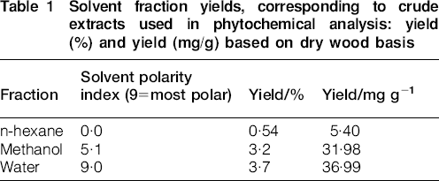
The original A. falcataria wood dust was extracted using increasing polarity solvents. The major fractions containing extractive compounds were found in the aqueous and methanolic fractions (Table 1). This corresponds with extractive analysis reported for other species, where there is a higher abundance of polar compounds as compared to non-polar compounds (Gutiérrez et al. 1999).
Solvent fraction yields, corresponding to crude extracts used in phytochemical analysis: yield (%) and yield (mg/g) based on dry wood basis
The phytochemical analysis of these extractives, collected in the n-hexane, methanol and water fractions show the presence of potentially bioactive compounds (Table 2), in some cases, with demonstrated toxicity effects if ingested (Harbourne et al. 1999; Ogunlesi et al. 2010; Pollack et al. 1990). While Table 2 summarises the phytochemical tests performed on A. falcataria sawdust, a detailed chemical characterisation of the extracted fractions is presented in Table 3, using GC/MS.
Presence of potentially bioactive compounds in A. falcataria: presence is represented by plus sign (+) and absence is indicated by a minus sign (−)
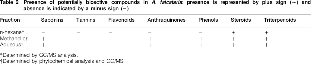
Determined by GC/MS analysis.
Determined by phytochemical analysis and GC/MS.
Chemical identification of individual volatile and semivolatile species, ordered by molecular weight (MW), composition and National Institute of Standards and Technology number (NIST#) isolated from Albizia falcataria sawdust
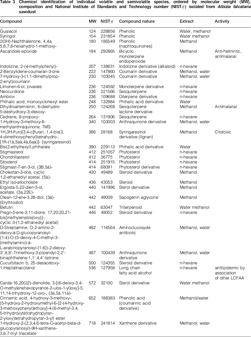
Table 3 identifies monoterpene derivatives, triterpenes and phytosterols in the n-hexane fractions analysed, with saponins, sterols, quinones and other phenolic derivatives as being present in the methanol and aqueous extracts. The elevated molecular weights of the compounds identified suggest that although there are bioactive compounds present in A. falcataria sawdust, these will not readily volatilise by environmental factors such as temperature or humidity. This may indicate that the route of exposure is through direct skin contact and exposure to mucous membranes during inhalation of sawdust particles, rather than by inhalation of gas phase compounds produced during wood processing.
Compound identification by GC/MS was complicated by overlapping signals and formation of artefacts in the chromatography (Fig. 2). Different techniques, including derivatisation, were used in an attempt to separate the overlapping signals (Halket et al. 2005), but artefacts were created (Muir et al. 2000), making the spectrum too complicated to analyse. Derivatisation compounds are employed in GC to increase the volatility and stability of organic compounds containing active hydrogens. Some compounds form additional unexpected derivatives or byproducts, so called artefacts. Artefact formation leads to multiple peaks for the same compound or unexpected components in the gas chromatographic analysis of mixtures. Some functional groups such as aldehydes, amides, carboxylic acids, esters, ketones and phenols under certain conditions form additional unexpected derivatives from silylation reagents and their byproducts. Techniques not available for the current study that may resolve some of the overlapping peak issues include solid phase extraction, before GC/MS (Mahato 2000). Developing a methodology to quantitatively isolate these bioactive compounds without carrying over artefacts and other extraneous compounds was beyond the scope of the current study.
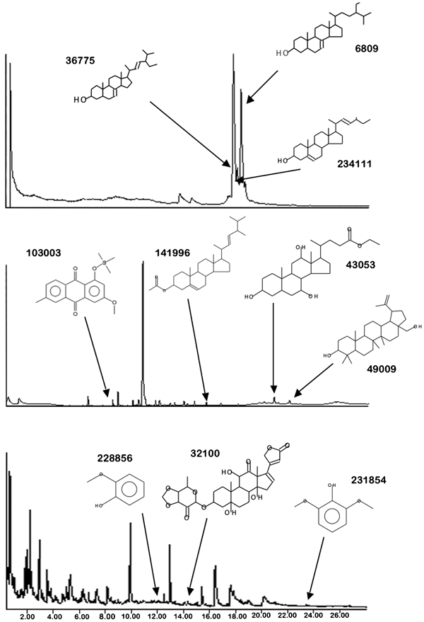
GC chromatograms of A. falcataria n-hexane extracts (top), methanol extracts (middle) and aqueous extracts (bottom): compound schematic and NIST# provided for reference
It is important to note that the evaluations conducted in this study were not quantitative, so extrapolating concentrations based on the crude extracts was not possible. Furthermore, many other compounds that were not bioactive either coeluted or coprecipitated with the bioactive compounds, resulting in additional challenges for quantifying bioactive components in A. falcataria sawdust.
Microscopy analysis
The microscopy analysis shows that A. falcataria sawdust is very irregular in shape, with individual fibrils extending in multiple directions (Fig. 3a). For comparison purposes, Fig. 3b shows sawdust particles of red alder (Alnus rubra), a locally available fast growing hardwood. The alder sawdust exhibits smoother surface geometry than A. falcataria.
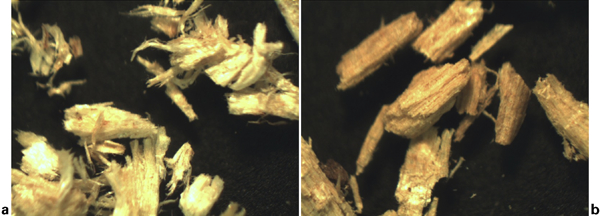
a Albizzia falcataria sawdust (×32) and b Alnus rubra sawdust (×32)
Unlike many hardwoods, the basic density of A. falcataria wood is directly related to fibre cell wall thickness rather than the per cent of vessels (water conducting cells) and fibres (Ishiguri et al. 2009). Additionally, in A. falcataria wood, the non-water conducting storage cells are scattered evenly among the wood fibres and arranged along the entire long axis of the plant (diffuse axial parenchyma cells). These cells were entirely crystal bearing (Ogata et al. 2008).
The high per cent of thick walled fibres, crystal bearing storage cells and the shredded wall configuration of the A. falcataria fibres in the sawdust particles (Fig. 3a) may expose the mucous membranes to wood cell bound extractable chemicals. In addition, once inhaled, the particles will lead to irritation and damage of delicate mucous membrane tissues. If the particles are rubbed in, as a result of scratching, particles may imbed in membranes, with the mucous moisture inducing phytochemical compounds to be released/solubilised, exacerbating the negative health effects and potentially resulting in the reported health effects (Tomioka et al. 2006).
Toxicity results
The concentration of wood dust in gram per litre of solid phase diluent required to produce a 50% measurable reduction (EC50) in light emission from the bioluminescent bacterium, V. fischeri showed a slight increase from a mean of 9800 mg L−1 at the five minute exposure (Fig. 4a) to 10 600 mg L−1 for the 15 min exposure (Fig. 4b) and 11 200 mg L−1 at the 30 min exposure (Fig. 4c). These mean EC50 data indicate that the A. falcataria wood dust extract was significantly less toxic than that reported for a standard zinc sulphate 7-hydrate, ZnSO4·7H2O of 3–10 mg L−1 at the 15 min exposure. Nonetheless, the results indicate that toxic effects were induced within 5 min of exposure to the A. falcataria sawdust extract. The cytotoxicity observed in V. fischeri was related to readily extracted A. falcataria compounds within 10 min of exposure to the water based solution.
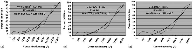
Per cent inhibition of V. fischeri bioluminescence produced at increasing concentrations of sawdust (mg L−1 SP diluent) after a 5 min, b 10 min and c 30 min exposures: mean EC50 indicates the concentration of sawdust (mg L−1 SP diluent) required to produce a 50% measurable reduction (EC50) in light emission; dark lines indicate mean per cent inhibition and light lines indicate the polynomial regression of the mean per cent inhibition; the regression formula and associated R2 value are listed on the graphic for each concentration
Based on the ease and speed of this water extraction, contact between A. falcataria sawdust with mucous membranes, eyes, mouth, respiratory or gastrointestinal tract membranes, as well as perspiration on an individual's exposed skin, would likely result in the release of bioactive compounds from the sawdust. It is reasonable to assume that the contact of sawdust and mucous membranes or moist skin, could create an aqueous extraction environment that would result in exposure of bioactive compounds that produced the observed cytotoxic effects in V. fisheri and that may account for the previous report of negative health effects of A. falcataria sawdust reported in the literature (Tomioka et al. 2006).
The naturally occurring, non-pathogenic bioluminescent bacterium, V. fischeri, has been used as a living single cell model for quantifying acute and chronic toxicity to airborne substances (Battelle, Inc., 2003), water and soil borne substances (Johnson 2005) as well as pure chemicals, including plant extracts (Layton et al. 1999; Jennings et al. 2001; Environment Canada 2002). Compared to the standard positive control zinc sulphate 7-hydrate and some plant derived compounds, A. falcataria sawdust extract is significantly less toxic as indicated by the EC50 values (Table 4).
Comparison of V. fischeri toxicity values (EC50) for aqueous extracts of A. falcataria sawdust to other water extractable pure compounds reported in literature: naturally occurring plant sources for pure compounds are included in parentheses after chemical name; toxicity to mammalian systems (LD50 values from Material Safety Data Sheet) is included for comparison
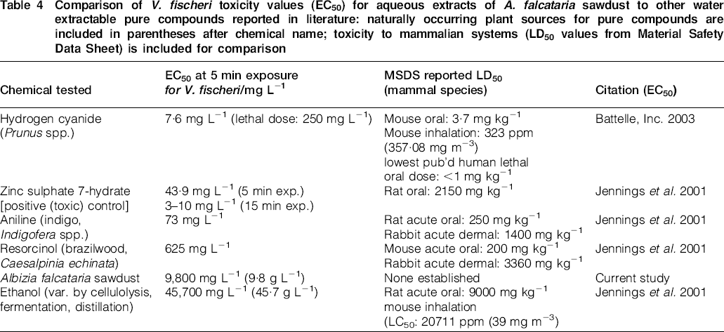
The current study does not directly evaluate toxicity in humans or other mammals. The potential for human toxicity can only be inferred from the results of other research investigating the toxicity of extractable plant compounds to both bioluminescent bacteria (V. fischeri) and to mammals (Table 4). Using this approach, the toxicity of A. falcataria sawdust extract to mammals (LD50, the dose that kills 50% of animals tested) would indicate that it could be more toxic than ethanol vapours, but over 10fold less toxic than other plant derived compounds such as resorcinol or aniline, or industrial compounds such as hydrogen cyanide based on LD50 (Table 4).
Reports from animal model studies (Määttä et al. 2006a, 2007) and cell tissue culture studies (Bornhaldt et al. 2007; Määttä et al. 2006b; Pylkkänen et al. 2007) indicated that both hardwood and softwood dust exposures were associated with increased inflammatory response in mammalian airways. Data for the health effects of A. falcataria sawdust at exposure levels duplicating the observed EC50 concentrations have not been reported in the literature. However, the current study's cytotoxicity data establishes its possible health effects within the context of the in vitro and in vivo results of other hardwoods already tested (Table 4).
Conclusions
The results from the experiments performed on A. falcataria sawdust confirmed the presence of many different types of compounds: saponins, anthraquinones, phenols, flavonoid, alkaloids, tannins and sterols, with some of them having bioactive responses, such as syringaresinol (Liswidowati et al. 2001) and phytosterols like stigmasterol and beta-sitosterol (Pascal and Segal 2006) among others.
These phytochemicals, however, are not volatile in nature, suggesting that the route of exposure to workers and people exposed to this tropical hardwood sawdust, is by direct ingestion or physical contact with A. falcataria sawdust particles in moisture rich conditions. Aqueous extracts of A. falcataria sawdust were found to induce cytotoxic effects in the bioluminescent bacterium, V. fischeri. Based on the ease and speed with which toxic components were extracted into aqueous solutions, contact of A. falcataria sawdust with an exposed individual's mucous membranes, eyes, mouth, respiratory or gastrointestinal tract or perspiration on skin would likely result in a release of bioactive compounds from the sawdust, resulting in exposure to the water extractable compounds that produced the observed cytotoxic effects in the test bacterial cells.
Reports on occupational asthma (Liswidowati et al. 2001; Tomioka et al. 2006) as well as mucous membrane irritation are indicative of a combination of bioactive compounds present in the sawdust as well as the sawdust physical shape, exhibiting a very irregular surface which can lead to persons exposed to scratch and damage further their mucous lining. Based on these findings, it is recommended that A. falcataria be handled as a toxic wood species, as delineated by the HSE toxic wood Information sheet (http://www.hse.gov.uk), and that personal protective equipment be used by people exposed to its sawdust, to prevent contact, inhalation or ingestion of A. falcataria sawdust particles.