
Abstract
The modification of wood with methylolated melamine formaldehyde resin belongs to the group of impregnation modifications. In the course of this study, Koto sapwood samples were impregnated with methylolated melamine formaldehyde resin solutions in a full cell vacuum pressure process. The samples were cured at a maximum temperature of 120°C for 24 h. To characterise the modification, the solution uptake and weight percent gain of the samples were calculated. The fixation of the melamine as parameter for the degree of curing was examined by C/N analysis. Areal UV-microspectrophotometry scans of ultra-thin transverse sections of an untreated control and samples impregnated with methylolated melamine formaldehyde resin at 240 nm were recorded. Additionally, photometric point measurements with a spot size of 1 μm−2 in the range 230 and 350 nm were conducted. UV-microspectrophotometry was proven as suitable technique for the quantitative analysis of wood modified with methylolated melamine formaldehyde resin.
Introduction
The impregnation modification of wood with methylolated melamine formaldehyde resin (MMF) has been scientifically investigated by various researchers during the last decades (Stamm 1964; Pittmann et al. 1994; Lukowsky 1999). MMF belongs to the group of amino resins and has been commercially used since the 1940s. The MMF is diluted with water to a given concentration, and then the wood is impregnated with the diluted MMF to be subsequently cured at temperatures between 80 and 120°C. MMF does not alter the original colour of the wood (Hagstrand 1999). It improves the surface hardness and dimensional stability of wood (Inoue et al. 1993; Miroy et al. 1995; Rapp 1999; Gindl et al. 2003), and from a concentration of about 7·5% MMF in the impregnation solution also the durability of wood in laboratory (Sailer 1995; Rapp et al. 1996; Rapp 1999) and outdoor exposure trials (Rapp 1999). Considerable embrittlement (Rosca et al. 2003) and low long term weathering stability (Rapp et al. 1999), however, are disadvantages of this modification approach. In order to quantify the cell wall penetration by amino resins, electron energy loss spectroscopy (Rapp et al. 1999) was used to assess MMF. UV-microscopy was employed to determine the concentration of melamine-urea-formaldehyde resin (Gindl et al. 2002). The techniques mentioned above allow for pointwise analyses of cell wall tissue only, whereas one UV-microspectrophotometry (UMSP) scan covers the entire cross-section of a cell. If suitable for the detection of MMF-resin in the cell wall, the use of UMSP would allow the investigation of larger sample areas in a shorter period of time.
Experimental methods
MMF modification
Koto (Pterygota macrocarpa) samples (25×50×50 mm, r×t×l) were conditioned at 20°C and 65% RH and end-grain sealed twice with the commercial coating Pyrotect Schutzlack 2 K (Rütgers Organics GmbH, Germany). Subsequently, the oven-dry density of the samples was determined. The samples were afterwards impregnated with a solution of the MMF Madurit MW840/75WA (Ineos Melamines GmbH, Germany). This resin was supplied as an aqueous stock solution with a solid content of approximately 75%, and diluted with tap water to obtain impregnation solutions with a solid content of 10 and 30%. These solutions were pH-stabilised by adding 1% of triethanolamine before pH 10 was adjusted by addition of NaOH. A full cell impregnation process (vacuum of 600 mbar for 30 min followed by a pressure phase of 120 min at 12 bar) was applied. Curing of the MMF was conducted in a drying oven at 120°C for 48 h. The oven dry mass of the modified samples was determined. Subsequently solution uptake (SU) with M and Mi as mass of sample before and after impregnation (equation (1)) and weight percent gain (WPG) with M1 and M2 as oven-dry mass of the sample before and after modification (equation (2)), were calculated. The calculations were based on 15 replicates per modification intensity


Nitrogen fixation
A melamine molecule contains six N atoms and the nitrogen content of untreated wood is negligible. Thus, C/N analysis allows the examination of nitrogen fixation (NF) (M1 and M2 as nitrogen content of sample before and after extraction) (equation (3)) in the wood. To examine NF, MMF modified wood was ground in a ball mill and one gram was extracted in 60 mL demineralised water at 85°C for 16 h. Extracted and non-extracted powder was oven-dried and subsequently analysed in a LECO CHN 2000-Analyzer (LECO Instrumente GmbH, Germany).

Cellular UV microspectrophotometry
Samples of 1×1×5 mm (r×t×l) from untreated controls and samples modified with 10 and 30% MMF were cut and embedded with Spurr's epoxy resin under mild vacuum. During hardening of the resin, several cycles of evacuation and ventilation were applied (Kleist and Schmitt 1999). Transverse sections (thickness 1 μm) of the embedded samples were cut with a diamond blade and subsequently transferred to non-reflective quartz-slides. After immersion in glycerine, these sections were covered with a quartz cover slip. A Zeiss UMSP 80 micro spectrophotometer (Carl Zeiss AG, Germany) set to a wavelength of 240 nm was used for areal scanning of the samples. The rectangular field of the examined tissue was digitised at a geometrical resolution of 0·25 μm−2 with the help of the software APAMOS (Carl Zeiss AG, Germany). The photometrical resolution amounted to 4096 grey scale levels which were converted to 14 colours for visualisation of absorbance intensities. Overall, more than 100 scanning-profiles and 150 UV-absorbance spectra were taken from the individual cell wall layers and cell types for the topochemical analyses. Photometric point measurements with a spot size of 1 μm−2 in the range 230 and 350 nm were conducted using an MSP 800 micro-spectrometer (J&M Spectralytics GmbH, Germany). Spectra were taken from the individual cell wall layers and were evaluated statistically. The data was evaluated with the software TIDAS-DAQ (J&M Spectralytics GmbH, Germany) and PANORAMA ProColorSearch (Analytical Software GmbH Co KG, Germany).
Results and discussion
MMF-modification and nitrogen fixation
With increasing MMF-concentration in the impregnation solution, WPG and nitrogen content increased as expected (Table 1). Since fixation is positively correlated with the thoroughness of curing (Wepner 2006), curing of the samples in the course of this study is considered sufficient. The fixation of MMF increased by about 12% at 30% MMF concentration compared to modification with 10% MMF-content. The authors assume that this difference in fixation can be explained by a higher degree of crystallisation in the sample impregnated with 30% impregnation solution.
Key data on MMF-modified Koto: solution uptake (SU) is unaffected by increase MMF-concentration in impregnation solution whereas weight-percent gain (WPG) and nitrogen fixation (NF) increase with increasing MMF-concentration
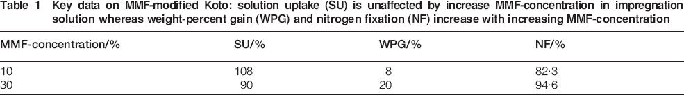
Given the same volume of impregnation solution in the vessels of samples treated with 10 and 30% impregnation solution, more MMF-monomers will be present in those vessels impregnated with the latter. Thus, during curing, supersaturation as critical initial state in the crystallisation process (Markov 2006) will be reached faster in the 30% solution compared to the 10% solution. Therefore, the onset of crystallisation is expected earlier in the former, allowing a longer and thus more complete crystallisation, resulting in a higher degree of crystallisation.
Cellular UV microspectrophotometry
Areal scans of fibre tissue of Koto are depicted in Fig. 1. Aromatic compounds of the lignified cell walls and constituents of extractives were detected in the untreated fibre tissue at a wavelength of 240 nm (Fig. 1a). A progressively increasing UV absorption was seen in the 10% (Fig. 1b) and 30% MMF treated tissue (Fig. 1c). UV absorbance reached a maximum of 0·64 in the CML of the fibre tissue, confirming the reported higher concentrations of melamine resin in the CML compared to the S2-layers (Rapp 1999). This is due to the fact that an impregnation agent diffuses from the cell lumen into the CML (Wardrop and Davies 1961; Wallström and Lindberg 2000; Zimmer 2012). Its main diffusion pathway begins in the pit chamber (Wardrop and Davies 1961). From there, submicroscopic spaces within the cell wall fibrils act as diffusion pathways into the CML (Bellmann 1955).
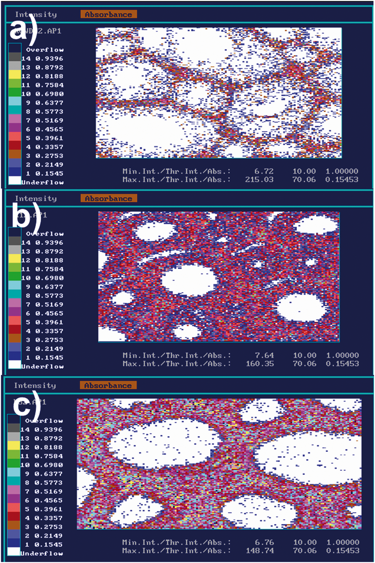
UV-microspectrophotometric scanning profiles of Koto fibres a untreated, b treated with 10% and c 30% MMF resin: absorption increases with increasing intensity of MMF-treatment
The increase in absorption of the cell walls with increasing intensity of the MMF-modification is caused by the increasing number of MMF-monomers. Each monomer contains an aromatic ring, and its delocalised π-electron contributes to the UV absorbance (Jaffé and Orchin 1962).
The photometric point measurements also show a tendency of increased absorption with increasing intensity of the MMF-modification (Fig. 2). The spectra of the control corresponds to the spectra of native wood as reported by Fergus and Goring (1970a, b) and Frankenstein et al. (2006), whereas the spectra of MMF-treated wood display elevated absorbances between the absorbance minimum at 250 nm and the maximum at 278 nm. The cell wall matrix is masked by the MMF and the microvoids are filled with the resin (Devallencourt et al. 2000). This contributes to the observed spectral behaviour.
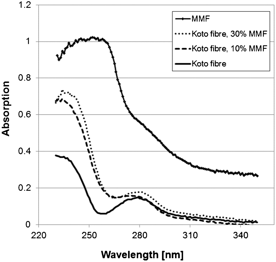
UV absorption as detected by UMSP point measurements. Increasing absorption was found with increasing treatment intensity. The maximum absorption was recorded when measuring a pure MMF-resin deposit (MMF)
The wavelength of 240 nm had been used for the detection of undisclosed MMF by UV-microspectroscopy by Gindl et al. (2003). The photometric point measurements of a MMF deposit conducted in this actual study verify this wavelength for the actual MMF-resin (Fig. 2), too.
Sint et al. (2013) applied UMSP to examine MMF-modified Bombax ceiba and Bombax insigne wood at a wavelength of 278 nm. Thus, they could only detect areas of the cell wall which were chemically similar to lignin, since lignin of hardwoods has a maximum absorption at this wavelength (Fergus et al. 1970a).
Conclusion
In this study, the suitability of UMSP for the examination of MMF-modified wood was examined. Therefore, samples of Koto were modified with impregnation solutions of 10 and 30% MMF-concentration. The different modification intensities were characterised by calculation of WPG and determination of the nitrogen-fixation. Subsequently, areal and point-photometric UMSP-scans of ultra-thin sections of the samples were recorded. The results derived from the UMSP-scans coincide with the results obtained from the supplementing investigations. Thus, the authors consider UMSP suitable for the detection of MMF in the cell walls of modified wood. Additionally, a tendency of increasing absorption with increasing modification intensity was observed.
Footnotes
Acknowledgements
The authors express their appreciation to Tanja Potsch and Karin Brandt for their valuable help during the sample preparation and the measurements conducted at the Thünen Institute of Wood Research in Hamburg, Germany.