
Abstract
Summary
Immune reconstitution disease (IRD) has been widely reported following the commencement of antiretrovirals. We report a case series from a cohort of HIV-1-infected patients of whom four developed acne vulgaris and one developed acne rosacea after the initiation of antiretroviral therapy. Acne vulgaris, as part of IRD, has been reported only once in the literature, whereas acne rosacea has not, to our knowledge, previously been described. This serves as a reminder not to overlook dermatological manifestations of disease in patients with HIV infection after starting antiretrovirals.
CASE REPORT 1
A 40-year-old black African man presented to hospital and was diagnosed as HIV-1 antibody-positive. He was found to have a CD4 count of 74 × 106 cells/L (normal range 350–1100) and a viral load (VL) >500,000 copies/mL. He was commenced on highly active anti-retroviral therapy (HAART) consisting of tenofovir, emtricitabine and efavirenz. Co-trimoxazole was started for pneumocystis prophylaxis. Three weeks after starting HAART, his CD4 count was 84 × 106 cells/L and VL was 105,920 copies/mL. A pruritic papular pustular facial eruption was seen two weeks after starting treatment. It was unsuccessfully treated with topical clotrimazole and oral minocycline, flucloxacillin and itraconazole for ‘folliculitis’, ‘seborrhoeic dermatitis’ and ‘eosinophilic folicultitis.’
At week 20 his CD4 count was 284 × 106 cells/mL with a VL <50 copies/mL. He was referred for dermatological evaluation due to the persistence of the rash.
A clinical diagnosis of acne vulgaris was made on the facial appearance of the rash by a consultant dermatologist (RCDS). Histology of a skin punch biopsy showed centrally dilated and keratin-filled follicles with mild surrounding chronic inflammation consistent with the diagnosis of acne vulgaris. Oral isotretinoin was prescribed in combination with low-dose oral prednisolone. A good clinical response with >50% resolution of the acneiform rash was achieved with three months of treatment.
CASE REPORT 2
A 33-year-old black African woman was commenced with HAART for HIV-1 infection one year after diagnosis. The patient had previous exposure to multiple antiretroviral combinations and switched regimens due to side-effects. The patient was taking a combination of tenofovir, lamivudine and nevirapine for 11 months until self-discontinuing treatment. CD4 count was 559 × 106 cells/L with a VL of <50 copies/mL while on treatment. She recalled a past history of acne during puberty.
Two weeks after discontinuing HAART, she was hospitalized with a generalized erythematous desquamating skin rash. A repeat CD4 count was 233 × 106 cells/L with a VL of >10,000,000 copies/mL. Histology from a skin biopsy revealed features in keeping with toxic epidermal necrolysis syndrome. This was attributed to HIV-1 viral rebound secondary to discontinuation of HAART. HAART was recommenced with tenofovir, emtricitabine and ritonavir-boosted saquinavir. Sixteen weeks after restarting treatment, she reported a worsening facial rash. Dermatological examination revealed a nodulo-cystic centrifacial acneiform eruption (RCDS). CD4 count at this time was 317 × 106 cells/L with a VL of 6018 copies/mL. A clinical diagnosis of acne vulgaris was made.
A course of oral minocycline and topical emollients was prescribed, which resulted in clinical improvement and resolution over a two-month period.
CASE REPORT 3
A 38-year-old HIV-1-positive Caucasian male was commenced on antiretroviral therapy (ART) consisting of tenofovir, emtricitabine and ritonavir-boosted atazanavir. CD4 cell count at the time of diagnosis was 550 × 106 cells/L with a VL of 6500 copies/mL. HAART was commenced because of declining CD4 cell count and worsening symptomatology. CD4 count was 361 × 106 cells/L and VL >500,000 copies/mL at the time of initiation of ART. The patient reported no previous acneiform or skin rashes.
Four weeks after commencing HAART, the patient developed a pustulo-papular eruption with comedones on his upper back consistent with a diagnosis of acne vulgaris (RCDS) (Figures 1 and 2). HIV VL was 4171 copies/mL.
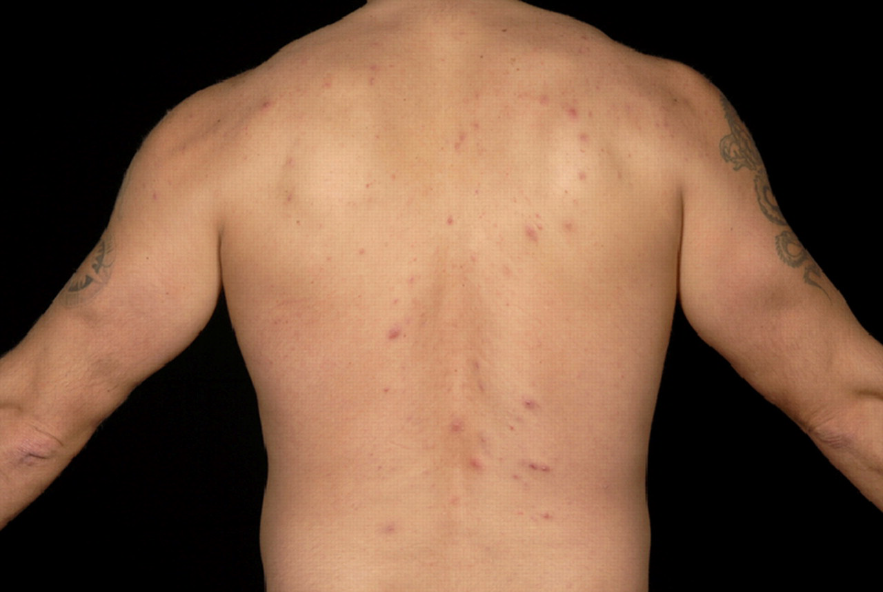
Acneiform rash on back
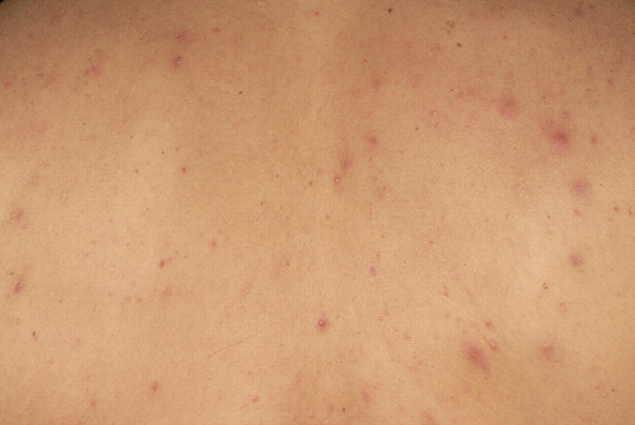
Papulo-pustular rash
CASE REPORT 4
A 37-year-old black African woman was diagnosed with disseminated Mycobacterium tuberculosis (MTB) infection and HIV-1. CD4 cell count was 6 × 10⁶ cells/L and VL >500,000 copies/mL. Abacavir, lamivudine and dose-adjusted efavirenz were started one month after MTB treatment consisting of rifampicin, isoniazid, pyrazinamide and ethambutol.
Twelve weeks after commencing ART, an itchy facial rash developed. HIV VL was <50 copies/mL. The eruption was consistent with acne vulgaris consisting of comedones, erythematous pustules and post-inflammatory hyperpigmented papules over the cheeks. The rash responded fully to topical erythromycin and oral cetirizine and clarithromycin.
CASE REPORT 5
A 38-year-old HIV-1-positive Ugandan woman restarted ART with tenofovir, lamivudine and ritonavir-boosted saquinavir after a six-month treatment interruption. CD4 count at restart was 14 × 106 cells/L. An itchy papular facial eruption developed on the forehead, cheeks and nasal bridge after two months on treatment when her CD4 count had risen to 237 × 106 cells/L and HIV VL was undetectable. This did not respond to topical erythromycin. The rash worsened with increasing papulo-pustular involvement over a four-month period. A diagnosis of acne rosacea immune reconstitution disease (IRD) was made by a consultant dermatologist (RCDS). The rash responded clinically to oral minocycline.
DISCUSSION
The patients in our case series were confirmed to have dermatological disease shortly after starting HAART. We believe this represents IRD.
After starting ART concomitant with suppression of HIV viraemia and recovery of immune function, a proportion of people will develop manifestations of other diseases – IRD. It is thought that the immunopathological response initiated by HAART restores immune response against pathogenic antigens. 1 Dermatological manifestations of IRD are common. Thevarajan et al. 2 had recently suggested the majority are due to dermatological problems in a cohort of HIV patients. 2
Acne vulgaris is a polymorphic eruption primarily of the face with the upper trunk and neck also affected, usually occurring in adolescents during puberty. It is characterized by the presence of seborrhoea, comedones, papules, inflamed nodules and pustular cysts. 3 It may be triggered by obstruction of the pilosebaceous duct, bacterial colonization of the pilosebaceous duct and inflammation. 4 The anaerobic bacterium, Propionibacterium acnes plays an important pathogenic role. 5 Acne vulgaris has been reported once previously as part of IRD. 6
The aetiology of acne rosacea is unknown. This condition presents as facial erythema, papules, pustules and telangiectasia. Comedones are absent. The role of Demodex species mites (that inhabit human hair follicles) is controversial. 7 Acne rosacea has not previously been described as IRD.
Factors that predispose to the development of IRD include the scale of HIV VL reduction. 8 All patients in our case series saw VL reductions >1 log10 copies/mL at the time of dermatological presentation. Rapid elevations in CD8 lymphocyte numbers after HAART are though to be implicated in IRD. 9 All patients in the case series experienced rises in CD8 cell counts after HAART. The skin manifestations seen in the patients of this case series may be related to an immune response directly against pathogens or Dermodex mites.
CONCLUSION
Acne vulgaris and acne rosacea should be considered as dermatological manifestations of IRD. Physicians should be aware of this (1) because symptoms and signs can mimic other diseases and hence can be misdiagnosed leading to delay in correct treatment (see Case 1); (2) because facial acneiform eruptions are distressing and can cause scarring and persistent facial post-inflammatory hyperpigmentation.