
Abstract
A 12-year-old Zimbabwean girl presented with tuberculous monoarthritis. She was moderately wasted, stunted and sexually immature. These clinical findings lead to the diagnosis of underlying HIV infection, which was thought to have been acquired from mother-to-child transmission.
INTRODUCTION
There is an increasing recognition of the substantial burden of HIV infection among older children and adolescents in southern Africa. 1 Recent UNAIDS estimates suggest that 17% of infants with HIV infection acquired from mother-to-child transmission will survive into adolescence in the absence of treatment of HIV infection. 2 Here, we describe a case of tuberculous monoarthritis in a 12-year-old Zimbabwean girl who was wasted, stunted and sexually immature. This presentation lead to the diagnosis of underlying HIV infection which was thought likely to have been acquired from mother-to-child transmission.
CASE REPORT
A 12-year-old Zimbabwean girl presented with a four-year history of right knee pain, swelling and an inability to walk. There was no history of antecedent trauma and no systemic symptoms. She had been treated presumptively for septic arthritis without improvement seven months earlier, following which she had been managed conservatively with analgesia and physiotherapy. There was a past history of hospitalization in early childhood with pneumonia on two occasions, subsequent lower respiratory tract infections requiring antibiotics every one to two years, and frequent skin complaints throughout childhood. There was no history of blood transfusions or sexual assault, and the patient denied being sexually active. A younger sibling had died in infancy from meningitis. The patient's mother had a history of recurrent respiratory infections, but the father was clinically well.
On examination, she was moderately wasted and stunted, (Z scores for height- and weight-for age were −2.28 and −2.41, respectively) 3 and sexually immature (Tanner Stage 1), with cervical and axillary lymphadenopathy, angular cheilitis and macular hyperpigmentation on the trunk, arms and face. The right knee was swollen with minimal joint line tenderness. There was wasting of the quadriceps and a fixed flexion deformity of 15° (Figure 1). Joint movement was minimal with positive varus and valgus stress tests; there was no effusion but the synovium was grossly thickened. The rest of the examination was unremarkable.
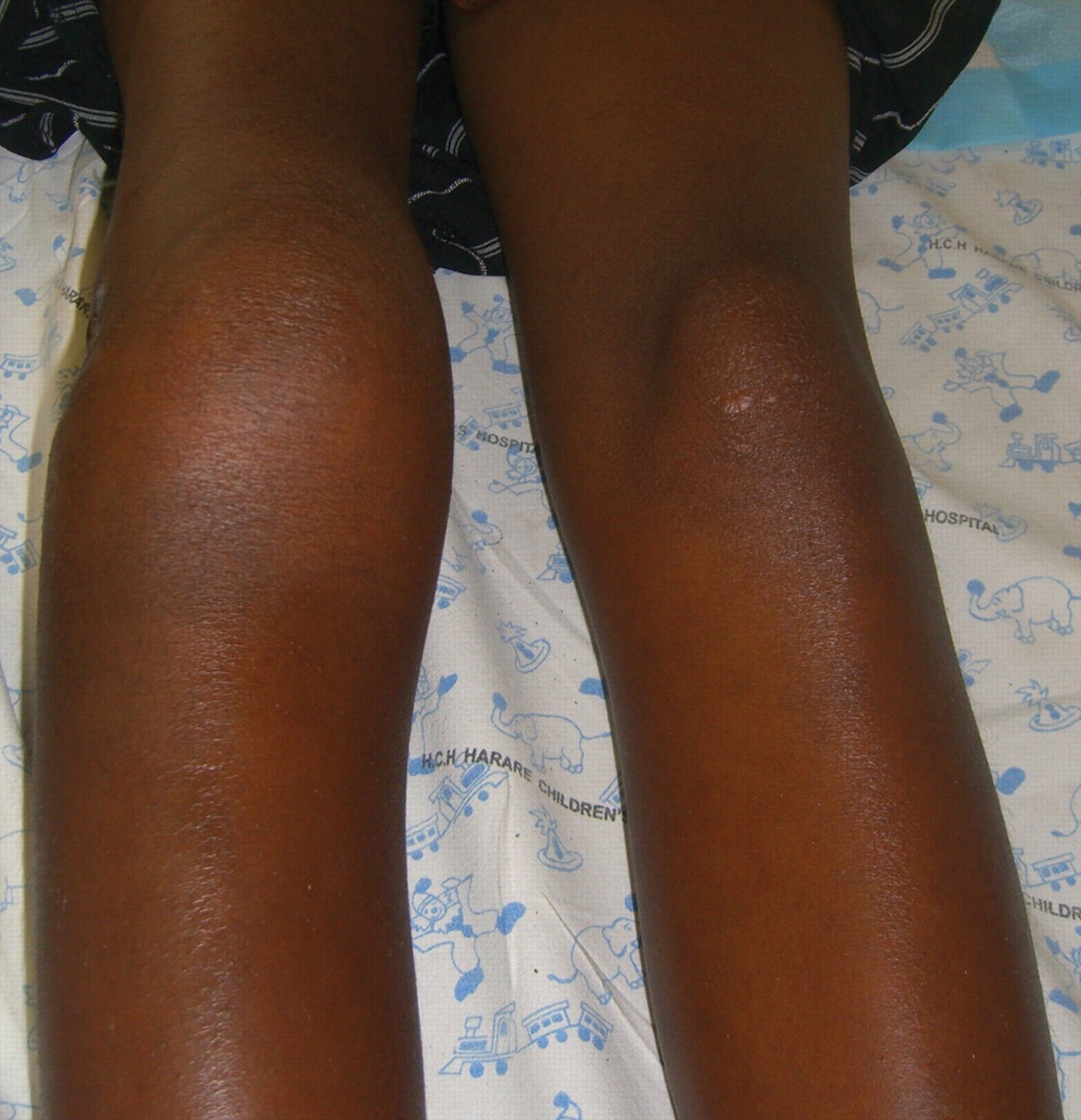
Swollen and deformed right knee with wasting of the quadriceps
A full blood count was normal and rheumatoid factor was negative. Plain radiology of the right knee showed narrowing of the joint space, subchondral erosive lesions along the femoral and tibial articular surfaces and total loss of the structure of the lateral femoral condyle (Figure 2). Synovial fluid analysis showed a mononuclear pleocytosis, but microscopy and culture for bacteria and mycobacteria were negative. Incisional biopsy of synovium showed caeseating epitheloid granulomata with Langhans giant cells, confirming a diagnosis of tuberculous osteoarthritis. An HIV-1 antibody test was positive and baseline CD4 count was 448 cells/µL. The patient's mother was found to be HIV-1 antibody positive, her father declined testing.

Radiograph of right knee showing joint deformity, loss of the femoral condyle (thick arrow), subchondral erosive lesions (thin arrow) and joint space narrowing
DISCUSSION
A worldwide re-emergence of tuberculosis has coincided with the advent of the HIV epidemic, with the risk of tuberculosis increasing with advancing immunosuppression. 4–6 However, monoarticular tuberculosis affecting the knee remains uncommon, 7,8 and its association with HIV is uncertain. 9 In resource-poor settings, late diagnosis of tuberculous arthritis is the norm due to the indolent nature of symptoms, absence of constitutional symptoms and need for surgical biopsy to establish the cause. The diagnostic delay of four years in this case is within the range previously reported, 10–12 and resulted in permanent disability. This patient had the characteristic triad of clinical features (stunting, delayed puberty, skin manifestations), past history (frequent respiratory and skin infections from early childhood) and family history (maternal death or HIV infection, sibling death) that suggests long-term survival with HIV infection acquired from mother-to-child transmission. 13 Also characteristic is the presentation with AIDS (extra-pulmonary tuberculosis), despite a relatively high CD4 count. The most recent UNAIDS estimates are that 17% of HIV-infected infants will survive to 15-years old without treatment, with long-term survival being potentially more common when HIV infection occurs postnatally from breast milk than when HIV infection is acquired at or before birth. 1,14 The resulting epidemic of adolescent AIDS is now a striking feature of clinical practice in countries, such as Zimbabwe, that have had severe adult HIV epidemics for more than a decade, 13,14 and has also been reported in children of African origin growing up in the UK. 15
Health-care workers may be reluctant to offer HIV testing to older children because of a perception that this age group is ‘low-risk’, their own lack of expertise in counselling and testing children, and concerns about the psychological impact of the diagnosis for the child and the family. However, this exposes patients to the ongoing risks of untreated immunosuppression. HIV should be considered as a possible underlying cause of growth failure, skin conditions or ill-health in older children born to mothers from high HIV prevalence countries.
Footnotes
ACKNOWLEDGEMENTS
Dr EL Corbett and Dr RA Ferrand are funded by the Wellcome Trust.