
Abstract
Circular frame treatment for limb reconstruction involves repeated follow-up visits, and a substantial number of these appointments are for pin site review only. We have encouraged our frame patients to take photographs of their pin sites when they carry out their weekly dressing changes. The photographs are taken with mobile phones or digital cameras by the patients themselves, and the images sent to us by email. We reply within 24 hours, with either reassurance or appropriate instructions as indicated. In the past 12 months, five patients have had their pin sites reviewed remotely using this method, and have expressed a high level of satisfaction. These early results are encouraging.
Introduction
Circular frames are useful for management of complex skeletal trauma (e.g. open fractures, infections, non-unions) and also for limb lengthening and deformity correction. Treatment often requires repeated follow-up visits for pin site reviews, radiographs, frame adjustments and physiotherapy. Pin site infection is a common complication after external fixation, the rates ranging from 0.5% to 30% in most series. 1 Clinical surveillance is fundamental to the treatment of pin infections, and has traditionally been done by repeat follow-up visits. This usually requires special arrangements in the fracture clinic, and most units have specialist frame clinics for this purpose.
This follow-up is not without considerable expenditure. For example, Patil et al. followed up 78 femoral and tibial non-unions treated with the Ilizarov technique, and reported a mean of 17 outpatient visits after the first postoperative check-up. Each visit cost £89 (£1 is approximately US$1.6) and the average cost over the treatment period was more than £1500 per patient for clinic visits only. 2 Similarly, Kanakaris et al.'s review found the average Ilizarov frame clinic visit and ambulance transport to cost £71 and £107 respectively. 3 A substantial number of visits, which therefore account for much of the costs, are dedicated only to pin site reviews in the first few weeks after surgery. Clearly, a quicker and cheaper mechanism for pin site surveillance would be advantageous. We advocate telemedicine as a suitable option for routine postoperative review of pin sites.
We perform 50–60 circular frame treatments in adults for trauma every year. For the past 12 months, we have been encouraging our frame patients to take photographs of their pin sites when they carry out their weekly dressing changes. This is done with mobile phones or digital cameras by the patients themselves, and the images are then sent to us by email. We reply within 24 hours. If the images are a cause for concern, further steps are taken, such as taking microbiological swabs, giving oral or topical antibiotics or recalling the patient for clinical assessment or admission. This arrangement is for pin site reviews only. Appointments for frame adjustment or radiological surveillance of fracture healing are made in accordance with existing practice. We have so far experienced a high level of patient satisfaction and significant benefits with pin site telesurveillance, albeit in a limited number of patients (five in a one-year period).
Case 1
A 19-year-old male had suffered an open fracture of his left tibia nearly three years earlier. The fracture had healed with intramedullary nail and skin grafts, but a concomitant common peroneal nerve injury predisposed him to hind foot equinovarus deformity. This was managed with percutaneous tendoachilles lengthening, and application of a Taylor spatial frame for gradual distraction. He developed a superficial pin site infection a few weeks after the frame was applied. He sent us pictures of his leg by email every week (Figure 1). He was started on a course of oral antibiotics, which cleared up the infection (Figure 2). He thus avoided making repeated three-hour round trips with his full-time working mother, just for the sake of routine pin site reviews.
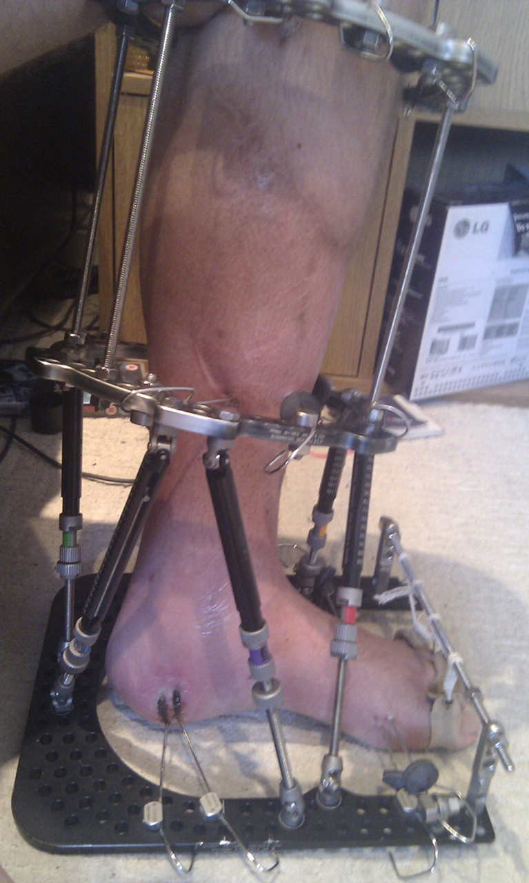
Case 1. Superficial pin site infection on the medial side. Antibiotics were started
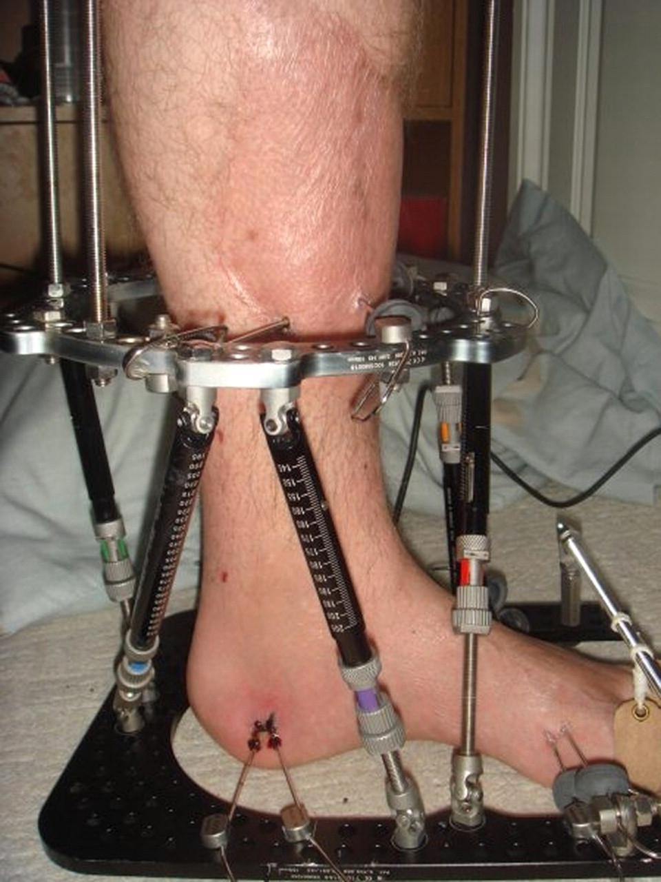
Case 1. The infection settled after the course of antibiotics
Case 2
A 39-year-old male was treated with an intramedullary nail for a tibial fracture. This went on to an atrophic non-union. He was treated with a Taylor spatial frame with excision and bone transport. Photographs were used to check wound healing, to monitor pin sites and to assess the overall alignment of the frame and limb (Figures 3 and 4).
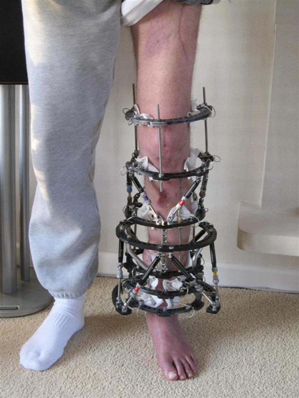
Case 2. A check of the overall alignment of the frame and limb length
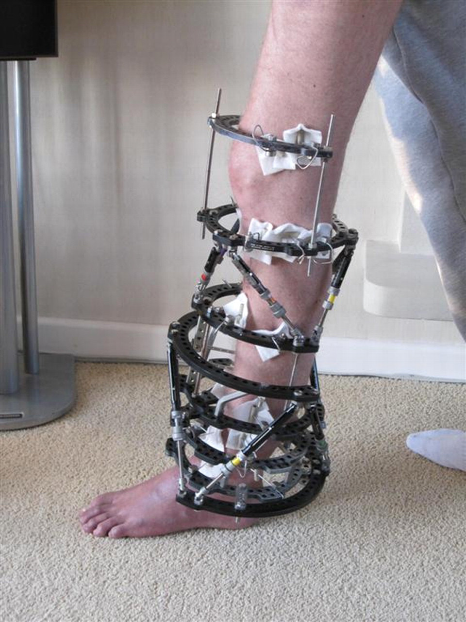
Case 2. A review of the pin-sites on the lateral side
Discussion
Numerous studies have investigated the clinical feasibility and cost-saving potential of telemedical wound care. 4–6 Telemedicine has also been applied to orthopaedics with encouraging resuts. 7,8 An Italian spinal unit found it beneficial in evaluating the status of halo brace pin sites, and initiating appropriate management when indicated. 9
Telemedicine is technically easy. The simplest model involves taking clinical images with a digital camera 10 or mobile phone, 11 attaching these as files to an email message, and sending it to the email address of a suitable specialist. About 70% of UK households have Internet access, and 92% of those aged 25–44 years are connected. 12 Many mobile phones have suitable cameras for taking high-resolution images and sending them by email. For patients without Internet access or digital cameras, imaging could be performed by a district nurse or perhaps at the GP surgery.
Circular frames tend to be tolerated without major psychological effects in most patients. 13,14 Teleimaging can be an empowering tool, and potentially further improve acceptance of and compliance with the treatment. This is especially pertinent in the case of patients treated with Taylor spatial frames, as they would have already been given computer-generated instructions for strut adjustment. There is very little patient-identifiable information on clinical images alone, so confidentiality and privacy become less of a problem than in teleradiology generally. 15 If these images are captured by patients themselves, consent is implied as long as they have been counselled by the responsible clinician. In addition, by storing the pictures, the images become available for comparison and assessment of progress. This is similar to the practice of entrusting patients with their own radiographs for fracture clinic visits. 16
Telesurveillance of pin sites has numerous time-saving benefits. Unnecessary appointments are avoided, thereby reducing the number and patient burden of specialist frame clinics, as well as the costs of ambulance transport. Patients can take and send these images in their own time and from their homes. Patients and their carers can avoid making trips in uncomfortable transport, over possibly long distances. They do not have to miss work or other commitments to make these visits. Increased patient satisfaction may result in fewer missed appointments, although we have not yet observed this in our practice.
The cost benefits to the patient and to the NHS are self-evident. Table 1 depicts the economic cost analyses of non-union treatment with circular frames, as described by Kanakaris et al. 3 and shows where we think that costs could possibly be reduced by telesurveillance of frame pin sites. This will need to be verified by a future health economics analysis.
Description of costs necessary for a complete health economics analysis of fracture non-unions (adapted from Kanakaris et al. 2007 3 ). The areas italicised are those where costs might be reduced by telesurveillance of frame pin sites