
Abstract
Aim
To illustrate the use of autologous femoral vein for grafting ilio-caval vein defects following abdomino-pelvic tumour resections.
Methods
Case report and literature review.
Results
Durable restoration of ilio-caval patency was achieved, with minimal morbidity from graft harvesting.
Conclusions
Autologous femoral vein presents a viable graft option for the immediate reconstruction of large intra-abdominal vein deficits.
Introduction
Invasion of the common iliac vein (CIV) by extra-adrenal pheochromocytoma has not been described, although reports of inferior vena cava (IVC) involvement by pheochromocytoma can be found in the world literature. 1 Effective surgical management of these tumours requires wide local resection that must include any infiltrated vein segment. Reconstruction of large venous defects is challenging; the optimal conduit for venous reconstruction remains to be defined.
Case report
A 35-year-old Caucasian woman, with three children aged eight, 12 and 15, presented with a three-year history of palpitations, sweating and hypertension. At the age of seven, she had undergone cardiac surgery for a septal defect. On physical examination, she had striking peripheral vasoconstriction. Average blood pressure was 156/122 mmHg falling to 130/108 mmHg. No mucosal neuromata, skin tags or fibromata and no axillary freckling were noted. Abdominal examination was unremarkable. Both lower limbs showed moderate diffuse swelling.
A meta-iodobenzyl guanidine scan showed increased uptake in a primary pelvic lesion, there was no tracer uptake in either bone or liver. Magnetic resonance venogram confirmed a highly vascular pelvic mass, compressing and invading the left CIV up to the commencement of the IVC with intraluminal extension up the IVC to the level of the renal veins. A complex network of para-aortic venous collaterals was also noted (Figures 1 and 2). During operation, a large vascular tumour was identified in the sacral hollow posterior to the rectum. The mass was infiltrating the left CIV and IVC origin but the intraluminal extension was free-floating beyond its base.
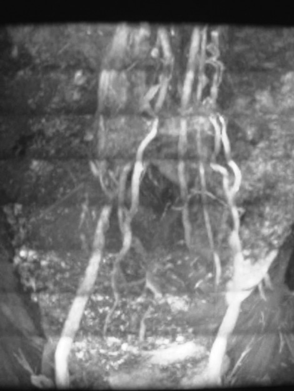
Magnetic resonance venogram demonstrating common iliac vein occlusion with multiple para-aortic venous collaterals providing drainage

Magnetic resonance arteriogram showing pheochromocytoma occluding the left common iliac vein and extending intraluminally up the inferior vena cava to the level of the renal veins
Vascular inflow to the tumour from the inferior mesenteric and internal iliac arteries was controlled, after which bleeding became manageable. Once the left CIV and tumour were dissected free, it was possible to withdraw the intracaval tumour intact and in continuity, using manual control of the juxtarenal IVC and with soft clamps controlling both CIVs. Once the intracaval tumour was removed, a caval clamp was applied distally. During the period of iliocaval clamping, we observed no major haemodynamic instability because of venous pooling, so that intraluminal venous shunting was not required. We attributed this to the well-established venous collaterals that had formed between the pelvis and retroperitoneum, as shown in Figure 1.
Excision of the mass resulted in a large venous defect which was bridged using an autologous left femoral vein (FV) graft, interposed between the left iliac venous convergence and the medial defect at the right CIV to IVC junction, using continuous polypropylene sutured anastomoses. The operative procedure was facilitated by the use of a cell saver that enabled 1.575 L of aspirated reconstituted red cells to be re-transfused and limited the transfusion of stored blood to two units.
The patient made an uncomplicated postoperative recovery and all antihypertensive medication was withdrawn prior to her discharge home after 10 days. Lower limb swelling reduced gradually and elastic hosiery was unnecessary after the first month. The patient returned to work after three months. Follow up with venous ultrasound scanning of the pelvic and lower limb veins had shown a patent FV graft at three, six and 24 months after operation.
Discussion
Reconstruction of deep veins is challenging and outcomes in comparison with arterial bypass procedures are generally poor. Factors that contribute to the high incidence of occlusion of deep vein grafts include the frequent presence of preformed venous collateral networks that compete with the graft for flow coupled with fact that reduced pressure and velocity of blood flow in veins when compared with arteries favours intravascular thrombosis. Consequently, the successful use of prosthetic grafts in large calibre arterial bypass procedures has not been reproduced in the context of deep venous reconstruction. 2
A limited number of autologous grafts are available for venous reconstruction: the internal jugular vein (IJV) has been widely used by Thomas et al. 3 in the reconstruction of the common femoral vessels following en bloc resection of tumours. However, its thin wall and limited length restricts the use of the vein in alternative sites. Harvesting of the IJV is not without complications and incurs visible scars. The calibre of the great saphenous vein (GSV) limits its usefulness as a deep vein substitute and although this can be enhanced by triple-panelling or spiral wrapping around a mandril, the long suture lines necessitated by this approach incur excessive labour and much potential for technical complication and thrombogenesis. 4 An alternative approach to increasing the calibre of the GSV is to expose it to arterial flow and pressure by creation of a distal arteriovenous fistula. Providing the vein is undamaged and satisfactory arterialization is achieved, its diameter should double after a period of three months, after which time the vein can be harvested for grafting. This approach is best suited to children, in whom growth potential is optional, and we have utilized it on two occasions (unpublished data). Because of time constraints its applications are limited.
The problems of low flow and pressure affecting deep veins has led to the use of adjunctive arteriovenous fistulas as a means of improving patency rates following prosthetic venous reconstruction. Steinman et al. 5 in 1966, achieved 86% patency rates using Teflon and Dacron grafts for IVC reconstruction with a temporary femoral arteriovenous fistula. The use of arteriovenous fistulas in clinical practice has been limited by a number of factors including: the additional operative time; the need for a secondary procedure to close the fistula; and the requirement of the graft to remain patent independently when the fistula is being created (early phase) or post closure (late phase).
Experience with the use of the FV as an autologous arterial graft in both aortofemoral and femoropopliteal reconstruction has confirmed that its harvesting produces little morbidity in terms of swelling of the donor limb and its calibre is comparable with that of the major intra-abdominal veins. 6,7 Our case illustrates the successful use of an autologous FV graft in reconstruction of the left CIV/IVC confluence. At two years scans confirm graft patency and minimal evidence of venous insufficiency in the donor limb.