
Abstract
We report three cases of stroke in association with peripheral venous interventions that each included foam ultrasound-guided sclerotherapy (UGS). All three female patients experienced a right middle cerebral artery (MCA) stroke causing dysphasia and left hemiparesis. A patent foramen ovale was found in each patient. The first incident occurred two days after foam UGS to treat small tributaries of a great saphenous vein (GSV). Paradoxical clot embolism was presumed in this case where concurrent deep vein thrombosis with non-occlusive thrombus in a medial gastrocnemius vein extending to the popliteal vein was detected on ultrasound. The second case occurred immediately at the completion of foam UGS and ambulatory phlebectomy to treat GSV tributaries. Paradoxical gas embolism was demonstrated in this patient confirmed by visualization of bubbles in the right MCA on CT angiography. The third case occurred one day after endovenous laser ablation (1470 nm) and foam UGS to treat both great and small saphenous veins. No specific cause could be confirmed in this patient. Sodium tetradecyl sulphate foam was used in all three cases (3%, 16 mL; 1.5%, 4 mL and 3%, 25 mL, respectively). All three patients recovered completely within a few days.
Introduction
The present report describes cerebrovascular events (CVEs) following peripheral venous interventions in three female patients with patent foramen ovale (PFO). The first patient was treated with foam ultrasound-guided sclerotherapy (UGS) alone while the other two underwent concurrent ambulatory phlebectomy (AP) and endovenous laser ablation, respectively. Sodium tetradecyl sulphate (STS, supplied as FIBRO-VEIN 3%, Australian Medical and Scientific, Chatswood, NSW, Australia) was used in all three cases.
Case reports
Patient 1: probable clot embolism
A 56-year-old female presented with recurrent varicose veins. External stenting of saphenofemoral junctions (SFJs) using Venocuff® and multiple avulsions were performed 15 years before. There was a history of migraines since the age of 12 but no personal or family history of venous thromboembolism (VTE), thrombophilia or CVE. She took tibolone, atorvastatin, omeprazole and calcium supplements. On examination, there were prominent varicosities in medial calves (C2). Duplex ultrasound (DUS) showed bilateral great saphenous vein (GSV) and left small saphenous vein (SSV) incompetence.
In February 2006, her left GSV, SSV and intersaphenous veins were treated with UGS (STS 3% foam, 15 mL) followed by a 40 mg subcutaneous injection (SCI) of enoxaparin. Foam was prepared with modifications to the original Tessari method 1 using STS 3% in a liquid-to-air ratio of 1:3 (1 in 4). Seven days later, she received 5 mL of STS 3% foam in a second UGS session. On one-week follow-up, she was asymptomatic-but DUS detected occluded segments of the left femoral (FV, 13 cm) and medial gastrocnemius veins (MGV, 9 cm), both associated with sclerosed perforators. Enoxaparin was administered for three weeks. Sequential DUS studies showed complete recanalization of the MGV but the FV remained occluded (11 cm) one year later.
In March 2006, her proximal GSV was treated with endovenous laser ablation (EVLA) (810 nm). This was followed by three sessions of UGS (STS 3% foam, 3, 3 and 6 mL per session) for the remaining segments with no complications. On six-week follow-up, DUS showed a lumen in the GSV and residual calf and thigh tributaries.
In November 2006, the residual veins of the right leg and small tributaries of the left popliteal fossa were treated with UGS (STS 3% foam, 16 mL) covered by enoxaparin 40 mg SCI. Full-length Class II graduated compression stockings (GCS II) were applied and she was instructed to walk for 30 minutes following the procedure. Two days later, she had a right middle cerebral artery (MCA) stroke causing dysphasia and left limb and facial paralysis but no visual disturbances. Magnetic resonance imaging (MRI) on the same day revealed ischaemic changes within the right lentiform nucleus and an incidental finding of a pituitary adenoma but no air bubbles were demonstrated. She made a complete recovery within one hour of the onset of symptoms. DUS showed normal carotid and vertebral arteries but there was non-occlusive thrombus in the right MGV extending to the popliteal vein. A trans-oesophageal echocardiogram (TOE) revealed a PFO measuring 25 mm. Detailed thrombophilia screening was negative. The patient was commenced on aspirin, enoxaparin and warfarin. Three weeks later and while on anticoagulants, she experienced transient left-sided numbness but no infarct or haemorrhage was detected on MRI. The PFO was closed percutaneously a week later. On one-year follow-up, she did not report any further neurological or thrombotic events.
Patient 2: Gas embolism
A 59-year-old female presented with right lower limb varicose veins. She had a history of classic migraines (with aura), for which she took ibuprofen intermittently. At the age of 39, she had suffered a stroke which presented with a left facial hemiparesis. She was treated with aspirin 100 mg daily, which was ceased three years following the event. She had no personal or family history of VTE or thrombophilia. Her current medications included atorvastatin 40 mg daily and fish oil supplements. On examination, she had prominent varicosities in the right posterior calf region, oedema and lipodermatosclerosis affecting the right medial ankle (C4b). DUS showed a right GSV incompetence, which was treated uneventfully with EVLA (810 nm) in March 2010.
Four weeks later, she underwent foam UGS (STS 1.5%, 4 mL) for the right distal GSV and posterior arch vein (PAV). This was followed by AP performed in the left lateral decubitus position, for the remaining calf varicosities. At the completion of AP, the patient was turned into the supine position and asked to lift her leg for application of GCS. Within seconds of lifting the leg, she became unresponsive and exhibited an altered mental state with slurring of speech, disorientation, a dense left arm and leg hemiplegia, and an extensor plantar response. She was transferred to hospital within 20 minutes where a computerized tomography (CT) angiogram of the brain revealed a right MCA air embolus (Figure 1). Two hours later, she was treated with tissue plasminogen activator (t-PA). Within several minutes of this treatment, her mental status returned to normal and there was a dramatic improvement in her neurological state abated with only a slight left facial droop and left-sided weakness (9/10 strength). Repeat CT several hours later confirmed complete resolution of the right MCA air embolus and no abnormalities of the right cerebral cortex. MRI the following day revealed small micro-infarcts of the right basal ganglia. She was monitored in hospital for three days and placed on clopidogrel 75 mg daily. TOE revealed a small PFO. The patient's residual facial and lower limb weakness fully resolved by discharge. DUS showed normal carotid arteries, occlusion of both GSV and PAV but no deep vein thrombosis (DVT). At three-month follow-up, she reported no further neurological or thrombotic events.
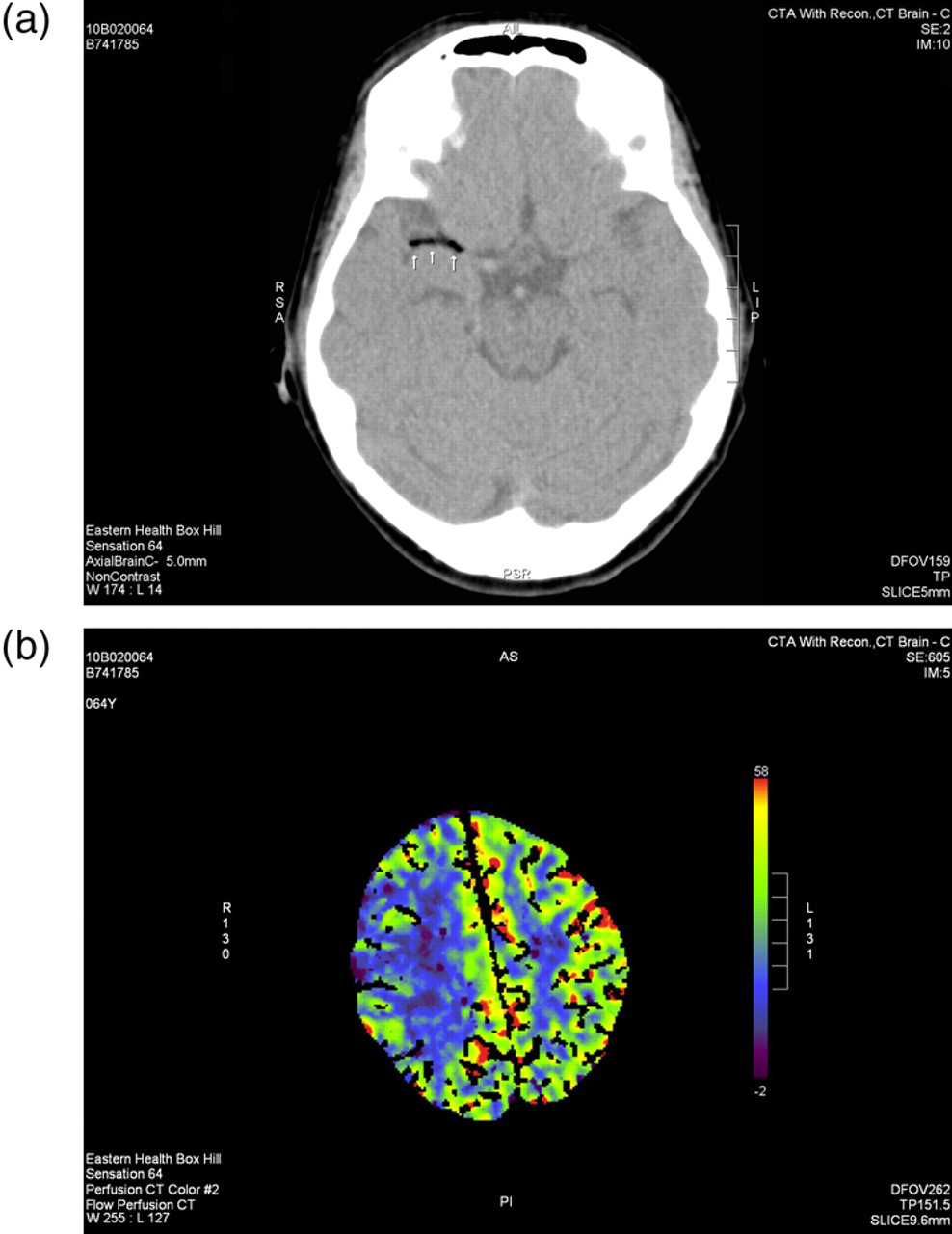
Patient 2: (a) Axial post-contrast computerized tomography (CT) of brain obtained within one hour of the onset of symptoms showing an air embolus in the right middle cerebral artery (MCA). (b) Altered perfusion on post-contrast CT perfusion imaging showing decreased flow in the right MCA territory
Patient 3: debatable cause
A 64-year-old female presented with bilateral skin signs of chronic venous hypertension. There was no personal or family history of migraines, VTE, thrombophilia or CVE. She took pentoxifylline 400 mg twice daily and aspirin 80 mg twice a week for prevention of travel-related thrombosis and thyroxine for hypothyroidism. On examination, she had prominent varicosities in medial calves, bilateral oedema and dermatitis, and lipodermatosclerosis affecting the left medial ankle (C4b). DUS showed bilateral incompetence of GSV and SSV.
In February 2008, her left GSV and SSV were treated with EVLA (810 nm) followed by two sessions of UGS (STS 1.5% foam, 15 mL per session) for the remaining segments with no complications. Foam was prepared using STS 1.5% in a 1:4 (1 in 5) ratio.
In July 2008, her right leg was treated with a combination of EVLA and UGS. One hour prior to the procedure, 30 g of lignocaine/prilocaine 5% (EMLA) cream was applied under occlusion along the length of both GSV and SSV and enoxaparin 40 mg SCI was administered. Proximal GSV was catheterized and the laser probe was placed 2 cm distal to the SFJ. A volume of 100 mL of tumescent anaesthesia (20 mL of xylocaine 1% with adrenalin 1 in 80,000, 7 mL sodium bicarbonate in normal saline) was infused into the peri-venous space. The thigh segment of the GSV was treated with a 1470 nm laser (Biolitec, Jena, Germany) at 14 W, continuous wave (CW), 63.5 J/cm linear endovenous energy density and 2.6 mm/second withdrawal rate. The proximal 2/3 of SSV was treated with EVLA (12 W, CW, 80 J/cm, 1.5 mm/second, 76 mL tumescent) and the remaining segments were treated with UGS (STS 1.5%, 25 mL). GCS II and two layers of short stretch bandages were applied. She was instructed to walk for 30 minutes.
Our patient felt unwell during that evening and was taken to the hospital following a fall the next morning. She was diagnosed with a right MCA stroke presenting with dysphasia and left limb and facial paralysis. A CT scan of the brain was normal but MRI performed several days later confirmed an infarct within the right frontal and temporo-parietal lobes. Neither modality demonstrated air bubbles in the cerebral circulation. TOE revealed a PFO measuring 18 mm. DUS showed a small plaque involving the left carotid bulb, occlusion of both GSV and SSV but no DVT. She made a full neurological recovery after four days. The PFO was closed percutaneously three weeks later and she was commenced on perindopril and atorvastatin. On two-year follow-up, she reported no further neurological or thrombotic events.
Discussion
The overall incidence of neurological complications of sclerotherapy is suggested to be less than 2%. 2,3 From 4059 foam UGS procedures performed in our Sydney practice during a six-year period, we have two known occurrences of stroke, giving an approximate annual incidence of 0.01%. A literature search revealed nine published cases of stroke following sclerotherapy since 1994. 4–12 These included four cases of stroke complicating liquid 4–7 and five following foam sclerotherapy. 8–12 Furthermore, four cases of transient ischaemic attacks (TIA) have been reported after foam 2,9,13,14 and one following liquid sclerotherapy. 15
We postulate that the stroke in our first patient was most likely due to paradoxical clot embolism (PCE). This event had a delayed onset of two days after the treatment with a concurrent finding of DVT involving the right MGV and the popliteal vein. This patient had developed a left FV occlusion following a previous sclerotherapy treatment. A pituitary adenoma was found on MRI. Intracranial tumours, including pituitary adenomas, are associated with an increased risk of VTE. 16 In such patients, VTE risk factors including genetic thrombophilias and the presence of a right-to-left shunt play an important role in the pathogenesis of stroke. 17 Small embolic clots, which would normally be lodged and lysed in the lungs without overt clinical sequelae, can paradoxically embolize to the brain causing a stroke. 18 Microemboli can also cause transient neurological dysfunction, 19 with recovery when the emboli have lysed and the circulation is restored. 20,21
In two cases described here, stroke occurred despite the prophylactic single dose of enoxaparin. The second patient was also on antiplatelet agents (aspirin and pentoxifylline). A recent study has shown that the single dose of enoxaparin is ineffective in prevention of DVT post-UGS. 22 We have since ceased this practice as a prophylactic measure.
We postulate that paradoxical gas embolism (PGE) was responsible for stroke in our second patient. This is based on the immediate onset of symptoms after the procedure and visualization of gas emboli on CT angiography obtained within the hour of the onset of symptoms. Bubbles have been visualized to enter the right heart within one minute of foam sclerotherapy despite all treatment modifications. 23 Five published cases including three cases of stroke 8–10 and two cases of TIA 9,14 had an immediate onset following foam sclerotherapy. In this group, gas emboli were visualized in the cerebral circulation of two stroke cases 9,10 and the neck arteries of one stroke 8 and one TIA patient. 9 Similarly, cases of stroke and cerebral ischaemia following the echocardiogram Bubble Studies (injection of saline froth) are all reported to occur during or immediately after the injection of the bubbles. 24
The cause of stroke in our third patient can be debated. The total volume of the sclerosant foam injected (25 mL) exceeded the volumes recommended by the European consensus (<10 mL). 25 This large volume may raise the possibility of PGE. No gas emboli were visualized on MRI but imaging for gas emboli can be negative as air and CO2 are absorbed rapidly. 26 We have subsequently limited the volume of the injected foam to 15 mL (<20 mL) per session to comply with the Australasian College of Phlebology Standards.
This patient underwent concurrent EVLA. Tongues of thrombus have been observed following EVLA of the GSV extending into the common femoral vein.
27,28
Furthermore, there has been a recent case report describing stroke following EVLA.
29
We used a 1470 nm laser with a relatively high power setting (12–14 W) compared with those currently recommended by the manufacturer (5–7 W). It is unknown whether the higher laser power can result in formation of loose clots with potential for embolism. DUS follow-up showed good occlusion of the GSV and no associated tongue of thrombus or concurrent DVT. These negative findings, however, do not rule out PCE from the segments treated with laser or sclerosants. Furthermore, EVLA also generates steam bubbles visualized on DUS generating a ‘ring down’ artefact. These bubbles have been observed to enter the cardiac chambers soon after EVLA (Dr Nick Morrison, personal communication via email to K Parsi,
Summary
In summary, we report three cases of stroke in association with peripheral venous interventions that each included foam sclerotherapy. All three patients recovered completely and had no residual neurological deficit. The common risk factor was the presence of a PFO.
Footnotes
Acknowledgements
We are grateful to Dr Chris Holden, radiologist with Imaging Independently, for providing the CT angiography images and Dr Nick Morrison for personal communication.