
Abstract
A variety of operative approaches and protective adjuncts have been used in thoracoabdominal aneurysm (TAA) repair to minimize the major complications of perioperative death and spinal cord ischemia. There is no consensus with respect to the optimal approach. We present 118 surgically treated patients over a 10-year period. The present study reviews our experience as a transition country (Serbia) in the treatment and problems we have encountered during open operative treatment of TAAs. Between 1999 and 2009, the authors reviewed 118 consecutive patients who underwent thoracoabdominal aortic resection using a variety of spinal cord protection. Clinical data collected prospectively were analyzed retrospectively. The purpose of the current study was to review the results of a large series of TAA repairs and to present some technical considerations and complications of open TAA repair. There were seven operative deaths (5.9%): two in the setting of ruptured TAAs, three myocardial infarctions and two due to hemorrhage. All 30 (25.4%) postoperative deaths occurred during the initial hospitalization. Postoperative complications included paraplegia in 11 patients (9.3%); renal failure in eight patients (6.8%), with four patients (3.4%) requiring hemodialysis; pulmonary complications in 75 patients (63.5%); bleeding requiring reoperation in two patients (1.7%) and coagulopathic hemorrhage in five patients (4.2%); cardiac complications in six patients (5.1%); stroke in five patients (4.2%); wound dehiscence in six patients (5.1%); and subdural hemorrhage in one patient (0.87%). Open TAA repair intrinsically has substantial complications, of which spinal cord ischemia and renal failure are the most devastating, despite major progress in our understanding of the pathophysiology and operative strategy. Our current review of data clearly proves that the surgical repair of TAAs remains a challenge even in the 21st century, especially in a country in transition.
Introduction
Thoracoabdominal aneurysm (TAA) repair is a technical challenge to the most experienced vascular surgeon and is associated with profound physiological insult to nearly every organ system. Although mortality rates have declined since the first repairs, 1,2 spinal cord injury manifesting as paraparesis or frank paraplegia remains the most devastating complication. Despite various surgical and adjunctive strategies applied in different centers to minimize overall operative morbidity, the state of the art in contemporary management of these patients still entails a 3–10% risk of perioperative mortality and/or morbidity in the form of renal, respiratory and spinal cord ischemic complications. 3–5 In contrast, endovascular stent-graft approaches to TAAs are still investigational, including open visceral and renal bypass grafting (extra-anatomical, ‘debranching’) followed by subsequent endovascular stent-graft coverage of the aorta and placement of branched or fenestrated endovascular stent grafts. 3 Although our civil war ended nearly 15 years ago, we are still fighting with the consequences and ghosts of our past. Here we describe the outcomes of thoracoabdominal aortic aneurysm open surgical repair. As a country in transition we have encountered numerous problems during open operative treatment and the whole picture is not as bright as you might think. The purpose of the current study was to review the results of a large series of TAA repairs and to present some technical considerations and complications of open surgical repair. An institutional board approved the study.
Patients and methods
From December 1999 to October 2009, 118 consecutive TAA repairs were performed by the senior author (LBD). Aneurysms were classified according to the Crawford/Safi scheme, with patients designated extent I/II having a proximal anastomosis immediately adjacent to the left subclavian origin and consequent resection of the entire descending aorta. Extent V, according to Safi et al., 6 considered TAAs below the sixth intercostal space to just above the renal arteries. Isolated descending thoracic aneurysms whose resection could be encompassed with an anastomosis proximal to the celiac axis were omitted. Clinical and demographic features are detailed in Table 1. Urgent operation was defined as either rupture or presentation with back/abdominal pain, necessitating observation in an intensive care unit and operation within 48 h of admission. Renal insufficiency was defined as serum creatinine exceeding 200 mmol/L or the need for hemodialysis. Chronic obstructive pulmonary disease was defined as the need for pharmacological therapy for the treatment of chronic pulmonary compromise or a forced expiratory volume in one second (FEV1) less than 75% of the predicted value. Cerebrovascular disease was defined as a history of transient ischemic attack or stroke, documented carotid stenosis more than 50%, or a history of carotid endarterectomy or other cerebrovascular operation. Coronary artery disease was defined as documented coronary stenosis more than 50% or a history of angina, myocardial infarction, or coronary artery angioplasty or bypass. Hypertension was defined as a history of high blood pressure (exceeding 140/90 mmHg) or the need for antihypertensive medication. Distal aortic perfusion methods were not used in patients with TAA type IV. Operations of TAA types I, II, III and V were carried out in 22 (64.7%) patients with a partial left-sided femorofemoral (FF) bypass; a Gott shunt was used in four (11.7%) patients, a left heart bypass (LHB) in three (8.8%) patients and a clamp and sew technique in four (11.7%) patients. The cerebrospinal fluid drainage was routinely used adjunctly, except in patients with type IV TAAs. A catheter was placed at L3 or L4 and advanced to 5 cm, maintaining a cerebrospinal fluid pressure of 10 mmHg or less. Cerebrospinal fluid was drained during the operation and 3–4 days after the operation, with the drainage catheter reinserted if a neurological deficit developed after this period. In all cases, the aorta was exposed using a left thoracoabdominal incision with circumferential, partial division of the diaphragm. For cases with type IV and V TAA, incision through the 10th and 11th intercostal space was used; for TAA I, II and III, incision through the sixth or seventh intercostal space was used. Full systemic heparinization (1.5 mg/kg) and mild permissive hypothermia (32–34°C, nasopharyngeal) were used. Patent intercostal vessels in the T8–L1 region were reimplanted by means of a separate inclusion button or were preserved with beveled anastomosis when technically feasible. Localized aortic endarterectomy was used whenever mural calcification interfered with obtaining a hemostatic anastomosis or when intercostal arteries in the critical zone could not be found; this method often allowed reattachment of important intercostal arteries located in a severely diseased portion of the aortic wall. 7 The drainage cannula was usually inserted into a left femoral vein, except in five cases when we had to use alternative approaches (iliac vein two cases, and left renal vein three cases) due to technical complications. After completion of the proximal anastomosis, distal aortic perfusion pressure was increased to 70–75 mmHg with the purpose of improving cerebrospinal perfusion pressure through hypogastric arteries. Backbleeding from the intercostal arteries is controlled with small balloon catheters to prevent steal phenomena. Perfusion of the left renal artery with 4°C crystalloid solution during intercostal and visceral/renal anastomoses was performed while the celiac, superior mesenteric artery and right renal arteries were reimplanted as one patch. Woo modification 8 was used in two patients. Based on the conservation of the principles of inertia and energy, anterograde flow is maintained within the side limb, allowing for a continuous flow loop and continuous flow to the intercostal arteries. Also, by avoiding direct anastomosis of the main tube graft to the intercostals, spinal cord ischemia is minimized. If the patient has had a previous infrarenal aortic graft due to prior aneurysm resection, the distal aortic anastomosis is made end-to-end to the previous graft. Once the blood pressure stabilized, the effects of heparin were reversed with an appropriate dose of protamine. The remaining portion of the aneurysmal wall was sewn over the graft for hemostasis. The left lung was then re-expanded, the chest was closed, and underwater sealed drainage was instituted.
Demographic and clinical data
COPD, chronic obstructive pulmonary disease; TAA, thoracoabdominal aneurysm
Results
The average aortic cross-clamp time was 68.1 ± 19.2 min for the overall group. There were seven intraoperative deaths (5.9%): three myocardial infarctions and four due to hemorrhage (two in the setting of ruptured TAAs). Perioperative mortality was 54.5% (6 of 11) in the spinal cord ischemia group. All 30 (25.4%) postoperative deaths occurred during the initial hospitalization. Overall mortality was 37 patients (31, 35%). The cause of death was multisystem organ failure/sepsis in four cases, hemorrhage in eight cases, coexisting coronary artery disease in four cases, postoperative cardiac failure in one case, acute respiratory distress syndrome in three cases, stroke in three cases and acute renal insufficiency in seven cases. Postoperative complications included paraplegia in 11 patients (9.3%); renal failure in eight patients (6.8%), with four patients (3.4%) requiring hemodialysis; pulmonary complications in 75 patients (63.5%); bleeding requiring reoperation in two patients (1.7%) and coagulopathic hemorrhage in five patients (4.23%); cardiac complications in six patients (5.1%); stroke in five patients (4.2%); wound dehiscence in six patients (5.1%); and subdural hemorrhage in one patient (0.87%). In five patients due to non-surgical hemorrhagic complications, we used rFVIIa (Novo Seven; Novo Nordisk A/S, Bagsværd, Denmark) in a single bolus dose (50–60 mg/kg), while in the last one an additional dose was necessary.
Discussion
Despite the evident and appropriate focus on spinal cord ischemia, a review of contemporary reports indicates that perioperative mortality remains a significant issue in TAA repair. Representative large clinical series, including the most recent publications, indicate that the mortality of TAA repair remains in the 5–10% range, identical to Crawford's series, which detailed patients treated between 1960 and 1991 9 and even up to 5% in the Cosseli group. 4 Without surgical repair, however, the outlook is grim, with less than 35% of patients surviving for more than two years after diagnosis. 10 Logically, more experienced thoracic aortic centers have seen improvement in outcomes over time, as reflected by a 55% reduction in mortality between high-volume and low-volume surgeons. Utilizing the National Inpatient Sample, low-volume surgeons (mean one TAA case/year) had a 23.8% mortality rate compared with 11% for high-volume surgeons (mean twelve TAA cases/year) (Figure 1). 11 At first look, our 30-day mortality of 31% seems very high compared with some of these studies; it is important to stress, however, that our results are a summary of our entire experience, which included the clamp and sew technique and use of the Gott shunt in the initial years. Furthermore, the number of patients in the group with very high operative mortality presents a small sample size, which gives us misleading data. For example, the group with a mortality of 66.6% has only three patients, so the definite drawback due to lack of patients could not be made. Another important weakness of our study is that it covers a long time frame during which operative techniques have changed from simple cross-clamping to FF bypass. We agree that, given the complexity of this intervention, familiarity within the surgical team is absolutely necessary to improve team performance. However, increasing individual experience in a certain surgical procedure may raise the acceptance threshold.
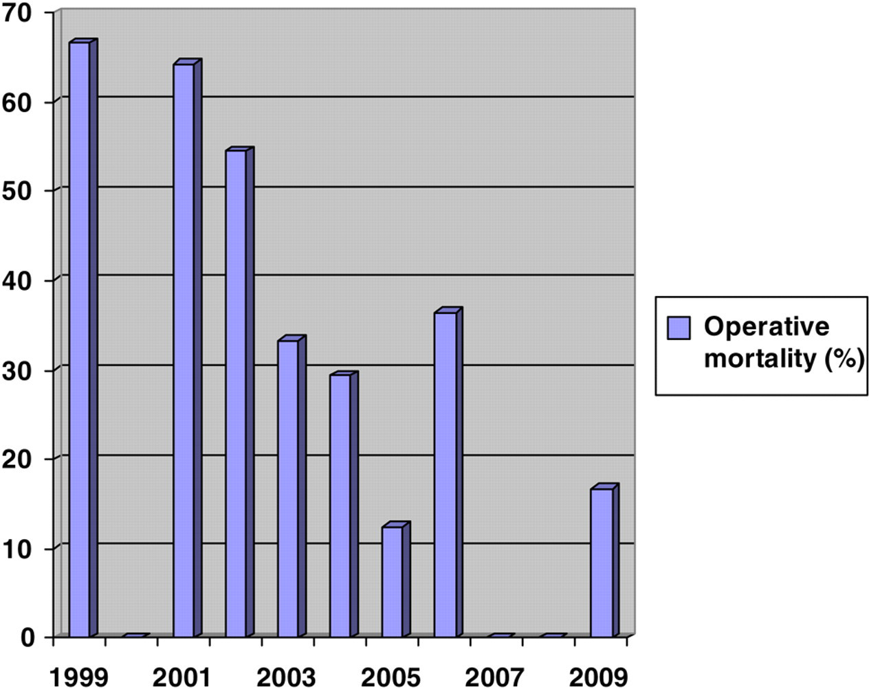
Annual in-hospital mortality
Paraplegia and paraparesis remain a major cause of morbidity after TAA repair and currently range between 5% and 16%, depending on the use of adjuncts and the extent of the aneurysm. 12 Our paraplegia rate is about 10%, but without type IV aneurysms even much higher. What can be the real reason for such a percentage of paraplegia in our group? By adjusting the pump flow rate, the distal aortic pressure (DAP) can be maintained between 60 and 70 mmHg, and by using a combination of partial exsanguination from a left atrium and retransfusion of blood, proximal pressures are maintained at 60–70 mmHg, which is the main principle of spinal cord protection based on the early work of Lascinger 13 during the mid-1980s. Lascinger concluded that maintenance of a DAP greater than 60–70 mmHg will uniformly preserve spinal cord blood flow in the absence of critical intercostal exclusion based on experiments performed on six dogs with somatosensory evoked potentials (SSEP). Since SSEP data reflect the conductive capabilities of white matter in the dorsal horn, which is less sensitive to hypoxia than the alpha motoneuron in the gray matter of the spinal cord, could the results of another measurement of DAP using motor evoked potentials (MEP) be different? Moreover, according to Jacobs et al. 7 after initial aortic double cross-clamping, MEP levels remained adequate in 77%, and in 23% significantly reduced MEP levels could be corrected with an increase in DAP. This raises again the question of the appropriateness of DAP. This is of special importance in transition countries like ours, where the use of MEPs and SSEP is not common practice and where the whole strategy is based on DAP. According to these results, we can calculate that our initial paraplegia percentage would be 23% (Table 2). Does distal aortic perfusion pressure have to be standardized for all patients? The majority of patients undergoing thoracoabdominal aortic reconstruction have hypertension as a traditional risk factor (80%). Vessel autoregulation mechanisms are at much higher levels and disturbed. After all, we cannot expect that a patient with hypertension over 160 mmHg or mean arterial pressure over 110 mmHg needs the same DAP for spinal cord perfusion as a patient with normal tension. Certainly, this observation needs further investigation. 14
Incidence of spinal cord ischemia in 118 thoracoabdominal repairs
Distal aortic perfusion, deliberate hypothermia, reimplantation of intercostal arteries, lumbar cerebrospinal fluid drainage, intraoperative neurophysiological monitoring and pharmacological approaches to protect the spinal cord from ischemic infarction have all been tried, but these techniques used alone or in combination have not been completely effective in preventing or treating this complication. Because it is difficult to identify which intercostal (segmental) arteries supply the anterior spinal artery, we adopted the strategy of reimplanting all intercostal arteries from T7 to L1 if feasible, particularly large, patent intercostal arteries with minimal or absent backbleeding. Although certain centers favor left heart bypass as the main method for distal aortic perfusion, we have chosen to use FF bypass because it provides optimal oxygenation and can be converted into full cardiopulmonal bypass for deep hypothermic circulatory arrest and also because we do not have a Biomedicus centrifugal pump. On the other hand, it reduces right heart strain and improves oxygenation in patients with pulmonary disease on single-lung ventilation. FF bypass reduces the risk of air embolism, and allows for incorporation of a heat exchanger and a reservoir. Someone might argue that a more complicated femoral vein to femoral artery circuit requires both an oxygenator and large heparin doses, and that the potential need for hypothermic circulatory arrest can be avoided on the basis of high-quality aortic arch preoperative imaging. The major problem we encountered was placement of the drainage venous cannula into the femoral vein (due to lack of the transesophageal echocardiogram), which was overcome in five cases by alternative placement through the iliac and left pulmonary vein. Problems with the arterial cannula were overcome by using cannulation through a previously placed Dacron graft over the femoral artery, a technique that minimizes the dreadful effect of products of lower-leg ischemia on renal function.
Hemorrhage was one of our main complications related to death. All eight cases died at the beginning in thoracoabdominal aortic surgery, in the period when rVII (Novo Seven) was not available. Moreover, our intraoperative and postoperative data in that time were insufficient in assessing hemorrhage type. Based on our experience 15 and worldwide experience, 16 we started to use Factor rVII-a in a patient with coagulopathic hemorrhage. According to all these publications and our experience, after complete vascular reconstruction was finished, we administered protamin, with all local hemostatic measures (fibrin glue, warming of the wide exposed area of the operative field, warming of the patients). In patients with a bad response after protamin administration during closing of the wounds, we used antifibrinolytic drugs such as aminocaproic acid, tranexamic acid and 1-deamino-8-d-arginine vasopressin. In extreme cases when all these measures are inadequate, we administered rFVIIa in a single bolus dose (50–60 mg/kg). Before administration of rFVIIa, we corrected platelet count (>50 × 109/L), fibrinogen level (>1 g/L) and electrolyte imbalance (pH >7.2). In the last two years we started using rotational thromboelastometry in order to define the cause of bleeding in a more precise manner in patients with great intraoperative blood loss. There is no consensus on the dosage of rFVIIa in vascular patients. 17 We now routinely use rFVIIa in these situations at our institution. Prospective randomized trials are needed to elucidate the true efficacy of rFVIIa in these patients.
These are our results. We are fully aware that they are not as promising as we want them to be. Someone might argue about the mortality and morbidity rates as well as the percentage of complications, but this is our reality and for the moment we cannot escape it.
Conclusion
Our current review of data clearly proves that the surgical repair of TAAs remains a challenge even in the 21st century, especially in a country in transition. The results of open surgical graft replacement of patients with descending or thoracoabdominal aortic aneurysms have improved with advances in technology and surgical techniques over the decades, but several problems including civil war, financial problems and poor technical equipment, have reduced and slowed down our learning curve.
Footnotes
Acknowledgments
Financial disclosure of authors and reviewers: none reported.