






Abstract
Objectives
To prospectively evaluate and reduce fistula rate after primary cleft palate repair in an academic setting.
Methods
After noting an institutional palate fistula rate of 35.8%, when a majority of palatoplasties were performed using the Furlow double-opposing Z-plasty, the decision was made to re-evaluate the surgical techniques used for palate repair. As part of our re-evaluation, Furlow and von Langenbeck repairs were limited to clefts less than 8 mm in width. Wider clefts were repaired early in the series with Veau-Wardill-Kilner and later with Bardach two-flap palatoplasties. Half of each palate repair was performed by the residents.
Setting
Multidisciplinary follow-up was obtained at the University of North Carolina Craniofacial Center.
Results
A palate fistula was noted in 2 (1.6%) out of 126 cleft palate repairs (both fistulas were located at the anterior hard palate). A split uvula was identified in 2 of 59 patients where the status of the uvula was reported (3.4%).
Conclusion
This study summarizes one of the lowest overall fistula rates reported in the literature. In a tertiary-care academic setting, plastic surgery residents can actively contribute to palatoplasty with a very low fistula rate. Technical keys to achieving low fistula rate include skeletonization of the vascular pedicle for medialization of the mucoperiosteal flaps, aggressive posterior repositioning of the levator muscle, and meticulous two-layer mattress-suture closure. We recommend Furlow repair for narrower clefts (less than 8 mm wide at the posterior border of the hard palate) and the Bardach two-flap palatoplasty for wider clefts.
The surgical goals for palate repair are complete palate closure, velopharyngeal competence, and normal maxillary growth. The latter two are long-term goals; whereas, avoidance of a palate fistula is a short-term goal that is established within a month following palatoplasty. Palate fistula rates reported in the literature range from 0% to 45%. In one of the largest series of palate repairs, Åbyholm reported an 18% incidence of palate fistulas in 1108 patients from 1954 to 1969 (Åbyholm, 1979). Salyer et al. (2006) reported a 10% fistula rate in 382 two-flap palatoplasties. The lowest fistula rates reported are by Wilhelmi et al. (2001), Van Demark et al. (1989), Agrawal and Panda (2006), and more recently, Losee et al. (2008). Wilhelmi et al. (2001) reported a 3.4% fistula rate using a two-flap palatoplasty in private practice. Van Demark et al. (1989) reported a 0% fistula rate; however, follow-up consisted of only a 3-day examination of 37 of 54 patients with complete unilateral cleft lip and palate. Not all patients were examined, and there was no indication whether secondary intervening surgeries may have been performed. Agrawal and Panda (2006) reported a 2.95% fistula rate in 678 palatoplasties.
In an institutional review in 2001, we noted a fistula rate of 35.8% in patients who underwent palatoplasty between 1996 and 2001 (Table 1). Evaluation of fistula rates according to various surgical procedures demonstrated that the highest fistula rates occurred when the Furlow repair was performed: 48% in Veau II, 41% in Veau III, and 87.5% in Veau IV patients. These rates were higher than those using other techniques. In the von Langenbeck procedure, for example, there was a 29% fistula rate in Veau II patients; when using the Bardach two-flap procedure there was a 25% rate in Veau III patients and a 75% fistula rate in Veau IV patients (Table 2). In response to these findings, a full re-evaluation of preoperative and postoperative care, as well as operative techniques, was initiated in order to decrease our institutional palate fistula rate. The following summary of 126 consecutive palate repairs from 2001 to 2005 with two craniofacial surgeons (W.H.L. and J.A.v.A.) reports the changes made in order to achieve a fistula rate of 1.6%.
Consecutive Palate Repairs Between 1996 and 2001 Performed by Two Craniofacial Surgeons (n = 109) and Occurrence of Fistulas Following Furlow Repair *
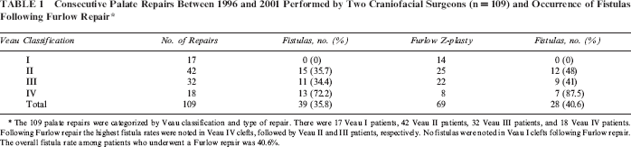
The 109 palate repairs were categorized by Veau classification and type of repair. There were 17 Veau I patients, 42 Veau II patients, 32 Veau III patients, and 18 Veau IV patients. Following Furlow repair the highest fistula rates were noted in Veau IV clefts, followed by Veau II and III patients, respectively. No fistulas were noted in Veau I clefts following Furlow repair. The overall fistula rate among patients who underwent a Furlow repair was 40.6%.
Consecutive Palate Repairs Between 1996 and 2001 Performed by Two Craniofacial Surgeons (n = 109) and Occurrence of Fistulas Following VWK, Bardach, and von Langenbeck Repairs *
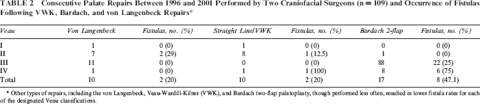
Other types of repairs, including the von Langenbeck, Veau-Wardill-Kilner (VWK), and Bardach two-flap palatoplasty, though performed less often, resulted in lower fistula rates for each of the designated Veau classifications.
Methods
The institutional review board approved this study in 2004 (IRB: 04-SURG-823). All consecutive palate repairs between 2001 and 2005 with at least 1 year of follow-up were evaluated. A total of 126 patients were identified and included in the study. Syndromic patients were included. Patients with Pierre Robin Sequence (PRS) were also included, and they were not considered syndromic. Retrospective review of these patients included examination of the yearly clinic notes of all craniofacial team services that performed an intraoral examination after palate repair, including plastic surgery, oral maxillofacial surgery, speech pathology, otolaryngology, pediatric dentistry, and the orthodontic service. Any documentation of a fistula was included in the group of patients with fistulas. The nonoperating craniofacial surgeon (H.W.L. or J.A.v.A.) examined the other craniofacial surgeon's patients in all cases. A palate fistula included both symptomatic and nonsymptomatic openings into the nasal cavity, but it did not include nasoalveolar fistulas.
Preoperative Care
Palate repairs were performed between 9 to 12 months of age. In children with PRS, palate repair was delayed until at least 12 months of age; if a tracheostomy was present, the repair was performed at the standard 9 to 12 months of age. In submucous cleft palates, the repair was only performed if significant velopharyngeal incompetence (VPI) was present. The Veau classification system was used to describe the extent of clefting and to aid in organizing repair techniques by cleft type.
Prior to surgery, families were instructed to change the child's mode of feeding from a Pigeon nipple to a sippy cup without a ball valve (to prevent suction-generated stress on the palate repair).
Techniques for Repair
Furlow Repair
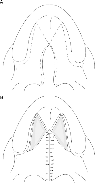
The Furlow double-opposing Z-plasty repair lengthens the soft palate while simultaneously mobilizing the levator muscle posteriorly: Oral mucosa and muscle from one side and nasal mucosa and muscle from the opposite side interdigitate (Fig. 1; Furlow, 1986). The palate is effectively lengthened in a narrow cleft and an incomplete cleft of the soft palate. Due to concerns that palate lengthening is not effective in a wider cleft and that there would be tension at the suture line (primarily at the junction of the hard and soft palates), the Furlow repair was limited to more narrow clefts (<8 mm). This distance was chosen based on discussions regarding the threshold for a moderate cleft and was arbitrary. In submucous clefts of the palate with VPI, a Furlow double-opposing Z-plasty repair was routinely used.
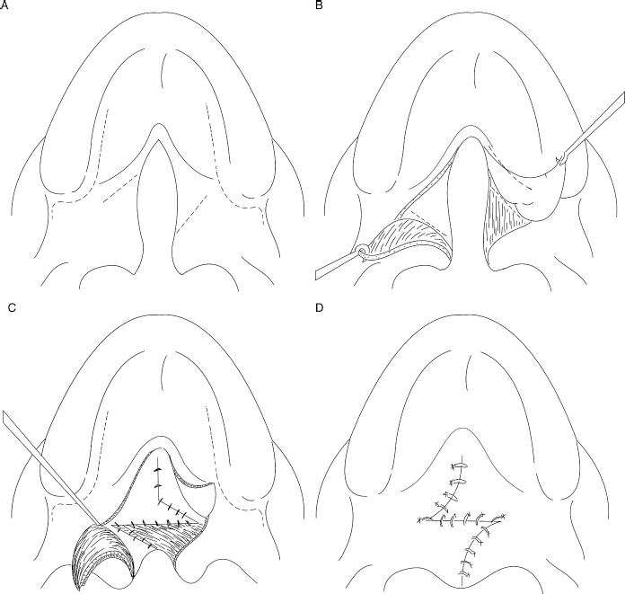
Initial markings for Furlow double-opposing Z-plasty. A: Markings for optional lateral releasing incisions. B: Underlying “opposing” Z-plasty. C: The flaps are then transposed with the levator palati muscle released from posterior edge of the hard palate and sutured posteriorly to reconstruct the velar sling. D: The oral mucosal closure has transposed the original flaps.
Von Langenbeck Repair
The von Langenbeck repair is a two-flap palatoplasty where the oral mucosa of the anterior hard palate is left intact, creating two bipedicle mucoperiosteal flaps (Fig. 2; La Rossa, 2000). This procedure was used in complete and incomplete clefts of the secondary palate but was not used in the presence of an alveolar cleft. The choice to use the von Langenbeck technique was at the primary surgeon's discretion; inclusion of this technique in the surgical armamentarium allowed residents to become familiar with an additional surgical procedure.
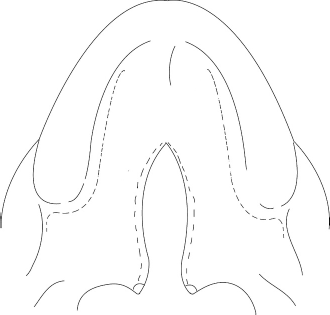
Von Langenbeck palate repair. Note the excision of mucosa on medial surface of uvula, with the incision line along medial edge of the cleft; the lateral releasing incision is marked along the medial aspect of the alveolar ridge, with extension of the incision posterior to the buccal sulcus.
Veau-Wardill-Kilner
In the Veau-Wardill-Kilner (VWK) repair, the two-flap palatoplasty was completely mobilized and the palate “pushed back” to achieve closure of the hard palate and maintain the length of the soft palate (Wardill, 1937). This repair is also referred to as the VY Pushback procedure (Fig. 3).
Veau-Wardill-Kilner repair. A: Initial markings show the preserved anterior, central hard palate mucosa. B: The palate is repaired with a pushback of soft palate, leaving bilateral raw areas of the anterior hard palate to heal secondarily.
Bardach Variation of Two-Flap Palatoplasty
The Bardach variation of the two-flap palatoplasty was used to repair both complete unilateral and bilateral clefts (Bardach et al., 1984; Bardach, 1999). The mucoperiosteal palate flaps were incised to the immediate posterior edge of the alveolar ridge, incorporating the entire oral mucosa of the anterior palate (Fig. 4), which allows the closure to extend into the alveolar cleft. In a bilateral cleft, the mucoperiosteal flaps were transposed medially to the lingual surface of the alveolus, and a vomer flap was routinely used (Fig. 5).
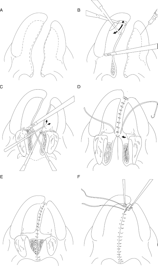
Bardach two-flap palatoplasty. A: Note the excision of mucosa on medial surface of uvula, incisions along the medial edge of the hard palate with extension anteriorly to include the full extent of hard palate mucosa. B: Marcaine is injected only at proposed incision sites. Dissection of the hard palate mucosa is from lateral to medial. C: The levator palati muscle is dissected free from posterior border of the hard palate; nasal mucosa is dissected radically along the underside of the hard palate. D: The nasal layer of the hard palate is sutured with mattress sutures inserted on the oral side with P-2 needle. When the nasal mucosal closure is tight at the junction of the hard and soft palate, the mucosa is released laterally. E: The soft palate muscles are freed from the nasal mucosa, and as needed, from the oral layer, and sutured to each other in a posterior position, consistent with normal anatomy. Vertical mattress sutures are inserted into the uvula. F: The soft palate is repaired with alternating mattress and simple sutures over the muscle; the remainder is reapproximated with mattress sutures. Note that the final anterior suture of the hard palate includes the nasal mucosa.
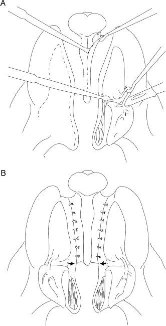
Repair of bilateral complete clefts. A: The vomer is incised at midline and extended laterally along the premaxillary segment allowing increased mobility of the vomer flaps. B: The nasal mucosa on the hard palate is repaired with vertical mattress sutures on the oral side. If the repair at the junction of the hard and soft palates is tight, bilateral releasing incisions in the nasal mucosa are made.
Technical Recommendations to Prevent Palate Fistulas
All markings should be made with Bonnie's Blue. The medial surface of the uvula is marked for excision; soft palate markings are made along the junction of the pale oral mucosa and the more-pink nasal mucosa (Figs. 2 through 4), then proceed along the medial edge of the hard palate. In a unilateral cleft, the markings on the hard palate are placed at the junction of the vomer and the hard palate. Markings for lateral releasing incisions are made posterior to the alveolus several millimeters onto the cheek mucosa, then anteriorly along the medial edge of the alveolus (Figs. 2 through 4). A mixture of 0.25% Marcaine with 1:200,000 epinephrine is used as the local anesthetic because it allows 6 to 8 hours of postoperative analgesia, which may improve initial feeding.
The medial surface of the uvula is excised with tenotomy scissors (Figs. 2 through 4). Incision is then made with a beaver blade along the junction of the oral and nasal mucosa then continued onto the hard palate. The lateral releasing incisions are made on the buccal mucosa extending along the medial alveolus, directing the cutting edge medially to avoid damaging the tooth follicles. A periosteal elevator is used to lift the anterior palate mucosal flaps (Fig. 4B). If the cleft of the hard palate is unilateral, the noncleft side is incised at the junction of the vomer and the hard palate. The hard palate flaps are dissected in a subperiosteal plane, avoiding damage to the bone. Abnormal attachments of the levator palati muscle to the posterior border of the hard palate are divided with tenotomy scissors (Fig. 4D). The tensor aponeurosis is released and freed from the superior constrictor muscle. The greater palatine vessels are skeletonized (Fig. 4C) to allow tension-free medialization of the mucoperiosteal flaps. The hamulus is not fractured, and the palate does not require an osteotomy at the greater palatine foramen.
The levator palati muscle is freed from the nasal mucosa of the soft palate, avoiding fenestration of the mucosa. If the muscle is firmly attached to the oral layer of the soft palate and interferes with adequate posterior mobilization of the muscle, it is dissected free of the oral mucosa; the muscle is moved posteriorly 20 mm (Fig. 4F). Nasal mucosa of the hard palate is freed with a Woodson elevator laterally for a tension-free closure (Fig. 4D). Closure of the nasal layer is performed using 5-0 Vicryl (P-2 needle) vertical mattress sutures; this everts the mucosal edges and avoids nasal mucosa on the oral side of the closure (Fig. 4D). In wider clefts, a longitudinal releasing incision in the nasal mucosa posterior to the greater palatine vessel may be required to allow tension-free closure at the junction of the hard and soft palate (Fig. 4D). Soft palate nasal mucosa is repaired with vertical mattress sutures using a 5-0 Vicryl on a TF needle. Initial excision of mucosa on the medial surface of the uvula ensures easy identification of the edges for more accurate approximation, reducing the incidence of a split uvula (Fig. 4E). The muscle is approximated using interrupted 5-0 Vicryl sutures; the oral mucosa is closed with 4-0 or 5-0 Vicryl vertical mattress sutures (Fig. 4F).
The anterior-most oral mucosal suture is inserted through both the oral mucosa and the repaired nasal layer, fixing the oral layer to the nasal layer (Fig. 4F). The lateral releasing incisions are closed only if they do not cause tension of the midline closure.
Postoperative Care
Following palate repair, the infant is admitted to the hospital overnight and hydrated with D5 Ringer lactate at a maintenance rate. Pedialyte is offered to the child in a sippy cup (without ball valve) and followed by formula or milk and level II baby food. Diet restrictions remain in place for 2 weeks. Arm splints are used to eliminate elbow flexion, preventing the child from inserting fingers and objects into his or her mouth and are worn for 2 weeks. All children are given Keflex elixir for 5 days postoperatively. At 2 weeks, the child is examined in the clinic. If the healing process is favorable, the arm splints are discontinued, and the diet is liberalized. The child is then seen at 1 month by the operating surgeon and again at 4 months by craniofacial team members including plastic surgery, otolaryngology, and speech pathology.
Results
Among the 126 patients, 20 had associated syndromes. According to the Veau classification, there were 17 Veau I clefts (clefts limited to the soft palate; seven of these patients had submucous cleft palates), 51 Veau II (clefts of hard and soft palates), 38 Veau III (unilateral complete clefts), and 20 Veau IV clefts (bilateral complete clefts) (Table 3). Cleft width exceeded 11 mm in 15 patients: two were 12 mm, three were 14 mm, six were 15 mm, two were 17 mm, one was 18 mm, and one was 20 mm.
Consecutive Palate Repairs Performed Between 2001 and 2005 (n = 126) and Occurrence of Fistulas Following Furlow Repair *

There were 17 Veau I clefts, 51 Veau II clefts, 38 Veau III clefts, and 20 Veau IV clefts. Using the Furlow repair, principally in Veau I and II patients, there were no fistulas.
Two of 126 (1.6%) patients developed anterior palate fistulas following palatoplasty. In one additional Veau III patient, where a VWK procedure was performed, a fistula posterior to the incisive foramen caused by the palate pushback was noted (Fig. 6). In this patient, there was complete healing of the palate flaps to the most anterior aspect of the flaps, and this was not included as a palate fistula. Of the two fistulas, one was in a patient with a Veau III cleft who had a VWK repair and the second in a patient with a Veau IV cleft who had a Bardach two-flap palatoplasty.
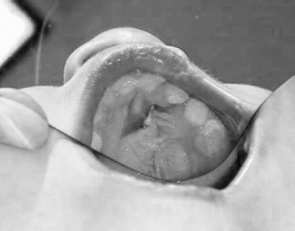
A palate fistula of anterior hard palate, posterior to the incisor foramen. Note the complete healing of the palate flaps. Consequently, this was not considered a complication of the palate repair.
The VWK pushback procedure was used in 16 patients at the beginning of the study. One (6.25%) of these patients developed a palate fistula at the anterior hard palate, and the technique was no longer used. One (2.1%) patient with a Bardach repair developed an anterior hard palate fistula (Table 4). In the last 94 patients, no fistulas were noted.
Veau Classification and Occurrence of Fistulas Following VWK, Bardach, and von Langenbeck Repairs *

One fistula was present following a Veau-Wardill-Kilner (VWK) repair in a Veau IV patient, and the second was noted following a Bardach two-flap palatoplasty in a Veau III patient. No fistulas were noted following von Langenbeck repair.
The status of the uvula was recorded in 59 of 126 patients. Of these, 2 of 59 developed split uvulas (3.4%). None of the patients developed hematoma or infection. None of the patients had to be taken back to the operating room for postoperative bleeding.
Surgical Procedures
Furlow double-opposing Z-plasty was performed in 36 patients, four of whom required lateral releasing incisions (Fig. 1A). There were 15 von Langenbeck repairs and 68 two-flap palatoplasties. Of the two-flap palatoplasties, 16 were of the VWK type and 48 were Bardach two-flap palatoplasties, where no pushback was performed.
Resident Participation in Cleft Palate Repair
All palate repairs were performed with resident participation. The educational goal of each surgery was to teach residents how to perform the operation and, under careful supervision, to have the resident perform half the surgery. The attending surgeon made the markings and dissected one side and the resident then dissected the second side. During closure, the attending surgeon and resident alternated placement of sutures.
Discussion
Multiple factors have been identified as contributing to the development of palate fistulas, including type of cleft (with width of cleft often directly related to the type of cleft), cleft width (the wider the cleft, the more likely a fistula), type of repair, number of cases performed by the operating surgeon (the fewer cases performed, the higher the fistula rate), and timing of repair.
Muzaffar et al. (2001) reported an 8.7% incidence of palate fistulas in 103 patients. One fistula was at the junction of the hard and soft palates and eight were located within the hard palate. All fistulas occurred in Veau III and IV clefts, suggesting that more extensive clefts are associated with higher fistula rates. Musgrave and Bremner (1960) reported on 780 cleft palate repairs performed from 1950 to 1959; these authors noted a 5% fistula rate in unilateral clefts and a 12.5% fistula rate in bilateral clefts. Similarly, Lindsay (1971) reported a 16% fistula rate in unilateral clefts and 23% fistula rate in bilateral clefts in 60 patients. A higher fistula rate is to be expected after repair of bilateral cleft palates as compared with unilateral cleft palates and incomplete clefts of the soft palate. In this series, we report one palate fistula in 38 unilateral complete clefts (2.6%) and one in 20 bilateral complete clefts (5%).
More direct evidence of cleft width playing a role in the incidence of palate fistula is provided in a recent study that suggests a statistically significant increase in fistula rate when cleft width is ≥15 mm and that a ratio of the cleft width to the sum of the width of the palatal shelves of ≥.48 is associated with fistula formation (Parwaz et al., 2008).
Location of palate fistulas is predictably either at the junction of the hard and soft palates or at the anterior hard palate (Musgrave and Bremner, 1960). Amaratunga (1988) noted that 42% of the fistulas were at the junction of the hard and soft palates. This location is problematic because it is generally the widest portion of a cleft, and it is associated with the greatest tension for both the nasal and oral mucosal layer closures. Keys to avoiding fistulas in this region are a two-layer, tension-free closure. This may require lateral release of the nasal layer on the underside of the hard palate for nasal layer closure and skeletonization of the vascular pedicle to minimize tension on the oral layer closure.
Fistulas in the anterior hard palate are best avoided by careful suturing techniques. In our repairs, the anterior-most suture on the hard palate mucosa incorporates both the oral and nasal layer. Bardach described suturing the oral and the nasal mucosa together along the full length of the hard palate (Bardach, 1999), but this may be unnecessarily time consuming.
The type of procedure performed for palate repair may also lead to differences in fistula rates. Cohen et al. (1991) reported a 23% fistula rate in 129 patients. In stratifying rates of fistula by technique, these authors noted a 43% fistula rate following VWK procedures, 10% following Furlow repairs, and 22% following von Langenbeck procedures (Cohen et al., 1991). Amaratunga (1988) reported a 21% fistula rate in 346 cleft patients. He found that von Langenbeck repair resulted in more palate fistulas than the VWK method of repair, contradicting the findings in Cohen and colleagues’ paper. Our institutional review of palate repairs from 1996 to 2001 suggested a high fistula rate associated with Furlow repairs: 48% in Veau II patients, 41% in Veau III patients, and 87.5% in Veau IV patients (Table 1). Subsequently, we limited Furlow repairs to narrower clefts (<8 mm), with no fistulas identified in 36 Furlow repairs. In clefts of intermediate width (5 to 7 mm), either a Furlow double-opposing Z-plasty or a von Langenbeck repair was used. In wider clefts (≥8 mm) a two-flap palatoplasty was used. We believe that the key to this repair algorithm lies in a tension-free midline closure of the palate.
Experience of the operating surgeon has been found to be a factor in the development of palatal fistulas (Cohen et al., 1991). This particular study examined the fistula rates of four surgeons: One had a 63% fistula rate; whereas, the remaining three had fistula rates of 18%, 15%, and 14%, respectively. The surgeon with the highest fistula rate performed only 15% of the cleft palate repairs, suggesting that the more occasional cleft surgeon will have a higher incidence of fistulas. In this series, patients operated on by a senior (W.H.L.) and junior (J.A.v.A.) craniofacial surgeon were included; fistula rates were similar when comparing the two surgeons.
Reports of the relationship between timing of palate repair and fistula rates show mixed results. Some studies have suggested that earlier repair may decrease fistula rates. Rohrich et al. (1996) reported on a longitudinal cohort of patients in whom a two-stage palate repair was performed; those whose hard palate closure was performed at 10 months of age had a 5% fistula rate compared with a fistula rate of 35 % when palate repair was delayed until 48 months. Van Demark et al. (1989), who followed a cohort of patients whose soft palates were repaired at 18 months and hard palates at 5 years of age, noted no hard palate fistulas, thereby suggesting that delayed repair of the palate may lead to decreased fistula rates. With improved speech results noted in earlier repairs of the palate and an uncertain effect on fistula rates based on timing of repair, most cleft practitioners would argue in favor of earlier repair of the palate (Rohrich et al., 1996; La Rossa, 2000).
The palate repairs reported in this series were performed at a teaching hospital with residents performing half of each of the cleft palate operations. To date, there have been no studies examining resident level of participation in outcomes after palate repair. Given the fact that residents perform as much as half of a particular operation, it would be expected that fistula rates would be higher when palate procedures are performed in an academic setting. Our fistula rate of 1.6% suggests that the same standard of excellence as in private practice (Wilhelmi et al., 2001) can be achieved in an academic setting with residents performing half the palatoplasty. Our goal at the outset of the study was to perform 100 palate repairs without a fistula; in this we failed, but no palate fistulas were noted in the last 94 patients.
The relative weighted contribution to fistula formation by level of surgical expertise and the technique used for palate repair is an intertwined argument. Both clearly contribute to fistula formation (inexperience and choice of inappropriate technique). Some experienced surgeons, using a single technique, manage to anticipate and avoid the pitfalls of that particular technique and report a low fistula rate. Others use the same technique and note excessive fistula formation. Choice of a particular technique alone does not guarantee avoidance of fistulas, but must go hand in hand with adherence to particular principles of repair, which include tension-free closure of the nasal and oral layers, particularly at the junction of the hard and soft palates. Whether using a Furlow or Bardach two-flap palatoplasty technique, if closure of either the nasal or oral layers is performed with tension, the possibility of fistula increases.
Our own institutional review suggested that Furlow repairs were associated with a high fistula rate. However, some authors have reported a low fistula rate using only the Furlow repair (Losee et al., 2008). The key pitfall to this technique is tension at the junction of the hard and soft palates (and hence an increased likelihood of fistula formation), which is avoided by incorporating a layer of Alloderm in the nasal layer repair, thereby eliminating tension. Other surgeons have exclusively used a two-flap palatoplasty technique with low reported fistula rates (Wilhelmi et al., 2001; Salyer et al., 2006). We accept that surgeons with extensive cleft experience may be able to use any of several surgical techniques with good outcomes. We recommend, however, that less experienced surgeons should consider doing the Bardach two-flap palatoplasty for wider clefts (including complete unilateral and bilateral clefts), and reserving the Furlow repair for narrower clefts (<8 mm wide at the posterior border of the hard palate).
Conclusions
Our experience delineates one of the lowest overall fistula rates (1.6%) following palate repair reported in the literature. This has been accomplished at a tertiary-care academic setting with plastic surgery residents doing up to half the cleft palate repair. This suggests that residents can actively contribute to palatoplasty with a very low fistula rate. We recognize that avoiding fistulas is a combination of choosing an appropriate technique, and as important, adhering to careful maneuvers at particular junctions during the palate repair. These key junctures are tension-free closure of both the nasal and oral mucosal layers (by completely freeing the nasal layer from the underside of the hard palate and skeletonization of the neurovascular pedicle), aggressive midline recruitment of muscle with an intravelar veloplasty technique, and meticulous two-layer mattress-suture closure extending into the area of the alveolar cleft. We recommend Furlow repair for narrower clefts (<8 mm wide at the posterior border of the hard palate) and the Bardach two-flap palatoplasty for wider clefts. This plan is strongly recommended for less experienced surgeons and is safe for experienced cleft surgeons.
Footnotes
Acknowledgment.
The authors would like to thank Justin Woodlief, a first year medical student at the University of North Carolina, Chapel Hill, for his work on the illustrations.