






Abstract
Objective
Six-year-old children with unilateral cleft lip and palate were examined to compare the prevalence of anterior crossbite and dental arch dimensions of those who later needed orthognathic surgery with the prevalence of those who did not.
Design
Retrospective longitudinal study.
Patients
A total of 68 consecutive nonsyndromic patients with unilateral cleft lip and palate (44 boys, 24 girls).
Main Outcome Measures
Children with unilateral cleft lip and palate whose palates had been closed in one stage by the Veau-Wardill-Kilner or Cronin-Brauer V-Y pushback techniques were analyzed from dental casts taken at a mean age of 6.1 years (range, 5.7 to 6.8 years) before orthodontic treatment or bone grafting. The need for orthognathic surgery in these patients was determined from hospital records at the mean age of 18.2 years (range, 15.6 to 20.2 years). Student's t test and chi-square test were used in statistical analyses.
Results
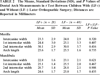
The prevalence of anterior crossbite was 62% (one or both central incisors in full crossbite). The prevalence was higher (75% versus 53%) in children later needing orthognathic surgery (28 of 68, 41%), but the difference was not significant. Nor were there significant differences in dental arch measurements between children who later needed osteotomies and those who did not or between the two modifications of the primary palatal pushback operations.
Conclusions
The prevalence of anterior crossbite and the dental arch dimensions did not differ between 6-year-old children with unilateral cleft lip and palate who later needed orthognathic surgery and those who did not.
Goals of treating patients with cleft lip and palate include palatal and velopharyngeal closure with good speech and hearing and without severe scarring or disturbance of dentofacial growth. However, maxillary retrusion with anterior and lateral crossbites and upper dental arch collapse are frequent findings in patients with unilateral cleft lip and palate (UCLP), regardless of the treatment approach. The reported prevalence of anterior crossbite in deciduous and transitional dentition ranges from 31% to 78% (Bergland and Sidhu, 1974; Turner et al., 1998; Garrahy et al., 2005; Reiser et al., 2010; Pegelow et al., 2011) and that of posterior crossbite, from 66% to 95% (Bergland and Sidhu, 1974; Mølsted et al., 1987; Turner et al., 1998; Garrahy et al., 2005; Reiser et al., 2010; Pegelow et al., 2011). The dimensions of the dental arches are smaller than in children without clefts (Athanasiou et al., 1988; Garrahy et al., 2005). In addition, hypodontia and dental abnormalities in the size and shape of teeth are common (Ranta, 1986).
In 1987, Ross stated that roughly one quarter of men with UCLP need orthognathic surgery to achieve adequate functional occlusion. Varying frequencies (22% to 48.5%) of Le Fort I osteotomies in patients with UCLP have since been reported (Rosenstein et al., 1991; Cohen et al., 1995; DeLuke et al., 1997; Molsted et al., 2005; Good et al., 2007; Daskalogiannakis and Mehta, 2009; Heliövaara and Rautio, 2011; Voshol et al., 2012). An early ability to diagnose deficient maxillary growth is important for optimal planning of type, timing, and length of orthodontic treatment. If orthognathic surgery is planned, dental compensation and excessive maxillary expansion to correct crossbites should be avoided.
It has been shown that it is possible to predict cephalometrically, at least to some extent, the later need for orthognathic surgery in children with UCLP. This study is a part of a larger series examining the later need for orthognathic surgery in 6-year-old children with clefts. The children in this study were earlier assessed by lateral cephalograms (Heliövaara and Rautio, 2011). It was shown that those children with UCLP with orthognathic surgery later in life already had smaller ANB (angle formed from Point A to Nasion to Point B, representing sagittal maxillomandibular relationship) at the age of 6 years than those without. None of the children whose ANB angle was greater than 4.5° needed orthognathic surgery; whereas, all the children whose ANB angle was less than -1° needed later osteotomies (Heliövaara and Rautio, 2011). Nollet et al. (2008) found that several cephalometric variables at the age of 9 years (s-n-ss, s-n-pog, sss-ns-sms, sss-ns-pgs) were significant predictors of the need for surgery at the age of 18 years. Oberoi et al. (2008) used midface length measurements, ANB, and Wits analysis to predict maxillary hypoplasia in children with UCLP. They concluded that maxillary hypoplasia can be determined as early as 10 years of age.
Another common approach to evaluate and plan treatment is to study dental casts. Several classifications have previously been described to assess occlusion, crossbite, and dental arch relationships in patients with clefts (Pruzansky and Aduss, 1964; Huddart and Bodenham, 1972; Mars et al., 1987; Atack et al., 1997; Fudalej et al., 2011). For comparison of treatment outcomes between centers, the GOSLON Yardstick and the 5-year-olds’ index are widely used (Mars et al., 1987; Atack et al., 1997). In the Eurocleft study, GOSLON Yardstick scores at the age of 17 years were used retrospectively to analyze dental arch relationships in five European centers. Based on these scores, the estimated rates for orthognathic surgery in these European centers varied from 4% to 50% (Mølsted et al., 2005). Whereas these indices are based on categorical scales requiring calibration, the dental arch dimensions can be measured with a continuous scale to evaluate dental arch size and constriction. Schwartz et al. (1984) established a method to predict eventual posterior crossbites in late primary and early mixed dentition in 35 children with UCLP. A discriminant function analysis identified four presurgical maxillary arch variables that could be used to predict posterior crossbite in approximately 90% of cases.
The purpose of this study was to assess retrospectively 6-year-old children with UCLP and to compare the prevalence of anterior crossbite and dental arch dimensions of those who later needed orthognathic surgery (LF+) with those who did not (LF–). We expected that children with later osteotomies would have more anterior crossbite and smaller dental arch dimensions than those who did not.
Material and Methods
The patients were 68 consecutive ethnic Finnish children with UCLP who had been operated on and followed until the end of the growth period at the Cleft Palate and Craniofacial Center, Department of Plastic Surgery, Helsinki University Central Hospital. The children with UCLP were born between 1980 and 1988. Since then, Finland has entered the Scandcleft intercenter study, and the surgeons as well as the timing and methods of operations have changed. Patients with syndromes were excluded.
More than half of the clefts of the lip were on the left side (39 of 68). Two thirds (n = 44, 65%) of the children were boys. Primary operations were performed by a total of eight surgeons between 1980 and 1989. The methods of lip repair at the age of 3 to 6 months were modifications of the Millard I and II procedures. Palatal closure had been done in one stage at the age of 1.0 to 1.9 years using the Veau-Wardill-Kilner V-Y pushback operation (n = 42) or the Cronin modification (n = 24). For two children, the Schweckendieck technique had been used. Fifteen children had secondary operations before 6 years of age. These included closure of palatal fistula (n = 7), pharyngeal flap (n = 5), and lip revision (n = 3). None of the children had bone grafting of the alveolar cleft or had been given orthodontic treatment before the age of 6 years. In our Cleft Palate and Craniofacial Center, bone grafting is done between 9 and 11 years of age.
Patients’ need for maxillary or bimaxillary osteotomies was determined from hospital records at the mean age of 18.2 years (range, 15.6 to 20.2 years). Patients were classified as needing maxillary osteotomy based on the opinion of the cleft team even if the patient had refused the surgical correction. The cleft team analyzes the need for orthognathic surgery by clinical assessment, facial photographs, dental models, orthopantomograms, lateral cephalograms, and posteroanterior x-rays. This has been explained in more detail in Heliövaara and Rautio (2011).
Alginate impressions of maxillary and mandibular dental arches of the cleft children were taken at the Cleft Palate and Craniofacial Center at the 6-year check-up. The mean age of the children at the time of dental casts was 6.1 years (range, 5.7 to 6.8 years). Anterior crossbites were registered by the deciduous or permanent central incisor teeth (excluding lateral incisors). Furthermore, an end-to-end relationship was recorded. The dental arch measuring methods and points are those reported by Moorrees (1959) and are shown in Figure 1. Dental arch dimensions that could not be properly measured were excluded. The measurements on the dental casts were made with a sliding digital caliper (Mitutoyo). Double measurements were taken for 24 patients, and Student's t test revealed no significant differences between the two measurements. The numbers of children with deciduous dentition and early mixed dentition were also recorded.
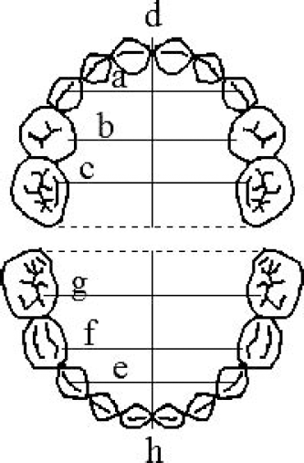
Maxillary dental arch measurements. a: Intercanine width: Distance between cusp tips of the upper deciduous canines. b: First intermolar width: Distance between the mesiolingual cusps or centers of the corresponding facets of the upper deciduous first molars. c: Second intermolar width: Distance between the mesiolingual cusps or centers of the corresponding facets of the upper deciduous second molars. d: Upper dental arch width: Distance between a line at a tangent to the labial surfaces of the upper central incisors and a line connecting the distal margins of the upper deciduous second molars. Mandibular dental arch measurements. e: Intercanine width: Distance between cusp tips of the lower deciduous canines. f: First intermolar width: Distance between the mesiolingual cusps or centers of the corresponding facets of the lower deciduous first molars. g: Second intermolar width: Distance between the mesiolingual cusps or centers of the corresponding facets of the lower deciduous second molars. h: Upper dental arch width: Distance between a line at a tangent to the labial surfaces of the lower central incisors and a line connecting the distal margins of the lower deciduous second molars.
A chi-square test was used to compare the prevalence of anterior crossbite in the LF+ and LF– groups. Student's t test was used to compare the dental arch dimensions. The dental arch dimensions were compared between children who later needed osteotomies and those who did not, between the two operative methods for primary palatoplasty, and between girls and boys. A chi-square test was also used to compare the number of patients operated on by the two surgical pushback methods and the number of patients with permanent incisors in the LF+ and LF– groups. Test statistics with P values equal to or less than .05 were considered significant. The research protocol was approved by the Helsinki University Central Hospital. Principles outlined in the Declaration of Helsinki were followed.
Results
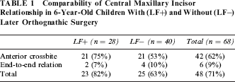
The prevalence of anterior crossbite was 62% (one or both central incisors in full crossbite). When incisors in an end-to-end relationship were counted, the prevalence was 71% (Table 1). The prevalence of anterior crossbite was higher (75% versus 53%) in children later needing orthognathic surgery (28 of 68, 41%), but the difference was not significant (χ2 = 3.06, df = 1). Nor were there significant differences in dental arch measurements between children later needing osteotomies and those who did not (Table 2) or between the two modifications of the primary pushback operation. Boys had significantly longer mandibular arch lengths than girls (P < .05). No other differences were observed between the sexes.
Comparability of Central Maxillary Incisor Relationship in 6-Year-Old Children With (LF+) and Without (LF–) Later Orthognathic Surgery
The Means, Standard Deviations (SD), and P Values of Dental Arch Measurements in t Test Between Children With (LF+) and Without (LF–) Later Orthognathic Surgery; Distances are Reported in Millimeters
No significant difference emerged in the later need for maxillary osteotomies between patients operated on by the Veau-Wardill-Kilner pushback approach (17 of 42) or those undergoing the Cronin modification (11 of 24; χ2 = 0.179, not significant, df = 1). Four of the five patients who had velopharyngoplasty, but neither patient who had been operated by the Schweckendieck technique (n = 2) needed later osteotomies. The data of secondary operations was too small for statistical analysis.
One characteristic that can affect the prevalence of crossbite is the stage of the dentition. Given that we assessed children at age 6 years, they may be in either the deciduous dentition stage or the transitional dentition stage. In our study 15 of the 28 children in the LF+ group and 20 of the 40 children in the LF– group were in the deciduous dentition at the time of sampling. The number of children in the transitional dentition (typically with one or more maxillary or mandibular permanent incisors present) did not differ (χ2 = 0.084, not significant, df = 1) between children who later needed osteotomies (LF+) and those who did not (LF–).
Discussion
Dental and skeletal relationships in patients with clefts can be influenced by genetic factors, the cleft itself, functional compensation, surgical technique, and additional therapy. It is interesting that the prevalence of anterior crossbite did not differ significantly between children later needing osteotomies and those who did not. The prevalence of anterior crossbite was anticipated to be higher in children who later needed osteotomies. However, on the basis of dental cast examination, it can difficult to judge whether anterior crossbite malocclusion is of dental or skeletal origin. Whereas a total anterior and bilateral posterior crossbite is likely to be related to deficient maxillary growth, a simple anterior crossbite of one tooth, especially on the cleft side, may be due to its malposition rather than to deficient maxillary growth. When classifying occlusion solely according to a central incisor crossbite, neither anteroposterior skeletal discrepancy nor incisor inclination is considered. The anteroposterior interarch relationship is also critical when grading the occlusion according to the 5-year-olds’ index and GOSLON Yardstick. Then, the apical base assessment is included. However, anterior crossbite may be found in all skeletal patterns and facial types. It is also noteworthy that in 6-year-old children with UCLP the eruption of rotated, palatally inclined central permanent incisors and the incisal wear of the primary incisors may distort the evaluation of anterior occlusion. In addition, in small dental arches, crowding may cause incisors to erupt more palatinally or lingually than if space was available in the dental arch. According to Moorrees (1959), the arch length in children without clefts increases markedly in the maxilla and slightly in the mandible during the emergence of the central incisors.
Several problems hamper comparison of studies dealing with crossbite and dental arches in children with UCLP. Few studies have comparable groups of patients with regard to type and timing of surgery, surgeon, initial extent of cleft, race, age of children, and developmental stage of dentition. Moreover, the sample size may be small and the determinations of crossbites may vary. If teeth in end-to-end relation are considered to be in crossbite, the prevalence of crossbites increases. Maxillary lateral incisors are usually excluded from the analysis in children with UCLP because they show great variation in number, shape, timing of formation, and eruption (Ranta, 1986; Pegelow et al., 2011). Garrahy et al. (2005) evaluated sixteen 3-year-old children with UCLP. The prevalence of a Class III incisor relationship was 31%. Reiser et al. (2010) used the crossbite score developed by Huddart and Bodenham (1972) in 27 children with UCLP at the age of 5 years. The frequency of anterior crossbite and/or end-to-end relation was 16 of 27 (59%). The developmental stage of the dentition is also important. There is a marked increase in the prevalence of crossbite in early mixed dentition. In 50 Norwegian children with UCLP (Bergland and Sidhu, 1974), the prevalence of anterior crossbite increased from 40% to 78% in early mixed dentition irrespective of the arch configuration in deciduous dentition. Thus, the age of the children and the transitional stage of the dentition in this study may partly explain the relatively high prevalence of crossbite (62%). In addition, the surgical techniques must be considered.
Although controversy exists about the relationship of the dental arch dimensions and maxillary growth and the type and timing of primary surgery, pushback palatoplasty has been reported to result in increased prevalence of crossbites relative to the two-stage nonpushback techniques (Friede et al., 1991). The pushback techniques may cause more scar tissue, which, in turn, may cause more scar contraction and affect the position of erupting teeth. The number of operating surgeons is a deficiency of this study and introduces uncontrolled variables. Nowadays, the number of operating surgeons at our center is lower, and the timing and methods of operations have changed. Surgical techniques as well as skill, individual tissue reaction to surgery, and healing capacity are important factors in this respect but are difficult parameters to measure.
Typical occlusal findings related to deficient maxillary growth include a narrow maxilla and a crossbite. We expected that the dental arches would be smaller in those who later needed surgery. However, no significant differences in dental arch measurements emerged between the children later needing osteotomies and those who did not. This finding underscores the importance of craniofacial growth and thorough cephalometric evaluation of patients with UCLP. Dental assessment of crossbite provides information about the occlusion but it does not give enough information about midfacial growth. In our previous cephalometric study with the same children as in the present study, it was possible to find significant differences in craniofacial growth and predict the need for orthognathic surgery at the age of 6 years (Heliövaara and Rautio, 2011). According to that paper those 6-year-old children with UCLP who had negative values of the ANB angle should be planned to have later orthognathic surgery. On the other hand, the children with UCLP with values of angle ANB larger than 4.5 did not seem to need orthognathic surgery, and conventional orthodontic treatment can be planned (Heliövaara and Rautio, 2011). The pattern of growth in UCLP is unique. Semb (1991) found almost no increase in the length of maxilla between 5 and 18 years of age in a mixed longitudinal cephalometric study of 257 patients with complete UCLP. In addition, a concomitant reduction in maxillary prominence and mandibular prominence over time was observed (Semb, 1991).
Most of the patients (66 of 68) in this series were operated on by two pushback techniques. No difference was present between these techniques in the dental arch dimensions or in the later need for maxillary osteotomies. This is in agreement with a previous finding. In an evaluation of the long-term results of one-stage closure of an isolated cleft palate with Veau-Wardill-Kilner V-Y pushback or the Cronin modification at 17 to 20 years of age, the results were similar with respect to dental arch dimensions (Heliövaara et al., 1993). Mølsted et al. (1987) studied 82 children with UCLP aged 3 to 8 years. No significant differences were observed in either maxillary dental arch width or frequency of malocclusion between the children operated on with a palato-vomer plasty or with a one-layer vomer flap. A minor decrease in the mandibular overjet and crossbite was seen in the vomer group. On the other hand, intercenter studies (Friede, 1991; Molsted et al., 2005; Nollet et al., 2005; Fudalej et al., 2011; Kozelj et al., 2011) have shown that different treatment protocols can result in different treatment results and affect dental arches and occlusion in patients with UCLP. Palatal surgery and the initial extent of cleft can be associated with morphological alterations during dentoalveolar and craniofacial growth, but the evaluation of the role of the postsurgical morphologic changes in the adult occlusion and facial form is difficult. In addition, multiple missing teeth, secondary palatal operations including pharyngeal flaps, and inconsistent team care with delayed orthodontic intervention have been found to be contributing factors to maxillary underdevelopment (Oberoi et al., 2008).
The decision for orthognathic correction may vary among centers. The decision to perform maxillary advancement is based on subjective assessment, usually without standardized criteria (Rosenstein et al., 1991). Facial and nasolabial balance, cheek contour, and nasal and lip aesthetics are essential components of facial attractiveness, but it is difficult to measure imbalance clinically. Dental casts and occlusion are one aspect when evaluating the need for orthognathic surgery. Other needed methods, besides clinical evaluation, include facial photographs, and cephalometric analysis. When using only dental casts, facial aesthetics, deficient smile line, skeletal midfacial growth, bimaxillary retrusion, and severe asymmetries with canting of the occlusal plane cannot be evaluated. Despite acceptable occlusions, patients may have midface deficiency. According to Good et al. (2007), the higher frequency of Le Fort I (47.4% in cleft lip and palate patients) in their unit may reflect their preference for operative correction for all patients with poor midfacial aesthetics despite their occlusal relationship. This is true in our unit also. Another factor that may vary considerably between the cleft centers is the cost of orthognathic surgery. In Finland, the costs are almost totally covered by the national health care, which can affect the patient's decision to undergo surgery.
Conclusions
The prevalence of anterior crossbite and the dental arch dimensions did not differ between the 6-year-old children with UCLP who later needed orthognathic surgery and those who did not. Other methods are needed to evaluate the need for later orthognathic surgery.