






Abstract
Objective
The aim of this work was to fine-map the region 6q23.1, which obtained suggestive linkage signal (logarithm of the odds [LOD] score = 2.22 under a recessive model) to cleft lip with or without cleft palate (CL±P) in our previous genome-wide linkage scan to identify possible genetic variants that may contribute to CL±P.
Design
We used densely spaced markers spanning the entire 6q23.1 region to test for association with CL±P in a family cohort sample.
Setting
Clinical information and DNA samples were obtained from families in the Philippines at their homes or primary health care clinics.
Participants
The study sample consisted of 477 subjects (224 females and 253 males), segregating isolated CL±P, from 72 living in the same area in the Philippines.
Main Outcome Measure
Overtransmission of alleles to persons born with CL±P.
Results
We found statistical evidence of association between a marker of TULP4 (rs651333) with CL±P (P = .00007).
Conclusions
Our results further support the linkage results for the chromosome 6q region and reveal a novel candidate gene for CL±P.
Cleft lip with or without cleft palate (CL±P) is genetically heterogeneous and is the most common craniofacial birth defect. To date, several candidate loci or regions, for example on chromosomes 1, 2, 4, 6, 14, 16, 17, and 19, have shown positive linkage or association results in CL±P, cleft palate only, or both (Marazita and Mooney, 2004). Among the established genes found in association with CL±P are IRF6, MSX1, PVRL1, RARA, TGFA, and TGFB3 (Lidral and Moreno, 2005).
The chromosome 6 has been consistently suggested as harboring genes that contribute to CL±P. In addition, CL±P has been mapped to 6p24 (OFC1; OMIM% 119530) and a genome-wide meta-analysis has mapped CL±P to 6q14.2–14.3 (Marazita et al., 2004). Fine-mapping of 6q14.2–14.3 suggested that PRSS35 is associated with CL±P and that this gene is expressed in mouse embryos at critical stages for palate development (ED12-ED15; Letra et al., 2010). We have also found a linkage signal at 6q23.1 (Vieira et al., 2008), which is approximately 6 million base pairs from the 6q14 region we fine-mapped previously. A logarithm of odds (LOD) score of 2.22 under a recessive model was found when only CL±P was considered in the analysis without dental anomalies. Here we report the fine-mapping of 6q23.1. Our results continue to suggest that there are one or more genetic variants in chromosome 6 that contribute to susceptibility to CL±P.
Materials and Methods
The study sample consisted of 477 subjects (224 female subjects and 253 male subjects) from 72 pedigrees segregating isolated CL±P and who were living in the same area in the Philippines. Information on dental anomalies outside the cleft area was collected from the subjects and all available relatives. Aside from tooth agenesis, which is the most common congenital anomaly in humans and the one we expected to see the most, other dental anomalies included supernumerary teeth, microdontia, macrodontia, missing cusps, and supernumerary cusps. In many instances, tooth agenesis needed confirmation by an X-ray exam, for which we used a portable X-ray system (MinXray P200D MarkIII, Toshiba, Tokyo, Japan). In addition, missing teeth due to tooth decay (caries) needed to be distinguished from congenitally missing teeth. We conducted careful exams and collected comprehensive caries data (data not shown) to aid in the differential diagnosis.
The University of Pittsburgh institutional review board (approval No. 0511198) gave approval for the study in conjunction with local approval in the Philippines. Appropriate informed consent was also obtained from all participants.
We selected 63 single-nucleotide polymorphisms (SNPs) covering the candidate 6q23.1 region between markers D6S474 and D6S1009 (spanning 29,443,509 base pairs) from the International HapMap Project (International HapMap Consortium, 2003) database. We used the approach devised by Carlson et al. (2004), selecting from a set of SNPs that maximally represent the linkage disequilibrium (LD) structure of a given region. Five markers were not informative in the study families (minor allele frequency lower than 1%) and were excluded from further analysis.
Single-Nucleotide Polymorphisms Studied
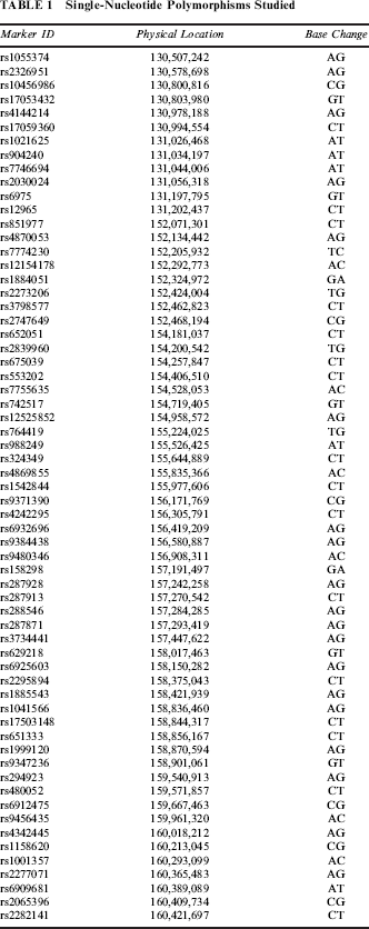
We used PedCheck (O'Connell and Weeks, 1998) to identify genotype inconsistencies and repeated experiments to confirm all genotypes that were inconsistent. Alleles at each marker were tested for association with CL±P with the use of the Family Based Association Test (FBAT) software (Horvath et al., 2001). Since the original genome-wide linkage result was under the recessive model, all analyses were performed under the recessive model, with no additional changes in the default statements of FBAT. We also tested whether adding dental anomalies in the definition of the affected phenotype would provide evidence for association with markers in 6q23.1.
We used chi-square calculations to assess Hardy-Weinberg equilibrium and in unrelated affected and unaffected persons. Bonferroni correction was applied considering the number of tests performed, and significance levels were established at α = 0.00079 (0.05/63).
Summary of the Results of the Family-Based Association Tests
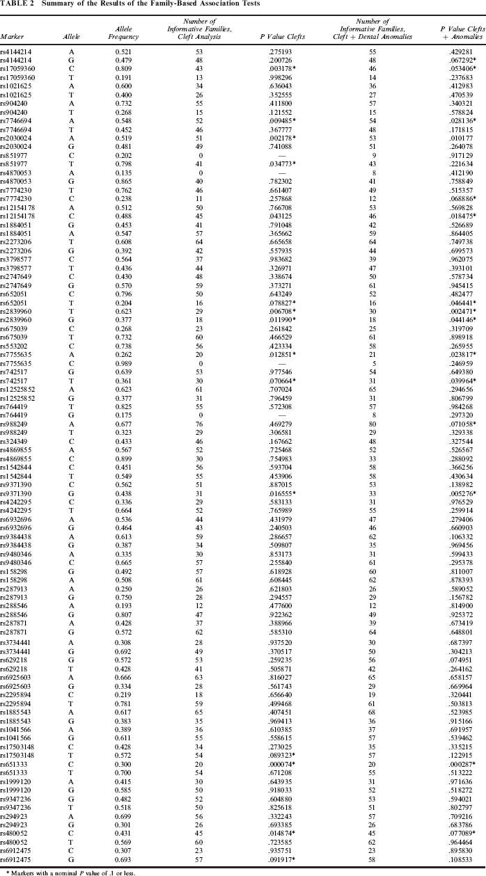
Markers with a nominal P value of .1 or less.
Results
We examined a region on chromosome 6q23.1 centered on the marker D6S1040, which showed suggested linkage with CL±P in our original genome-wide search (Vieira et al., 2008) and generated the LD plot of the region to select densely spaced SNPs to be tested for association with CL±P. We selected 63 SNPs spanning the entire region. Table 2 summarizes the results of the association tests under the recessive model. We found an association with marker rs651333 located in the intron of TULP4 (P = .00007). Evaluation of the LD patterns of the markers close to rs651333 and TULP4 suggest weak to moderate LD, with the exception of the closest marker to rs651333 (rs1999120) (Figure 1). Adding dental anomalies when defining affection status did not improve this result, confirming the original linkage findings, which were stronger for CL±P than for CL±P plus dental anomalies (Vieira et al., 2008). However, several additional markers showed a trend for association, although these were not formally statistically significant, which may indicate that dental phenotypes may also be influenced by genetic variation in the locus. These signals were scattered across the region (see Table 2), and some flanked EPB41L2, SAMD3, and TMEM200A.
LD patterns of the markers closest to TULP4. The relationship between two SNPs is represented by the intersection between the two squares and may present different shades (or shade intensity) based on the value obtained for each pair. Darker indicates D'= 1 and LOD ≥ 2. Light indicates D'= 1 and LOD < 2. Shades of dark indicate D'< 1 and LOD ≥ 2. Numbers inside squares give the actual value of D'.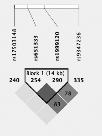
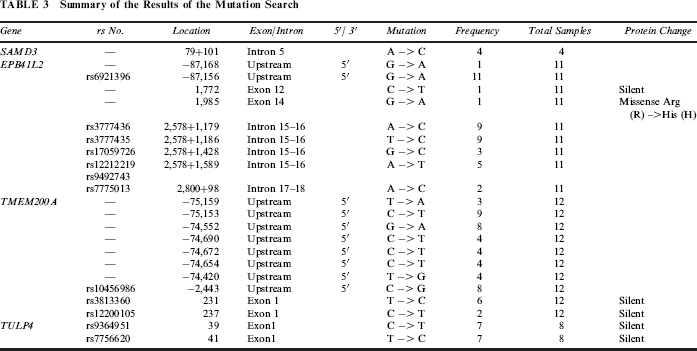
Summary of the Results of the Mutation Search
Discussion
Chromosome 6 has been long considered a candidate region for the etiology of oral-facial clefts. A series of studies have reported the presence of cleft lip and/or palate (CL/P) in persons with chromosomal aberrations, including deletions and translocations affecting chromosome 6 (Hopkin et al., 1997; Yu et al., 2005). A meta-analytic approach of 13 genome scans revealed multiple cleft lip/palate genes and novel loci, including chromosome regions 1p12-13, 6p23, 6q23-25, 9q21, 14q21-24, and 15q15, all with heterogeneity logarithm (base 10) of odds score ≥3.2 (Marazita et al., 2004). Posterior probability of linkage calculations over the reported results on chromosome 6q revealed a region at 6q14.2–14.3 between markers D6S1031 and D6S1056, presenting an approximately 90% chance of harboring a new cleft gene and suggesting that PRSS35 and SNAP91 are involved in CL/P, but these data are yet to be replicated (Letra et al., 2010). However, genome-wide association analyses have not identified associations between CL/P and markers in chromosome 6 (Grant et al., 2009; Beaty et al., 2010; Mangold et al., 2010).
Genotyping random SNPs across the linked chromosome 6q region revealed the association of one novel gene under a recessive model, TULP4, with CL±P in Filipinos. TULP4 in humans interacts with cullin5 and elongins B and C, generic components of the E3 ubiquitin ligases. TULP4 is thought to be a TULP family member and is characterized by a large amino terminus containing WD (tryptophan-aspartate) repeats and E3 ubiquitin ligase binding motifs. This protein is conserved in evolution, the Drosophila TUSP (tubby domain superfamily protein) and Caenorhabditi elegans TUB-2 are related homologs, and the mammalian TULP4 is distantly related to the intraflagellar transport complex A (IFT-A) protein subunit WDR35 (Mukhopadhyay and Jackson, 2011). TULP4 is hypothesized to be involved in apoptosis, and ethanol-exposed mouse embryos show repression of TULP4, which could result in cell death (Hard et al., 2005). Mutations in WDR35, the TULP4 homolog, cause Sensenbrenner syndrome (cranioectodermal dysplasia), which is thought to be a ciliary disorder (Gilissen et al., 2010) and includes craniosynosotosis and ectodermal and skeletal abnormalities.
It is intriguing that ethanol-exposed mice have TULP4 expression repressed. Accumulated evidence in humans suggests that maternal alcohol drinking increases the risk of having a baby with CL±P (DeRoo et al., 2008; Grewal et al., 2008; Leite and Koifman, 2009). Furthermore, an association between genetic variation in chromosome 9 and maternal alcohol consumption is suggested in cases of isolated cleft palate (Beaty et al., 2011). We had no data on gestational alcohol consumption in the families that participated in our study, but the annual alcohol consumption among Filipinos 15 years or older in 2012 was 6.1 L of pure alcohol per person (World Health Organization, 2012). We did not find published data on the scientific literature regarding the frequency of maternal alcohol consumption in the Philippines, but a survey conducted by the church-run radio station Veritas in the Philippines showed that 66% of the 2,500 respondents were alcohol drinkers, and among the drinkers, 52% were women (Philippine Daily Inquirer, 2012). These data suggest that one can expect some infants in any Filipino population sampling may have been exposed to maternal alcohol consumption during pregnancy. However, we have no evidence that mothers of affected children in the 72 families included in this study drank alcohol or smoked during their pregnancies.
One of the largest studies of the high rates of clefting in Filipinos (1.94 per 1,000 live births) suggested that babies born with isolated clefts are primarily from indigent families who exhibit signs of malnutrition (Murray et al., 1997). It is also common knowledge that disenfranchised people have higher risks for several negative health outcomes and for domestic violence, and they are more often exposed to teratogens, including alcohol drinking and cigarette smoking.
In summary, the accumulated evidence for linkage of the chromosome 6q with nonsyndromic CL/P has driven us to pursue additional research within that particular region. Fine-mapping of the 6q23.1 region allowed us to verify the association of a novel gene, TULP4, with CL±P. Further studies are warranted, therefore, to further confirm TULP4 involvement in human clefts.
Footnotes
Acknowledgments
We thank the individuals and families who collaborated with this study. Sarah Vinski provided administrative support.