






Abstract
Objective
The objective of this study was to determine whether patients with isolated microtia or aural atresia have an increased prevalence of renal or cervical vertebral anomalies.
Design
The study design was a retrospective medical record review.
Setting
The setting was the following four distinct institutions: an urban tertiary care children's hospital, two urban academic medical centers, and a staff-model health maintenance organization.
Participants
Patients diagnosed with microtia, aural atresia, or oculoauriculovertebral spectrum were identified. Patients with facial asymmetry, craniofacial microsomia, and other craniofacial abnormalities or syndromes were excluded.
Main Outcome Measures
Main outcome measures were the number of patients with isolated microtia or aural atresia who underwent a renal ultrasound or cervical spine X-ray, the results of those studies, and further evaluation or treatment for any abnormalities found.
Statistical Analysis
A binomial analysis using a one-sided 95% confidence level was performed.
Results
A total of 514 patients with isolated microtia and/or aural atresia were identified. Of these patients, 145 (28%) had undergone a renal ultrasound and 81 (16%) had undergone cervical spine X-rays. A total of 3 patients (2%) had minimal renal pelviectasis, all of which had resolved on repeat ultrasound and required no treatment. There were no structural renal abnormalities identified, and there were no cervical spine abnormalities identified.
Conclusions
The data suggest that there is no increased prevalence of structural renal or cervical vertebral anomalies in patients with isolated microtia and/or aural atresia. Therefore, these patients do not require routine screening renal ultrasound or cervical spine X-rays.
Keywords
Microtia is a congenital anomaly of the auricle that can range in severity from a mild structural abnormality to complete absence of the auricle and external auditory canal (aural atresia). Microtia can occur as an isolated anomaly or as part of a spectrum of anomalies or syndrome. The reported prevalence of microtia ranges from 0.83 to 1.74 per 10,000 births and is the second most common congenital craniofacial condition seen at most craniofacial centers (Luquetti et al., 2012). Controversy exists whether patients with isolated microtia (not associated with other craniofacial anomalies or syndromes) have Oculoauriculovertebral Spectrum (
Materials and Methods
A retrospective medical record review of patients diagnosed with microtia (International Classification of Diseases [
This study was approved by the human subjects institutional review boards of all four institutions at which the study was conducted. Waiver of informed consent was granted by the institutional review boards of all four institutions.
Results
A total of 514 patients with isolated microtia or aural atresia were identified (Table 1); 329 (64%) at CHLA, 59 (11%) at LAC, 64 (13%) at UCD, and 62 (12%) at KP. Of the patients, 500 (97%) had microtia; 303 (61%) with right microtia, 145 (29%) with left microtia, and 52 (10%) with bilateral microtia. A total of 494 patients (96%) had aural atresia; 308 (62%) with right atresia, 143 (29%) with left atresia, and 43 (9%) with bilateral atresia. Of the patients, 14 (3%) had aural atresia without microtia. The presence or absence of aural atresia was not documented in 8 (1%) of the 500 patients with microtia. The absence of aural atresia was documented in 12 (2%) patients with microtia.
Patient Demographics∗
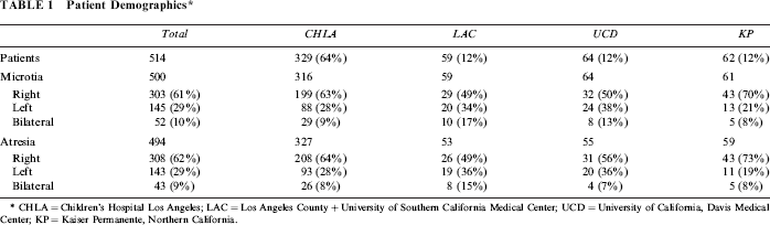
CHLA = Children's Hospital Los Angeles; LAC = Los Angeles County + University of Southern California Medical Center; UCD = University of California, Davis Medical Center; KP = Kaiser Permanente, Northern California.
A total of 145 (28%) patients underwent renal ultrasound (Table 2); 59 (18%) at CHLA, 11 (19%) at LAC, 37 (58%) at UCD, 38 (61%) at KP. Age at ultrasound ranged from 1 day of life to 17 years, with a median of 5.5 months. No patients had any structural renal abnormalities. A total of 3 patients (2.1%) demonstrated minimal renal pelviectasis, which resolved on repeat ultrasound and required no treatment. An exact confidence interval of 0-0.0204 was calculated using binomial analysis with a one-sided 95% confidence level.
Patients With Isolated Microtia or Aural Atresia Who Underwent Renal Ultrasound and/or Cervical Spine X-Rays *

No structural renal or cervical spine abnormalities were identified. US = ultrasound; CS = cervical spine; CHLA = Children's Hospital Los Angeles; LAC = Los Angeles County + University of Southern California Medical Center; UCD = University of California, Davis Medical Center; KP = Kaiser Permanente, Northern California.
A total of 81 (16%) patients underwent cervical spine X-rays (Table 2); 42 (13%) at CHLA, 8 (14%) at LAC, 21 (33%) at UCD, 10 (16%) at KP. Of these patients, 58 (72%) underwent more extensive spine X-rays. Age at X-ray ranged from 1 day of life to 41 years, with a median of 2 years. No patients had any cervical spine abnormalities, and no thoracic, lumbar, or sacral spine abnormalities were identified on those patients who underwent more extensive spine X-rays. An exact confidence interval of 0-0.0346 was calculated using binomial analysis with a one-sided 95% confidence level.
Discussion
Microtia, the second most common congenital craniofacial abnormality after oral clefts, can occur as an isolated anomaly or as one feature of a spectrum or syndrome with other congenital abnormalities. An estimated 20% to 60% of patients with microtia have associated anomalies or a recognizable syndrome (Castilla and Orioli, 1986; Kaye et al., 1989; Mastroiacovo et al., 1995; Shaw et al., 2004; Luquetti et al., 2012). Syndromes that may contain microtia include Treacher Collins, Branchio-Oto-Renal, CHARGE, Nager, Miller, and Townes Brocks. Other congenital anomalies associated with microtia include congenital structural renal anomalies (such as agenesis, hypoplasia, crossed ectopia, horseshoe kidney, pelvic kidney, cystic kidney, duplicated ureters, and megaureter); cervical vertebral anomalies (such as hemi-vertebrae, butterfly vertebrae, hypoplastic vertebrae, vertebral fusion, scoliosis, platybasia, and occipitalization of the atlas); cardiac defects; oral clefts; and OAVS, which includes craniofacial microsomia as one of its characteristic features. The clinical spectrum of craniofacial microsomia varies widely in phenotypic severity and may incorporate craniofacial hypoplasia, macrostomia, cranial nerve palsies, epibulbar dermoids, and other ocular deformities (Heike et al., 2013).
The occurrence of microtia and aural atresia is of public health importance because of the psychological sequelae resulting from the stigma associated with ear malformations, hearing impairment, and the burden of undergoing multiple surgeries for ear reconstruction and hearing restoration. Patients with microtia require long-term, multidisciplinary care that is specific to the individual's requirements. The diverse clinical spectrum of patients with microtia makes it difficult to establish clear, evidence-based criteria for screening evaluations in these patients. However, it has been recommended that patients with microtia undergo a routine screening renal ultrasound at the time of diagnosis and cervical spine X-rays at 3 years of age (Cohen et al., 1989; Heike et al., 2013).
Congenital abnormalities of the kidney and urinary tract occur in 0.3% to 0.6% of live births (Ramanathan et al., 2016). Congenital abnormalities of the axial skeleton occur in approximately 0.1% of live births, with vertebral defects among the most common (Alexander and Tuan, 2010). Studies have shown an increased prevalence of structural renal and cervical vertebral abnormalities in patients with microtia associated with other craniofacial anomalies or a recognizable syndrome (Gosain et al., 1994; Manaligod et al., 1999; Healey et al., 2002; Tsirikos and McMaster, 2006; Wang et al., 2001). Cervical vertebral abnormalities occur in 40% to 60% of patients with OAVS, a condition that is etiologically and pathogenetically heterogeneous and frequently includes microtia (Cohen et al., 1989). Although controversial, isolated microtia is often considered to represent the mild end of OAVS because of the presence of microtia as a common feature of OAVS and the difficulty in recognizing subtle facial asymmetry (craniofacial microsomia) in infants (Rollnick and Kaye, 1983; Bennun et al., 1985; Keogh et al., 2007). This debate remains unresolved, as many continue to consider microtia as a distinct entity from OAVS (Luquetti et al., 2012).
Studies have demonstrated that renal ultrasonography is not indicated in patients with minor auricle deformities, such as preauricular sinuses and skin tags, in the absence of other malformations (Wang et al., 2001; Deshpande and Watson, 2006). What is not clear is whether there is increased risk for structural renal or vertebral anomalies in patients with isolated microtia or aural atresia, which represent 40% to 80% of all patients with microtia. In this study, no patients with isolated microtia had any structural renal anomalies on renal ultrasound or cervical vertebral anomalies on cervical spine X-rays. A total of three patients (2.1%) demonstrated minimal renal pelviectasis, which resolved on repeat ultrasound and required no treatment. Renal pelviectasis, which occurs in 0.5 to 4.5 of fetuses on routine second trimester ultrasound, is usually self-limited and most commonly represents a transient physiologic state (Norton, 2011). The renal pelviectasis in these three patients is therefore likely unrelated to the microtia.
Interestingly, considerably fewer renal ultrasounds were obtained at CHLA and LAC then were obtained at UCD and KP. This may be explained by increased knowledge of recommended screening evaluations in patients with microtia because the study was conducted at CHLA and LAC 10 years prior to UCD and KP. However, considerably more cervical spine X-rays were obtained at UCD than at CHLA, LAC, and KP. There is no obvious reason for the disparity in cervical spine X-rays obtained between UCD and KP. Knowledge of recommended screening evaluations should not be a factor because the rate of renal ultrasounds obtained at each institution was similar. Many patients with isolated microtia may not be evaluated by a multidisciplinary cleft/craniofacial team, a genetics provider, or another provider with experience treating these patients, which may at least partially account for the fact that more screening studies were not performed. Furthermore, the number of screening renal ultrasounds and cervical spine X-rays actually requested may be underrepresented for several reasons: insurance companies may not have approved the studies, patients did not keep the appointment to obtain the studies, or the studies were performed at another facility and the results were not incorporated into the medical record. Furthermore, paper medical records, which are more likely than electronic medical records to be incomplete, were in use during the time period when the study was being conducted at CHLA and LAC.
In conclusion, the data suggest no increased prevalence of structural renal anomalies or cervical vertebral anomalies in patients with isolated microtia and/or aural atresia. Therefore, these patients do not require routine screening renal ultrasound or cervical spine X-rays.