
Abstract

To the Editor,
The diagnosis of Hirschsprung disease is often based on a suction rectal biopsy, which samples mucosa and submucosa. Essential to the diagnosis is the absence of ganglion cells despite adequate sampling. The presence of hypertrophic nerves is helpful but not always present. The complexity in making such a diagnosis and the gravity of the consequences has led to the quest for ancillary tests to increase diagnostic confidence. It is now widely accepted that calretinin-positive nerve fibers are present in the muscularis mucosa and lamina propria of ganglionated intestine but are absent in aganglionic segments. We sought to continue searching for markers that would help distinguish ganglionic from aganglionic segments. Growth-associated protein 43 (GAP-43) is involved in neural development and repair [1] and has been shown to be expressed in the human enteric nervous system [2].
With institutional review board approval, we retrieved 6 consecutive endorectal pull-through specimens and 3 controls (colonic resections performed for reasons other than Hirschsprung disease) from our archives. All slides were reviewed and blocks were selected to correspond to ganglionic and aganglionic segments.
Immunoperoxidase staining for GAP-43 was performed on a series of 6 endorectal pull-through specimens from patients with Hirschsprung disease as well as control cases. For the pull-through specimens, staining was carried out on representative blocks of ganglionic and aganglionic segments. The authors can be contacted directly for detailed methods.
We found abundant GAP-43–positive fibers within the lamina propria and muscularis mucosa of controls, as well as ganglionic and aganglionic segments (see Fig. 1). GAP-43 also stained neural cell bodies within ganglionic segments in both plexuses as well as hypertrophic nerves. In addition, GAP-43 marked the nerve fibers within the mucularis propria of both ganglionic and aganglonic colon.
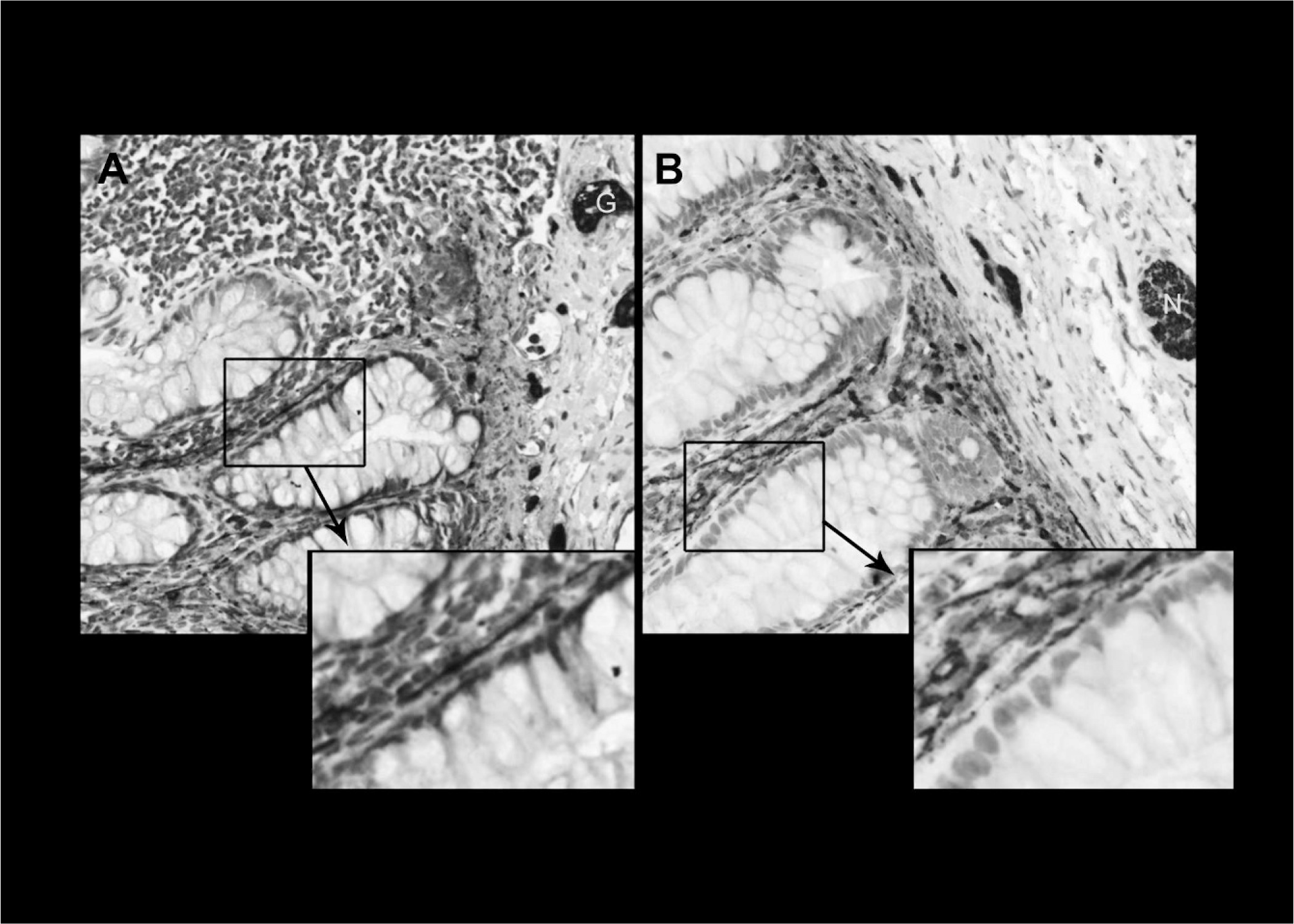
Composite images showing photomicrographs of immunoperoxidase for GAP-43 in ganglionic segment (
Thus, we found that immunostaining for GAP-43 was relatively similar in quantity and pattern in ganglionic versus aganglionic colon. A recent RNA expression profiling study showed a 5.81-fold increase in GAP-43 expression in ganglionic versus aganglionic colon [3]. Nonetheless, our results suggest that immunoperoxidase for GAP-43 cannot be relied on as an adjunctive marker in the assessment of biopsy specimens for Hirschsprung disease.
Footnotes
ACKNOWLEDGMENT
The authors gratefully acknowledge Rhonda Kimmerly, Medical Imaging Specialist, for her help with the composite image.