
Abstract

Until recently, a significant obstacle to the acceptance of computed tomography (CT) for the investigation of non-suspicious deaths relates to the diagnosis of cardiac death and the failure of current post-mortem imaging to yield detailed information concerning the coronary arteries. This is due to the lack of an active circulation to deliver suitable contrast agents via an intravenous route. Currently there are two approaches reported within the literature to assist with overcoming this problem; firstly the use of whole body angiography using a modified heart-lung machine (1–4) and secondly the more recently reported approach of targeted cardiac angiography (5–7). Saunders et al reported a novel double contrast imaging methodology where air is used as a negative contrast medium with a traditional positive contrast medium to enable the examination of the coronary arteries and chambers of the heart (6). Air has been found to be an ideal contrast medium for this purpose (unpublished data), as it improves imaging of vessels with severe calcification or stents, where luminal pathology can be obscured by the use of positive contrast alone. The natural process of putrefaction with the formation of air within the vessels can also allow one to examine the vessels for luminal pathology in decomposed bodies without invasive procedures.
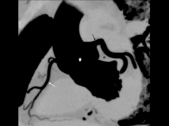
The image arises from an adult male who was 178 cm in height and 87 kg in weight. He had suffered blunt trauma to his chest resulting in sternum and rib fractures, lacerated lungs and a pericardial hematoma. Prior to autopsy a postmortem computed tomography scan was undertaken within 48 hours of death. The figure shows a multiplanar reconstructed (MPR) image in minimum intensity projection (MIP) following injection of air into the aorta using the method for PMCT targeted angiography as reported by Saunders et al. (6). It shows the normal Right Coronary Artery (white arrow) and Left Coronary System (black arrow) arising from the sinus of Valsalva (star). The patency of the ostea, the vascular systems and the myocardial wall thickness can all be assessed using this simple approach. As the protocol reported by Saunders et al (6) images the heart prior to the introduction of air, the air seen in the vessels is known to be as a result of the PMCT angiography procedure rather than the chest trauma. There was no evidence of decomposition to the body to account for image appearance.
Footnotes
Acknowledgment
This paper presents independent research funded by a grant from the National Institute of Health Research (NIHR) under its Research for Innovation, Speculation and Creativity (RISC) Programme (Grant Reference Number RC-PG-0309-10052). This image was acquired with appropriate consent under the local research ethics committee (LREC 04/Q2501/64, UHL 09523) approval for the study.