
Abstract
Adequate photographic documentation of important autopsy findings, both positive and negative, is vital for serving as a record of the pathologist's examination, as well as for a tool in the courtroom, for consultations, or for educational endeavors. Examination of the eye, especially the retina, is an important part of the investigation of suspicious infant deaths; however, obtaining good photographs can be difficult due to many factors. The method described herein allows for the development of a cost-effective, technically simple, and mobile way to take outstanding photographs of the retina.
Gill et al. recommend that when a forensic pathologist is investigating the death of an infant or young child with either known or suspected inflicted traumatic brain injury, the eyes be removed and examined (1). To assist investigators in this task, a specific protocol exists that outlines a detailed examination of the eye for infant death investigations (2). One purpose for the extensive examination of infant eyes is to assess and subsequently document the presence or absence of retinal hemorrhages. Although they occur in other situations, including accidental injury and natural disease processes (3–5), retinal hemorrhages are a commonly listed feature of child abuse (6), and, like other autopsy findings related to child abuse, must be adequately photographed to allow for exhibition in court, or for other uses such as educational activities. However, given the vitreous body, the pigmented epithelial layer of the retina, and the curved nature of the inner surface of one-half of a sectioned eye, obtaining these photographs can be difficult when using only a light box with direct illumination of the retina (7). To assist in producing good quality photographs that adequately demonstrate the retina and any retinal hemorrhages, transillumination of infant eyes was introduced as a technique by Nolte (7) and is endorsed by other authors (8); however, set-ups that allow for transillumination can be relatively expensive, complicated, and space-occupying. For example, the set-up diagrammed by Nolte (7) requires either a dedicated light stand, or must be regularly moved to allow for other photographs to be taken. Also, a specialized transillumination light source can cost $105 to $329 dollars (9), and, potentially more, if a colonoscope or microscope light is used, such as advocated by Nolte (7).
To provide for a more cost-effective and simple set-up, which is easily mobile, another method can be used. The light source is a headlamp, with a cost of $6–10 (9), with its back surface on the light stand and the lights oriented upward
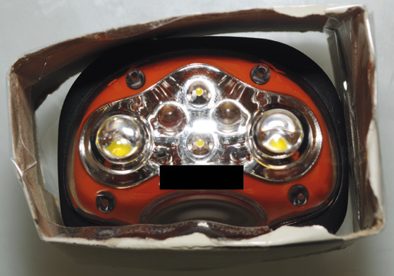
Headlamp on photo stand. The headlamp is on the photo stand, with its base down, and lights oriented upward.
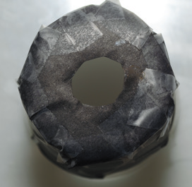
Base of specimen cup, previously wrapped with black paper. At the base, a hole is cut upon which the one-half of the eye globe is placed.
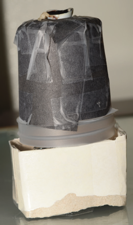
Complete set-up. One-half of the eye globe rests upon the base of the specimen cup (top), with the specimen cup in turn resting upon a rectangle of heavystock paper (bottom), which encircles the headlamp.
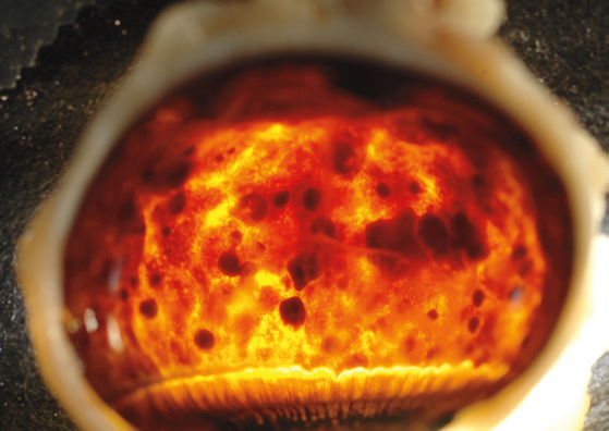
Retinal hemorrhages.
In summary, the technique of transillumination of infant eyes for the purpose of photographing the retina, and, if present, retinal hemorrhages, is not novel, having been described in 1997 by Nolte (7); however, the above described technique is a novel variation, which can be accomplished easily and with inexpensive equipment. Both direct lighting and backlighting should be used when photographically documenting the retina and retinal hemorrhages in a bisected globe, as each technique has its own merit and can produce photographs that highlight different aspects of the pathologic findings; however, technical difficulties with direct lighting of the retina (e.g., lack of contrast between retinal hemorrhages and surrounding retina, the concavity of the bisected globe, and reflectivity of the vitreous body) can impair this method, and, as opined by Nolte, “…transillumination of a bivalved globe with a bright external light source such as a colonoscope or microscope light yields high contrast superior photographs” (7). Transillumination of the bisected globe is not itself, without pitfalls, as a dense retinal pigmented epithelium or thick layers of retinal hemorrhage can impair passage of light; however, the brightness of a headlamp assists with overcoming these difficulties.
Footnotes
The author, reviewers, editors, and publication staff do not report any relevant conflicts of interest.