
Abstract
Amyloidosis, deposition of improperly folded insoluble proteins, may affect one organ or may be systemic. Although plasma cell dyscrasia is frequently implicated in etiology and is due to immunoglobulin light chain production (AL amyloidosis), age-related amyloidosis is believed to be secondary to transthyretin production (ATTR), chronic inflammation-related amyloidosis is thought due to acute phase reactants (AA amyloidosis), and dozens of others are also described. Clinical presentation is dependent upon the organ(s) involved and those associated with unexpected death are expected to involve the cardiovascular system. All cases received for forensic postmortem examination at the Medical University of South Carolina from 2008 to May 15, 2015 were searched to identify any in which amyloidosis was listed as a final diagnosis. Seven cases were identified that met the criteria and were reviewed for demographic information, presentation, cause and manner of death, and assessment of pertinence of the diagnosis of amyloidosis to the cause and manner of death. Interestingly, gross examination of the heart was suggestive of amyloidosis or other infiltrative process in only two of the cases reviewed and a history of myeloma was only noted in one individual. Common gross and microscopic findings are described and relevant medical history and toxicology findings are compared.
Keywords
Introduction
Amyloidosis results from the deposition of improperly folded insoluble proteins in the extracellular space. These insoluble proteins may interfere with the normal function of organs either due to physical disturbance of organ function or through direct toxicity of circulating proteins. The protein can be identified histologically because of its chemical nature (nonbranching fibrils arranged in a cross-β-pleated sheet) via Congo red staining and characteristic “apple-green” birefringence with polarized light. Formalin-fixed paraffin embedded sections stained with Congo red will also demonstrate positive red emissions filter fluorescence by ultraviolet light examination. In fact, the fluorescence assay provides higher sensitivity (1, 2). Electron microscopy demonstrates nonbranching 7.5-10 nm diameter rigid fibrils (3).
Plasma cell dyscrasias with consequent immunoglobulin light chain accumulation are frequently implicated in the etiology of amyloidosis (AL amyloidosis). Other causes include age-related amyloidosis secondary to normal or abnormal transthyretin (ATTR) and chronic inflammation-related amyloidosis due to acute phase reactants (AA amyloidosis). In fact, dozens of precursor proteins are described (4). Direct toxicity is more common in AL amyloidosis (3), particularly lambda subtype, which is significantly more common than the kappa subtype. AA amyloidosis and ATTR amyloidosis may be more difficult to identify clinically due to lack of known concurrent disease process (e.g., plasma cell dyscrasia in AL amyloidosis). Since symptoms are not specific, a high index of suspicion is needed and appropriate biopsies of affected tissue pursued.
More than 100 transthyretin mutations have been identified and may be pathogenic, neutral, or even protective (5). However, age-related accumulation of wild type transthyretin appears to be more common than any inherited event (6). In the absence of an inexpensive panel assay or positive family history with a known variant, identification of specific defects via molecular analysis remains low yield. However, the African-American population, which is reportedly more vulnerable to amyloidosis after age 60, frequently demonstrates the V221I TTR mutation. In one autopsy series, more than 20% of African-Americans with cardiomyopathy due to amyloidosis demonstrated this mutation (7, 8). Variable penetrance of amyloidosis and significant differences in population prevalence of familial variants compounds the difficulty of the molecular approach to diagnosis.
Clinical presentation is dependent upon the organ(s) involved and may be localized or systemic. The kidneys are most commonly affected and renal failure may occur. Spleen and liver involvement may result in organomegaly. Cardiac amyloidosis is classically described as a restrictive process but may also affect the conduction system and precipitate arrhythmias. Rapezzi et al. described several physical diagnostic clues:
… carpal tunnel syndrome in ATTR (particularly if bilateral in a male), history of unexplained neuropathic pain, orthostatic hypotension, and a diagnosis of 'hypertrophic cardiomyopathy' after the sixth decade (6).
At autopsy, if medical records are available, the presence of these findings may increase the pathologist's index suspicion for ATTR.
Amyloid fibril typing may be accomplished by mass spectrometry, which is commonly performed clinically on endocardial biopsies that have been shown to be Congo red-positive. However, the utility of subtyping amyloid in the postmortem setting is unclear unless a heritable form is identified. The costs of this reflex testing would be prohibitive for most forensic pathology offices.
The forensic autopsy database at the Medical University of South Carolina was searched for cases citing amyloid in the final diagnosis, and these cases were reviewed for demographic information, presentation, autopsy findings and cause and manner of death.
Methods
The electronic database housing all forensic autopsies performed at the Medical University of South Carolina (MUSC) from 2008 to May 15, 2015 was searched using keywords including “amyloid” and “amyloidosis” within either the cause of death statement or the final diagnosis section. Seven cases out of a total of 5055 forensic autopsies were identified and reviewed.
Results
Case 1
A 74-year-old black male presented to the hospital following a fall down three stairs. Multiple rib fractures, Klebsiella oxytoca urinary tract infection, elevated troponin I, decreased urinary output and an elevated creatinine of 2.3 mg/dL (which progressed to 4.4 mg/dL) were noted upon evaluation. Respiratory distress and arrest ensued and he died on hospital day three despite resuscitation attempts. Medical history was significant for congestive heart failure with an ejection fraction of 30%, mitral regurgitation, systemic and pulmonary hypertension, and chronic obstructive pulmonary disease.
Findings at autopsy included a calculated body mass index (BMI) of 26.8 kg/m
2
, cardiomegaly (650 g) with chamber dilation and diffuse pale brown coloration, and increased ventricular wall thicknesses (left ventricle, 1.7 cm; right ventricle, 0.8 cm; and interventricular septum, 2.3 cm). No significant coronary artery atherosclerosis was present. The lungs weighed 660 g together and bilateral pleural effusions were noted. Microscopy demonstrated cardiac myocyte hypertrophy and patchy, amorphous pink material in the myocardial interstitium
Case 1, heart with patchy, interstitial amyloid pattern (H&E, x40).
Case 2
A 63-year-old white female presented to the hospital with loss of appetite and shortness of breath. Laboratory data demonstrated acidosis, acute renal failure with a serum creatinine of 16.2 mg/dL, urea nitrogen of 142 mg/dL, and anemia for which a red blood cell transfusion was provided. She died within 24 hours of hospitalization.
Findings at autopsy included a BMI of 30.2 kg/m 2 , cardiomegaly (560 g) with normal appearing parenchyma on sectioning, fibrinous exudate on the pericardial and epicardial surfaces, no significant coronary artery atherosclerosis, and combined lung weight of 1540 g. The ventricular wall thicknesses were increased (left ventricle, 1.7 cm; right ventricle, 0.6 cm; and interventricular septum, 2.0 cm). Additional findings included pleural and peritoneal effusions, cholelithiasis, and colonic diverticula. Histologically, numerous organs sampled, including kidney, heart, liver, thyroid, and lung, demonstrated pink globular material in the blood vessel walls and interstitium. No Congo red stain was performed. Toxicology was not performed due to the length of hospitalization and lack of admission blood sample. The cause of death was acute renal failure due to systemic amyloidosis and the manner was natural.
Case 3
A 64-year-old black male was found dead on the floor of his residence bathroom. Medical history included hypertension, gout and back pain. Autopsy showed a BMI of 28.3 kg/m
2
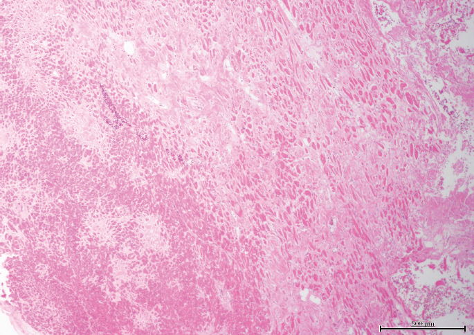
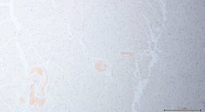
, cardiomegaly (640 g) with a normal heart appearance (no wall measurements documented), no significant coronary artery atherosclerosis, edematous and emphysematous lungs with a combined weight of 1490 g, and, cystic “end-stage” kidneys. Small pleural and pericardial effusions were present. Histologically, serofibrinous acute pericarditis, peripheral pulmonary thromboemboli, and cardiac myocyte hypertrophy and interstitial fibrosis were identified
Case 3, heart with interstitial amyloid pattern (H&E, x100).
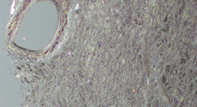
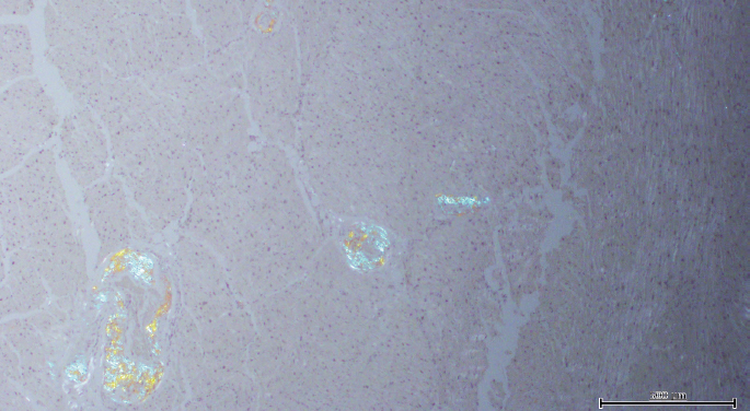
Case 3, heart under polarized light (Congo red, x100).
Case 4
A 72-year-old white female presented with severe fatigue, sweats, body aches, and light-headedness. Upon hospitalization, an abnormal electrocardiogram was noted and troponin was elevated at 0.38 ng/mL. B-type natriuretic protein concentration was 955 pg/mL and the serum creatinine was 1.0 mg/dL. Medical history was significant for hypertension, hypothyroidism, new onset atrial fibrillation, recent rotator cuff surgery, and remote multiple myeloma. She died within 24 hours of admission.
Gross findings at autopsy included a BMI of 18.9 kg/m
2
, heart weight of 300 g with a normal appearance of the parenchyma, and no significant atherosclerotic coronary artery disease. The combined lung weight was 1430 g. Pleural and peritoneal effusions, pulmonary edema, and severe spinal scoliosis were present. Histologically, amyloidosis confirmed by Congo red stains was in all organs examined, including heart, kidneys
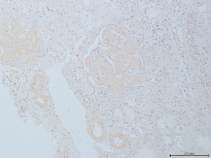
Case 4, renal amyloidosis with vascular and glomerular involvement (Congo red, x100).
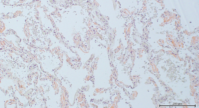
Case 4, lung with alveolar wall involvement (Congo red, x100).
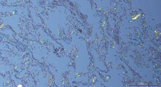
Case 4, lung under polarized light (Congo red, x100).
Case 5
A 67-year-old black male was found in an early state of decomposition in his residence yard. Medical history was significant for dyspnea upon exertion, hyperlipidemia, and hypertension, and a continuous positive airway pressure machine or CPAP was at the scene. Gross findings at autopsy included a BMI of 29.6 kg/m
2
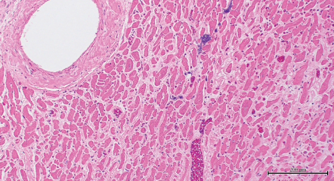
, cardiomegaly (810 g), dark red brown and markedly firm and rubbery heart parenchyma, thickened heart wall thicknesses (left ventricle, 1.9 cm; right ventricle, 0.5 cm; and interventricular septum, 1.8 cm), no significant coronary artery atherosclerosis, and a combined lung weight of 1450 g. Bilateral pleural and pericardial effusions recorded. Histology demonstrated amorphous pink substance in the cardiac interstitium
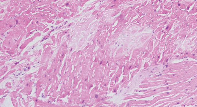
Case 5, heart with patchy interstitial amyloid pattern (H&E, x100).
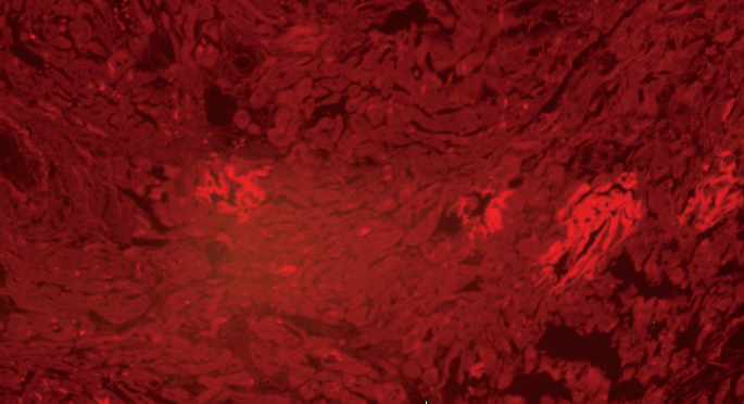
Case 5, heart under rhodamine fluorescence (Congo red, x100).
Case 6
A 70-year-old black male with was found dead in a bathtub without immersion of his face. Medical history included hypertension, diabetes mellitus, and congestive heart failure as well as cocaine use. At autopsy, the BMI was 21.7 kg/m2 and the heart weighed 450 g and appeared normal on sectioning. Coronary artery atherosclerosis impeded the left anterior descending coronary artery lumen by 20% and the right coronary artery lumen by 60%; no heart wall measurements were documented. The combined lung weight was 1260 g and bilateral pleural and peritoneal effusions were noted. Histology of the heart demonstrated perivascular and interstitial fibrosis and granulation tissue as well as patches of pale pink fibers with amorphous pink material in vessel walls that was Congo red-positive
Case 6, cardiac amyloidosis with vascular pattern (Congo red, x40).
Case 6, heart under polarized light (Congo red, x40).
Case 7
An 81-year-old white male was found dead after a nine foot fall off of a residence balcony. Medical history was significant for hyperlipidemia. Autopsy demonstrated a BMI of 25.8 kg/m 2 , blunt head trauma with subdural and subarachnoid hemorrhages, cerebral contusions and skull fracture, 300 mL hemopericardium, a superficial myocardial laceration, and sternal and rib fractures. The heart was 430 g and had a normal appearance. Mild coronary artery atherosclerosis and a 2380 g combined lung weight were recorded. Histology showed cardiac myocytolysis and multifocal areas of Congo red positivity in the myocardium surrounding myocytes. Vitreous creatinine was 0.4 mg/dL. Toxicology was positive for caffeine. The cause of death was blunt force trauma to the head due to a fall from height. The manner of death was accident. After review of the scene and circumstances surrounding the death, cardiac amyloidosis was considered contributory in that it presumably precipitated an arrhythmia resulting in the fall. No other pathology, intoxication, psychiatric disorder, or alternative explanation accounted for the decedent's fall from the balcony.
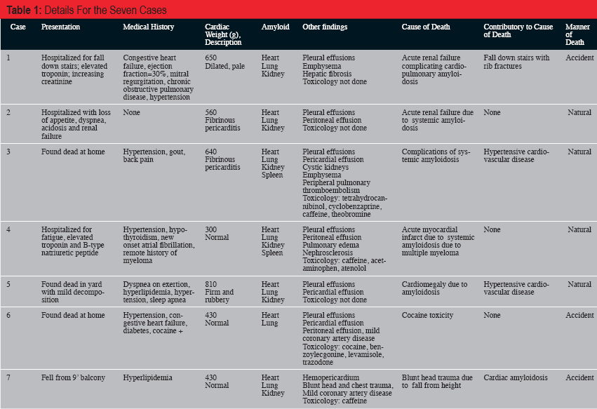
The details for the seven cases regarding presentation, medical history, autopsy findings, and cause and manner of death are summarized in
Details For the Seven Cases
Discussion
During a 7.4-year period, only seven cases of amyloidosis were identified in our forensic autopsy population (0.1% of cases). Of these, amyloid was the cause of death or contributory to cause of death in 86% of cases (six of seven) and all demonstrated cardiac involvement. Not surprisingly, the manner of death was classified as natural in the majority of cases. However in the remaining 43% (three of seven), the manner of death was classified as accident. Two accidental deaths were at least partially attributable to blunt trauma sustained in falls and the third accidental death was due to cocaine toxicity with amyloidosis an incidental and noncontributory finding.
The majority (five of seven) of the deaths presented with apparent collapse or a fall with trauma. Sudden collapse is consistent with acute cardiac compromise that may be attributable to cardiac amyloidosis in these cases. Of the two cases presenting with a fall, one resulted in trauma that was nonfatal but considered contributory to the cause of death, and one resulted in fatal trauma that was thought to have been precipitated by a cardiac event secondary to amyloidosis. Cardiac amyloidosis precipitating an arrhythmic event clarified the context of the fall, as other circumstances did not adequately explain such (i.e., case 7).
All decedents were more than 60 years of age. The average age was 71 years with a range from 63 to 81 years. There was a slight African-American predominance (57%) and a male predominance (71%). None of the individuals had a medical history of amyloidosis and only one had a history of multiple myeloma. Only two decedents showed systemic symptoms such that medical attention was sought immediately prior to death. Creatinine levels showed renal dysfunction in three of the six for whom this datum was available.
The average heart weight was 548 g and the range was from 300 to 810 g. Surprisingly, the cardiac parenchyma appeared grossly normal in 71% of cases. No significant atherosclerotic disease was noted in the coronary circulation of any. However, histopathologically, all cases demonstrated involvement of the heart by amyloidosis. The pattern was variable with interstitial, patchy, and vascular deposition patterns noted. All patients showed multiple organ amyloid deposition including lung, kidney, and spleen when these organs were sampled.
Limitations of this series include the small number of cases, lack of toxicological analyses in three of seven cases, and the inability to distinguish a purported cardiac arrhythmia as hypertensive in nature or due to cardiac amyloidosis.
Conclusion
Amyloidosis is a rare cause of sudden unexpected death. Cardiac involvement classically results in a restrictive cardiomyopathy, but patients may also present with a hypertensive, ischemic appearance, or arrhythmia. Renal dysfunction is common. Since the organs may look grossly normal, microscopic sections and special stains may be justified, even in traumatic cases, to fully elucidate the circumstances surrounding the death. Despite the rarity of amyloid in the forensic setting, proper death certification is essential for epidemiological purposes. Additionally, diagnosis may have genetic implications for the family. The authors hope this short series increases the awareness of amyloidosis as a cause or contributory cause of death in a subset of the forensic population.
Footnotes
The authors have indicated that they do not have financial relationships to disclose that are relevant to this manuscript