





Abstract
The intestinal epithelium is continuously renewed through a balance between cell proliferation and apoptosis. We identified genes of which expression profiles showed significant modulation, and we investigated the cellular mechanisms of this gene regulation in rat intestine after ventromedial hypothalamic (VMH) lesions. Total RNA was extracted, and differences in the gene expression profiles between rats at day 3 after VMH lesioning and in sham-VMH lesioned rats were investigated using DNA microarray analysis and real-time polymerase chain reaction (PCR) methods. DNA microarray analysis revealed that VMH lesions regulated the genes that were involved in functions predominantly related to neuronal development, cell proliferation and apoptosis. Real-time PCR also confirmed that gene expressions of Efnb1 were downregulated. Meanwhile, expression of Casp3 was similar. It is noted that the signaling networks of many gene families, including neuron-specific genes and apoptosis genes in the intestine were changed after VMH lesioning. VMH lesions may suppress mainly the caspase independent type II pathway for apoptosis and induce cell proliferation in the intestine.
Introduction
A focus of attention among researchers has been the pathways that connect the nervous system and the gastrointestinal tract. Knowledge of these mechanisms might help to develop strategies for therapy of neuronal abnormalities, which cause various gastrointestinal diseases. It has been established that activities in the gastrointestinal tract such as inflammation, infection, hypermotility, or changes in visceral sensitivity affect the nervous system. Conversely, psychosocial stressors such as stress, anxiety, and expression that manifest in the central nervous system are transmitted to the bowel by various pathways (1). Morphological homeostasis of the small intestinal epithelium is precisely regulated by both cell proliferation and apoptosis (2). The mucosa has three types of detectors: neurons, endocrine cells, and immune cells (3). The detecting systems in the intestine are more extensive than those of any other organs: the enteric nervous system contains on the order of 108 of neurons, the gastro-enteropancreatic endocrine system uses more than 20 identified hormones, and the gut immune system has 70–80% of the body’s immune cells (3). The enteric nervous system in the mammalian gut is histologically and to some extent functionally similar to the central nervous system. Structural and functional similarities between these systems are evident. Earlier observations, based largely on the influence of vagotomy, indicate that the vagus nerve plays a role in intestinal protection in rats (4). The enteric nervous system is comprised of many functionally different types of neurons: sensory neurons, interneurons and secretomotor neurons. The hypothalamus is composed of a complicated set of regulatory neurons that in most cases cannot be identified by traditional means of cell segregation, i.e. location, soma size, or dendritic arbor. Ventromedial hypothalamus (VMH) lesions have been shown to induce hyperphagia and obesity in rats, and they enhance cell growth in the gastrointestinal tract through efferent vagal nerves (2, 5, 6). It has been reported that the VMH plays an important role in apoptosis in the gastrointestinal tract (7). Consistent with this, apoptosis in the intestine may be controlled by the VMH through the sympathetic nerves in part (7).
DNA microarray analysis is a powerful tool for detecting the characterization of mRNA expression pattern of a large number of genes. In the present study, we used DNA microarray analysis to identify genes for which expression profiles showed significant modulation and to investigate the cellular mechanisms of gene regulation in the rat intestine at day 3 after VMH lesions, because it has been reported that cell proliferation in the intestines increases and reaches a maximum at day 3 (2), and real-time polymerase chain reaction (PCR) also confirmed a part of the results obtained by DNA microarray analysis.
Materials and Methods
Animals.
Female Sprague-Dawley rats weighing 230–250 g were used in this study. They were maintained in a constant-temperature environment (23 ± 2°C) in light-controlled cages with a 12-h light-dark cycle (lights on 7:00 AM) and were given free access to food and water. Tissue samples were taken from the small intestine of VMH-lesioned rats and sham VMH-lesioned rats at day 3 after the operation (n = 2 in each group for DNA chips and n = 3 in each group for Real-Time Polymerase Chain Reaction).
VMH Lesions.
VMH lesions or simulated operations were performed as previously described (2, 8). The stereotaxic coordinates were at bregma anteriorly, 0.75 mm lateral to the midsagittal line, and 1.0 mm upwards from the base of the skull, according to the atlas of De Groot (9). Sham operations were performed in an identical manner except that no current was applied. After the operations, the rats were returned to their cages and given free access to food and water. Localization of the VMH lesions was verified by microscopic examination of the brain at the end of the experiment.
Total RNA Preparation and DNA Microarray Analysis.
In two individual rats at day 3 after VMH lesioning and two sham-VMH lesioned rats, in order to circumvent RNA lysis by RNases that may be released in the rat intestine when the animal is stressed, all procedures were conducted as swiftly as possible after each rat was sacrificed. The abdominal and chest cavities were opened. The samples of intestine were quickly placed in 10 volumes of RNAlater® (Ambion, Inc., Austin, TX) at room temperature. The distance between the tissue surface, which is exposed to preservative, and the innermost regions of the fragment was minimized. We did this by cutting the tissues into 2 mm thick slices, thereby reducing the diffusion distance to 1 mm or less. RNA was isolated from the rat intestine, using a commercially available kit (RNA easy Mini Kit, QIAGEN GmbH, Hilden, Germany). The RNA was quantified spectrophotometrically at 260/280 nm, and the quality of the isolated total RNA was determined by electrophoretic separation on anthodium bromide-containing 1% agarosegel. The preparation of cRNA was carried out by Ambion’s MessageAmp® II-Biotin Enhanced and the target hybridization was performed according to the instructions provided in the Affymetrix GeneChip® technical manual. The double-stranded cDNA was synthesized from 5 μg of total RNA and hybridized Affymetrix GeneChip® arrays (Rat Genome 230 2.0, Affymetrix Japan Co., Tokyo, Japan) for 16 h at 45°C in GeneChip® Hybridization Oven 640. After washing and staining in GeneChip® Fluidics Station 450, hybridized cRNA was detected by GeneChip® scanner 3000. The digitized image data were processed using the GeneChip® Operating Software 1.4. The amount of probe-specific transcripts was determined based on the average of the differences between the perfect-match and mismatch intensities. As replicate assays were not performed, a very stringent cutoff point was selected for the detection of significant upregulation or downregulation of the genes in the mRNA amount between the arrays. Using the signal intensity of selected genes that were upregulated or downregulated compared to the sham-VMH lesioned control group, the analysis was performed using GeneSpring GX 7.3.1 (Agilent Technologies, Santa Clara, CA) and Ingenuity Pathway Analysis (http://www.ingenuity.com/) (Redwood City, CA). Using a computational tool, Ingenuity Pathway Analysis, we were able to build networks of interacting genes from protein-related genes lists.
Real-Time Polymerase Chain Reaction.
Total RNAs of three VMH lesioned rats and three sham VMH lesioned rats were extracted using a Qiagen RNA-easy column. The concentration of RNA was determined by absorbance at 260 nm in relation to absorbance at 280 nm. RNA was stored at −70°C until real-time PCR was performed. An aliquot (1 μg) of extracted RNA was reverse-transcribed into first-strand complementary DNA (cDNA) at 42°C for 15 min, using 200 U/μl reverse-transcriptase (Takara Biochemicals, Shiga, Japan) and 10 mM of oligo (dT)-adapter primer (Takara Biochemicals) in a 2.0-μl reaction mixture.
Real-time PCR was carried out with a Terminal Cycle Dice TP800 (Takara Biochemicals) using the DNA-binding dye SYBR Green I for the detection of PCR products. The reaction mixture (RT-PCR kit, Code RRO43A, Takara Biochemicals) contained 12.5 μl SYBR Premix Ex Taq (2x) (Code RRO41A, Takara Biochemicals), 10 μM PCR Forward Primer (0.5 μl), 10 μM PCR Reverse Primer (0.5 μl), and cDNA (2.0 μl) to give a final reaction volume of 25 μl. The sequences were obtained using Perfect Real Time support system (http://www.takara-bio.co.jp/prt/intro.htm). The PCR settings were as follows: the initial denaturation for 10 s at 95°C was followed by 40 cycles of amplification for 5 s at 95°C and 30 s at 60°C, with the subsequent melting curve analysis increasing the temperature from 60°C to 95°C. Relative quantification of gene expression with real-time PCR data was calculated relative to GAPDH.
In the present study, three representative genes related to neural development or cell proliferation were investigated by real-time PCR: 1) Adra2a (alpha 2A adrenoreceptor), that plays a key role in regulating neurotransmitter release from sympathetic nerves in the central nervous system (10); 2) Efnb1 (ephrin-B1), one of the Ephrin families that is involved at various stages of neuronal development, including axon guidance, neural crest migration, and cell positioning (11); and 3) Casp3 (Caspase-3), that plays a central role in the execution-phase of cell apoptosis (12).
Statistical Analysis.
Results are expressed as the mean ± SEM. The mRNA levels were analyzed by the Mann-Whitney U test. Statistical analysis was conducted with SPSS version 11.0 statistical software. The differences between the groups were considered significant if the P value was < 0.05 (two-tailed).
Results
Gene Expression Profile by DNA Microarray Analysis.
Among 31,099 probes, the expression of 235 probes (0.8%) showed at least a 2-fold upregulation (37 probes) or downregulation (198 probes) at day 3 after VMH lesioning as compared with sham-VMH lesioning. Table 1 shows three main gene networks associated neuron-specific genes and apoptosis genes after VMH lesioning identified in Ingenuity Pathways Analysis. Table 2 shows the upregulated (> 2.5 folds) and downregulated (> 10 folds) genes identified by DNA microarray analysis. VMH lesions upregulated Acetoacetyl-CoA synthetase, Runx1, T cell factor (Tcf) 3 and ATPase inhibitor factor (Atpif) 1, and downregulated alpha 2A adrenoreceptor gene (Adra2a), ephrin-B1 (Efnb1), LIM and SH3 protein (Lasp) 1, insulin-like growth factor-binding protein (Igfbp) 1, protein phosphatase (PPP) 3R1 and amino-terminal enhancer of split (AES).
Real-Time PCR Results.
The expression of Adra2a, Efnb1 and Caspase-3 (Casp3) was examined by real-time quantitative analysis (Fig. 1). The expression of Efnb1 was downregulated at day 3 after VMH lesions (P = 0.05); and the expression of Adra2a was also downregulated at day 3 after VMH lesions, but not significantly (P = 0.13). Meanwhile, the expression of Casp3 in VMH lesioned rats was similar to those in sham-VMH lesioned rats.
Discussion
In the present study, we used the DNA microarray technique for mRNA expression profiling of rat small intestinal cells to investigate cellular responses in response to VMH lesions. The DNA microarray analysis revealed that VMH lesions regulated some genes that are involved in neural development, cell proliferation and apoptosis (Table 1). In the present study, DNA microarray analysis and real-time PCR results showed that VMH lesions downregulated the expression of Efnb1 in the intestine (Table 2 and Fig. 1). Ephrin and Eph receptor signaling are involved at various stages of the neuronal development, including axon guidance and neural crest migration, and cell positioning in the intestinal epithelium (11). Therefore, there is a possibility that the autonomic dysfunction due to VMH lesions may affect the expression of neuron-specific genes in the intestine. Consistent with this, the change of some neurotransmitters signals by VMH lesions may be required for the activation of intraintestinal neuron-specific genes. Adra2as are known to have a critical role in regulating neurotransmitter release from sympathetic nerves in the central nervous system (10). The present study suggests that VMH lesions could not significantly downregulate the expression of Adra2a in the intestine (Fig. 1). Therefore, there is a possibility that the other neurotransmitter release-regulating genes, except for Adra2a, may be involved in this mechanism.
The intestinal epithelium is continuously renewed through a balance between cell proliferation and apoptosis. VMH plays an important role in apoptosis in the gastrointestinal tract (7). Although many apoptotic stimuli and signal transduction pathways have been demonstrated, only two principal apoptosis pathways are well recognized: the type I pathway activated by extrinsic stimuli and the type II pathway activated by intrinsic stimuli (13). Mitochondrial release of cytochrome c into the cytoplasm is involved in type II pathway apoptosis (13). Cytochrome c is released from mitochondria following the formation of a pore in the mitochondrial membrane called the permeability transition pore. This pore is thought to form through the action of the pro-apoptotic members of the Bcl-2 family (13). DNA microarray analysis showed that VMH lesions downregulate expression of Bcl2l1 in the intestine. Therefore, there is a possibility that the type II apoptosis pathway may be inactivated in the intestine after VMH lesioning, indicating that VMH lesions induce cell proliferation in the intestine. Consistent with this, the real-time PCR results confirmed the fact that the expression of the Casp3 in VMH lesioned rats is similar to that in sham-VMH lesioned rats.
In conclusion, this study suggests that parasympathetic hyperactivity and sympathetic hypoactivity based on VMH lesions might induce expression of neuron-specific and apoptosis-specific gene families in the intestine (Fig. 2), although the networks of these genes involved in this process have not yet been elucidated sufficiently. Moreover, our results also suggest that the autonomic dysfunction caused by VMH lesions may suppress mainly the type-II apoptosis pathway and induce cell proliferation in the intestine. Although further investigation is needed to clarify the relationships among the neural factors, cell proliferation and apoptosis, this study is the first report to demonstrate that VMH lesioning causes changes of expression of neuron-specific genes and apoptosis genes in the rat intestine.
The accession numbers for information regarding the microarray analysis are: 1367984_at; 1368126_at; 1368680_a_at; 1369152_at; 1369476_at; 1369684_at; 1370485_a_at; 1383030_at; 1387043_at; 1387708_at; 1387844_at; 1388116_at; 1388231_at; 1389270_x_at; 1395363_at; 1395394_at; 1395409_at; 1396152_s_at; 1398114_at.
Three Main Gene Networks Associated Neuron-Specific Genes and Cell Proliferation-Regulating Genes at 3 Days After VMH Lesioning Identified in Ingenuity Pathways Analysis a
Upregulated (> 2.5 Folds) and Downregulated (> 10 Folds) Genes Identified by DNA Microarray Analysis at 3 Days After VMH Lesioning a
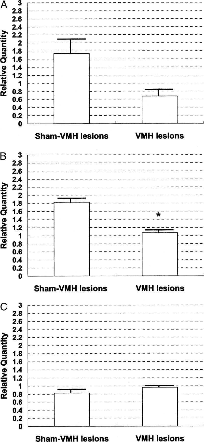
VMH lesion-induced gene expression in real-time PCR analysis. Real-time PCR analysis of total RNA extracts was described in Materials and Methods. A: Adra2A, alpha 2A adrenoreceptor; B: Efnb1, ephrin-B1; C: Casp3, Caspase-3. Values are the means ± SE of 3 different experiments. * < 0.05 compared with sham-VMH lesioned rats.
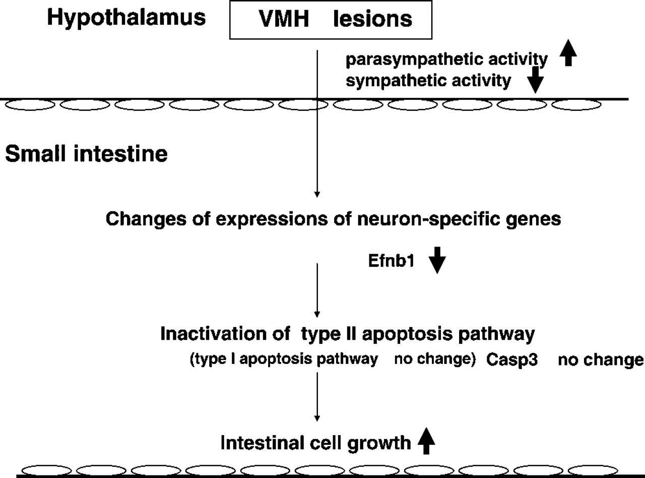
The hypothetical schema of the relationships between autonomic dysfunction by VMH lesions and intestinal cell growth. Efnb1, ephrin-B1; Casp3, Caspase-3.