
Abstract
Background:
Polycaprolactone (PCL)-biphasic calcium phosphate (BCP) scaffolds fabricated using Melt-Stretching and Compression Molding (MSCM) can release calcium and phosphate ions, which are essential for bone formation.
Objective:
Responses of the osteoblasts seeded on three groups of scaffolds including group A; PCL-20% BCP (%wt), group B; PCL-30% BCP and group C (control); pure PCL (100% PCL) were evaluated.
Methods:
The cell-scaffold constructs were made by seeding osteoblast cell lines at
Results:
The cells attached and grew on the scaffolds of all groups. The cell number and the differentiation markers of groups A and B were remarkably higher than those of group C over the observation periods. Slow proliferation of the cells of group A and B in the PR medium in the first 7 days corresponded to the maximum increases in alkaline phosphatase activities (ALP). The maximum levels of ALP of those groups in the OS medium were not detected until day 14. The levels of osteocalcin of those groups were not statistically different when cultured in both mediums.
Conclusions:
The MSCM scaffolds are suitable for supporting attachment and growth of the osteoblasts. Additional BCP into the PCL-based scaffolds accelerate early differentiation of the cells in the constructs even without osteogenic-inductive condition.
Introduction
Over the last several decades, using autogenous bone grafting for reconstructing large bone defects in the maxillofacial region has been the gold standard, nevertheless, it requires donor site operations that sometimes increases patient morbidity. Therefore, some types of ceramic materials such as hydroxyapatite (HA) and beta-tricalcium phosphate (TCP) and biphasic calcium phosphate (BCP) are often selected for replacing the autogenous bone. Our research team successfully developed the technique of modified Melt Stretching and Multilayer Deposition (mMSMD) for fabricating a three-dimensional (3-D) scaffold [1], which is now named as “Melt Stretching and Compression Molding” or “MSCM”. This technique is easier to process allowing any surgeon to instantly build up the 3-D scaffolds on the chair-side of surgical operations (Fig. 1). Therefore, time spent for fabrication processing is remarkably reduced. In addition, building the scaffolds within glass molds is a closed environment that can prevent contamination. The materials used for fabricating the composite scaffold includes Poly ε-caprolactone (PCL) as a major component and BCP as a filler. Food and Drug Administration (FDA) have approved PCL as a medical and drug delivery device that extensively supported by both in vitro and in vivo studies [2–7]. Biphasic Calcium Phosphate is a combination of a stable phase of HA and a soluble phase of TCP in different concentrations. Bioactivities, mechanical properties and degradation behaviors of BCP are controlled by varying the ratios of the HA and TCP. With higher ratios of TCP, higher degradation rates can be obtained. On the other hand, with more composition of HA, BCP has better mechanical strength. In addition, BCP can release calcium and phosphate ions that are considered very important factors for enhancing new bone formation [8–16]. The calcium ion is essential for migration and maturation of pre-osteoblasts, and it can induce expression of osteoinductive growth factors. The phosphate ion is a signaling molecule that regulates the proliferation rate of the cells and bone matrix mineralization. Moreover, the calcium ion is able to neutralize the adverse acidic by-products during degradation of the polyester-based scaffolds [17–19]. Several studies [20–23] demonstrated successful results of the BCP containing high ratios of TCP in terms of supporting osteo-progenitor cells to produce bone formation. In regard to our previous study [1], BCP particles with HA/TCP at 30/70% were used as the filler in the PCL-based scaffolds at the ratio of PCL/BCP (w/w%) = 80/20% and 70/30%. The results showed that the scaffolds could sustain release of calcium and phosphate ions over 30 days when immersed in double distilled water (dH2O) and in culture medium. In addition, proliferation of osteoblasts, when cultured in medium that immerged with the scaffolds, would relate to the profiles of the releasing ions. Therefore, the results supported that the release of those ions have indirect effects on behaviors of the bone forming cells. Nevertheless, responses of the cells, which directly seeded on the surfaces of the scaffolds, had not yet been evaluated. In this study, bioactivities of BCP fillers in the scaffolds were assessed by measuring proliferation and osteogenic differentiation markers of the cells in the cell-scaffold constructs. The behaviors of the cells were assessed when the constructs were cultured in standard medium and in osteogenic induction medium. The schematic drawing demonstrates instant building of 3-D MSCM scaffolds for an operation of socket preservation. (a) After tooth extraction, morphologies of the extraction socket is recorded using polyvinyl impression material. (b) The diameter of the registry impression is measured for selecting sizes of glass molds. Afterwards, a 3-D scaffold is made by putting a stocked scaffold-monofilament into the mold and compressing. (c) The scaffold is inserted into the extraction socket and the wound is closed by platelet rich fibrin (PRF) membrane.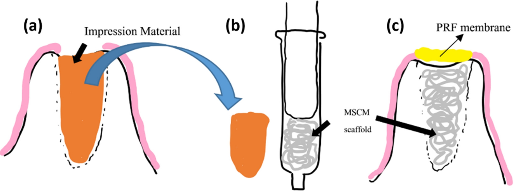
Scaffold fabrication
The scaffolds were fabricated for the experiments as follows: group A with PCL-20% BCP, group B with PCL-30% BCP and group C with pure PCL (100% PCL) using the MSCM technique [1]. In brief, PCL pellets (Mn 80,000 PC, Sigma-Aldrich, USA) and BCP particles (HA/TCP = 30/70%, particle sizes < 75 μm, MTEC, Pathumthani, Thailand) were mixed together in the ratios of PCL: BCP at 80:20 and 70:30 by weight and melted in the melting-extruding machine. The PCL-BCP monofilaments were made by extruding the PCL-BCP blend through the nozzle tip of the machine. Afterwards, the filaments were stretched to decrease their diameters, and then they were stocked for fabricating the scaffolds. To fabricate a 3-D scaffold, a single filament was cut into 50 cm in length, put into a 5-ml glass syringe, then the plunger of the syringe was pushed until reaching the reference point 3 mm above the bottom of the syringe. The tip of the syringe was sealed by polyvinyl siloxane (3M ESPE, USA) and then immersed into warm dH2O. By using this technique, contacted surfaces of the filaments could be fused together and a scaffold (diameter: 11 mm, height: 3 mm) was built. The pure PCL scaffolds were fabricated by using 100% PCL pellets, and their processing was the same as described above. Morphologies of the scaffolds were demonstrated in Figs 2 and 3. They were sterilized using ethylene oxide gas 2 weeks prior to the experiments.
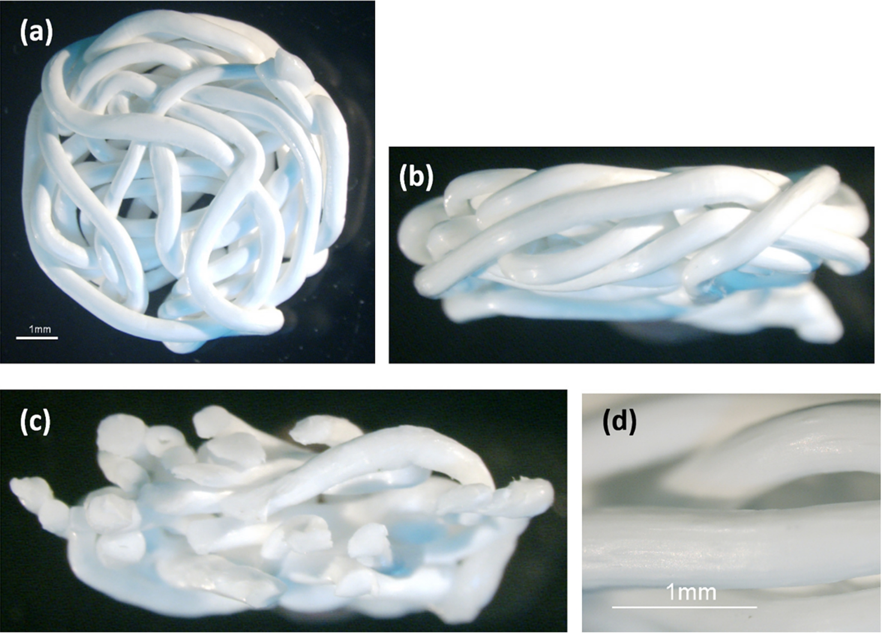
The stereomicroscope images demonstrate the morphologies of the PCL-BCP scaffold: (a) shows the superior view, (b) shows the lateral view, (c) shows the cross-sectional view, and in (d) the magnified picture focuses on the rough surface architecture of the scaffold.
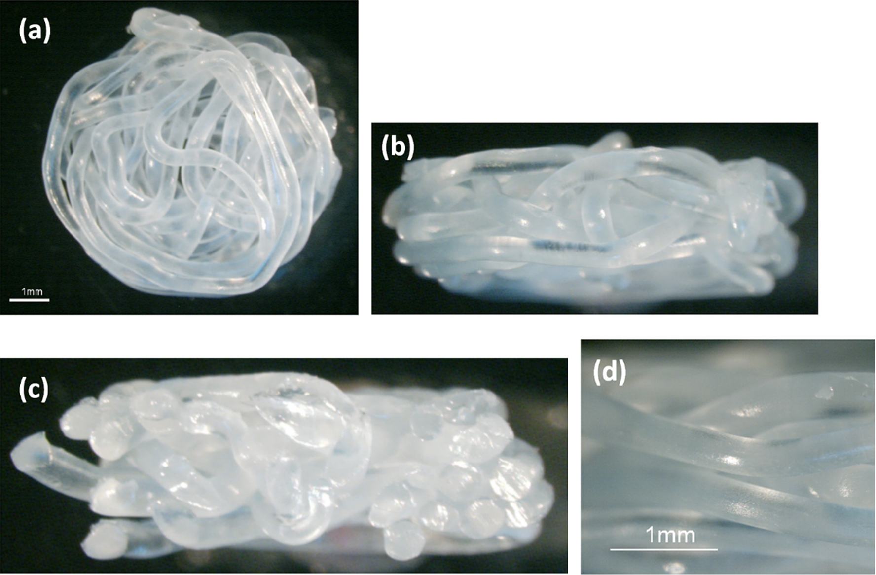
The stereomicroscope images demonstrate the morphologies of the pure PCL scaffold: (a) shows the superior view, (b) shows the lateral view, (c) shows the cross-sectional view and in (d) the magnified picture focuses on its surface.
The scaffolds of group A and B were left in 500 μL per well of proliferation medium [PR, Alpha-Minimum Essential Medium (α-MEM; Gibco, Invitrogen, USA) containing 10% fetal bovine serum (Gibco, Invitrogen, USA), 10,000 units/ml penicillin/streptomycin (Gibco, Invitrogen, USA), and 250 μg/mL fungizone (Gibco, Invitrogen, USA)] for detecting release of calcium and phosphate ions over 30 days. The same volumes of PR medium and osteogenic induction medium [OS, the PR medium supplemented with 10 mM β-glycerophosphate (Sigma, USA), 10−7 M dexamethasone (Sigma, USA) and 50 μM ascorbic acid-2 phosphate (Sigma, USA)] were used as negative and positive control respectively. The well plates were incubated at a constant temperature of 37°C for the entire experiments. On day 3, 7, 14, 21 and 30 thereafter, the scaffolds were moved into next wells and the fresh mediums were added. The solution of each previous well was collected for measuring calcium and phosphate ions using a Calcium and Phosphate Colorimetric Assay Kit (Biovision, USA) (n = 5/group/time point/testing). At the same time points, the solutions of the control groups were collected for the testing, and then they were replaced by fresh mediums (n = 5/group/time point/testing). To detect the calcium ion, 90 μL of the Chromogenic Reagent and 60 μL of the buffer solution were added into 50 μL/well of the sample solutions, and mixed gently in a 96-well plate. The plate was incubated away from light for 5 min at room temperature. The absorbance (OD) of the chromophore was read at 575 nm using a microplate reader (Thermo Fisher Scientific, USA). To detect the phosphate ion, 200 μL/well of the sample solutions were placed in a 96-well plate. Thirty microliters of phosphate reagent were added to each well and mixed gently. The plate was incubated at room temperature for 30 min. The absorbance of Malachite Green and Ammonium Molybdate formed a chromogenic complex with phosphate ions, which was read at 700 nm. The levels of OD were compared with a standard curve to calculate the amounts of calcium and phosphate ions in the solutions.
Preparing the cell-scaffold constructs
Prior to cell seeding, the scaffolds were immersed in PR medium for 24 h. On day 3, 7, 14, 21 and 30 days prior to the experiments, the cell-scaffold constructs were made by seeding
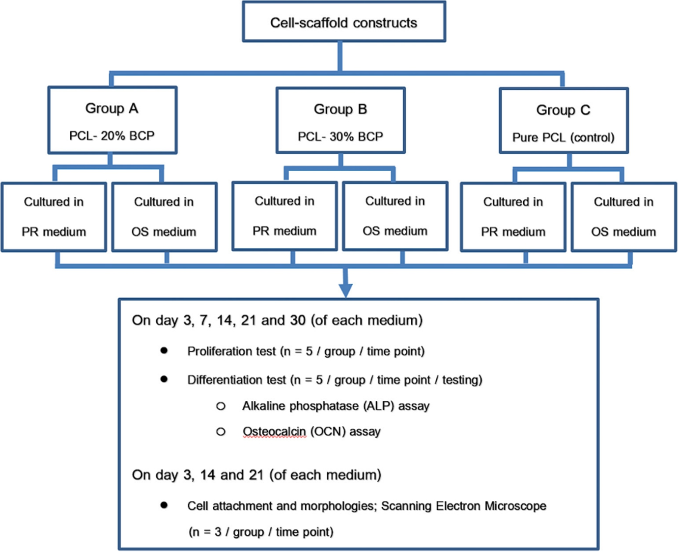
The diagram shows an overview of the cellular experiments.
The constructs of day 3, 14 and 21 were removed from the culture plates and rinsed with phosphate buffer saline (PBS), afterwards they were fixed in 2.5% glutaraldehyde (Sigma-Aldrich, USA) in PBS for 2 h. The specimens were dehydrated in the 50–100% ethanol series and coated with gold-palladium. Characteristics of the cells in the constructs were descriptively assessed using a scanning electron microscope (SEM, JOEL Ltd, Japan).
Cell proliferation
On the day of the experiment, a cell proliferative reagent (WST-1; Roche, Germany) was used to measure activity of mitochondrial dehydrogenases reflecting the number of viable cells according to the following protocol. Each construct was moved to a new well, rinsed with PBS, and then 200 μL of the fresh culture medium with 20 μL of WST-1 solution was added. The well plates were incubated for 4 h in 5% CO2 at 37°C. After that, 100 μL of the solution from each well was transferred to a 96-well plate in duplicate and the absorbance of the formazan product of each well was measured at 440 nm using the micro-plate reader. The levels of OD were compared with a standard curve to infer the amounts of the cells.
Cell differentiation
On the day of the experiment, the constructs were moved to the new wells and washed with PBS. Afterwards, 200 μL of 1% Triton X-100 in PBS was added to each well and then the constructs were minced into small pieces. The cells were lysed by freezing and thawing for three cycles (1 cycle: at −20°C for 15 min and at room temperature for 15 min). The mixtures were transferred into the micro-centrifuge tubes and centrifuged at
Statistical analysis
The data was analyzed using statistics analysis software (SPSS, version 21, USA). One-way Analysis of Variance (ANOVA) followed by Tukey HSD was applied to compare the differences of all parameters among the groups and time points. Dunnett’s T3 was performed when equal variances were not assumed. The level of statistical significance was set at a
Results
Release of calcium and phosphate ions
The amounts of the calcium and phosphate ions in the sample solutions were demonstrated in Fig. 5. Both ions of groups A and B significantly released on day 7 when compared with day 3 (
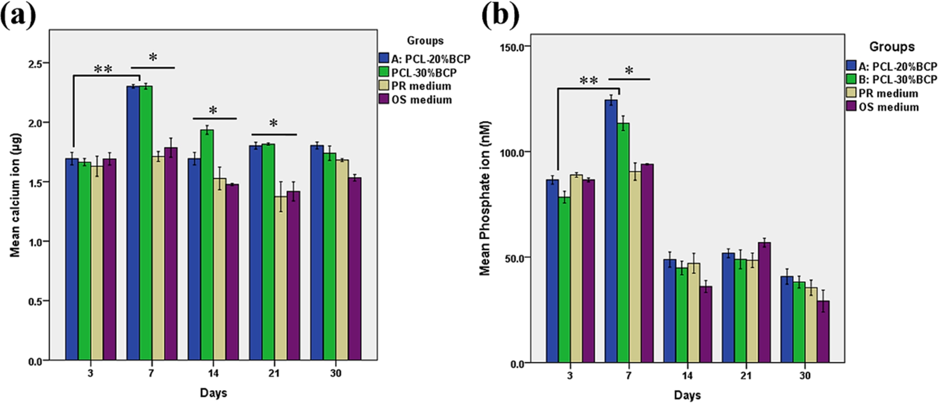
The graphs demonstrate the amounts of the calcium ion (a) and the phosphate ion (b) in the solutions of group A and B. The ions measured in the PR and OS medium were served as control. The ions of both groups significantly increased on day 7 (
The SEM pictures in Figs 6–8 demonstrate the behaviors of the osteoblasts in the cell-scaffold constructs when they were cultured in PR and OS mediums. It was found that the cells could attach and grow well throughout the scaffold surfaces of all groups. Since day 14, the cells formed multilayer cell-sheets covering the entire surfaces. Mineralization nodules could be observed in all groups when cultured in both medium from day 14.
The table shows the average concentrations (mM) of calcium and phosphate ions of the groups at each time-point
The table shows the average concentrations (mM) of calcium and phosphate ions of the groups at each time-point
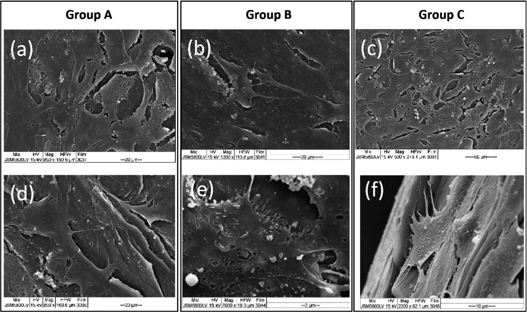
The SEM images demonstrate morphologies of the cells on culture day 3: (a)–(c) in PR medium and (d)–(f) in OS medium. The osteoblast cells spread their cytoplasmic processes attaching well to the scaffold surfaces of all groups.

The SEM images on culture day 14: (a)–(c) cultured in PR medium and (d)–(f) cultured in OS medium. Dense multilayer cell-sheets throughout the scaffold surfaces of all groups were seen. Mineralized nodules were detected in some areas of the constructs, which cultured in the OS medium (arrows).
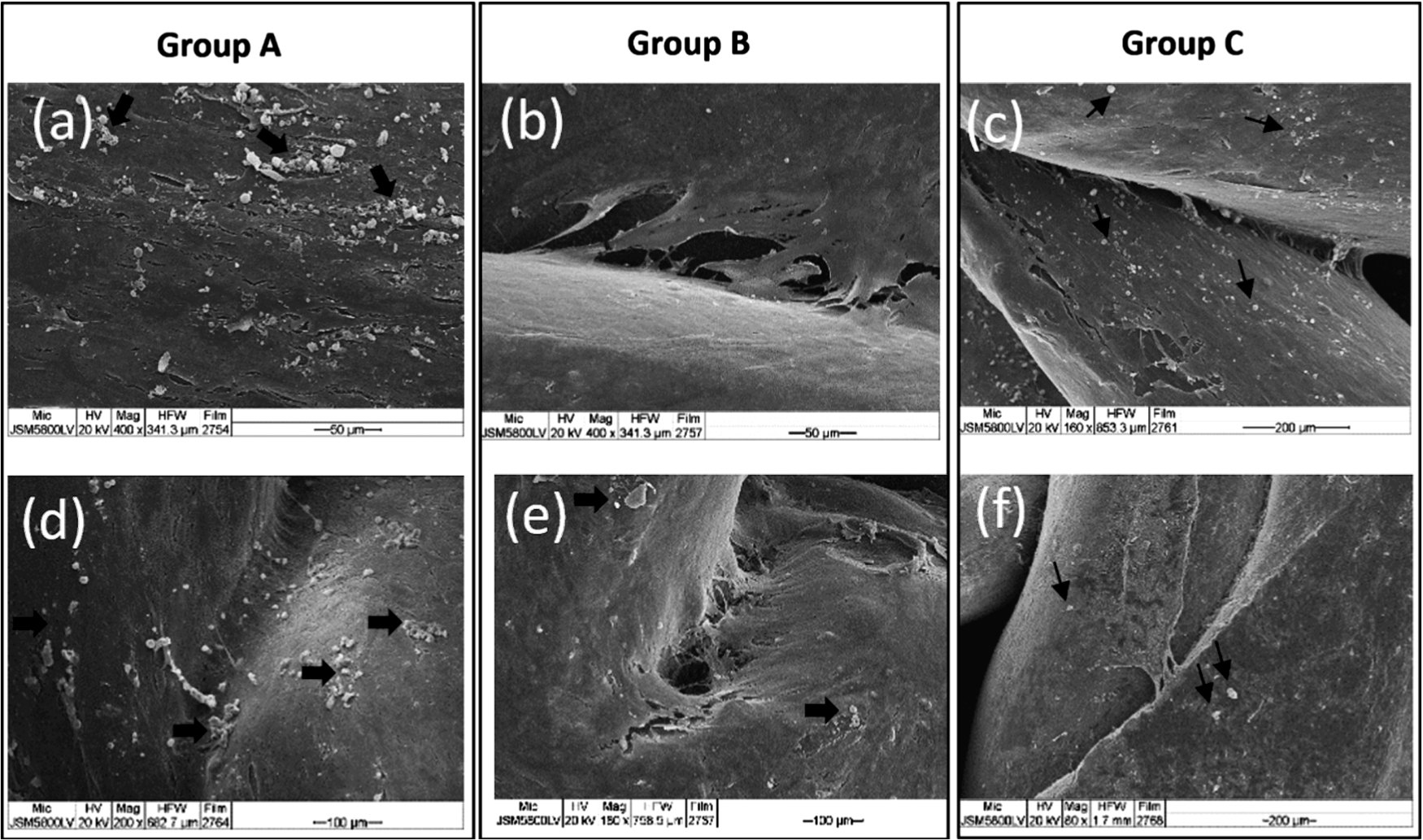
The SEM images on culture day 21: (a)–(c) cultured in PR medium and (d)–(f) cultured in OS medium. Denser cell-sheets covered the entire scaffold surfaces of all groups, and the morphologies of the cells were difficult to identify. General mineralized matrix formations were seen in the constructs of all groups (arrows).
The amounts of the viable cells in the cell-scaffold constructs are demonstrated in Fig. 9. In the PR medium, the amounts of the cells of group A increased with time and reached their maximum growth on day 30 (
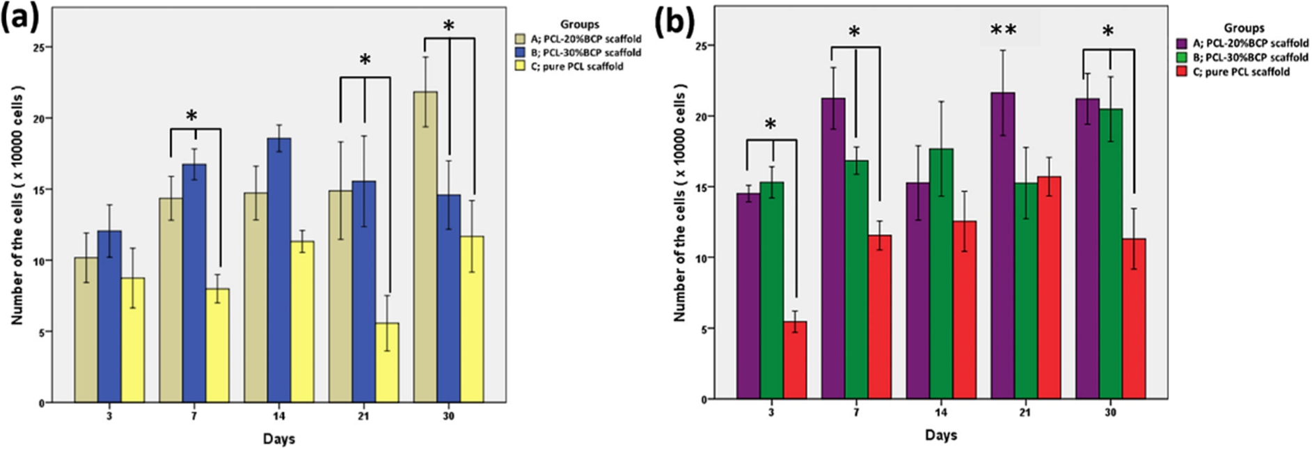
The graphs demonstrate the growth profiles of the cells in the constructs. In the PR medium (a), the viable cells of group A and B were greater than group C at every time point (significantly different on day 7, 21 and 30;
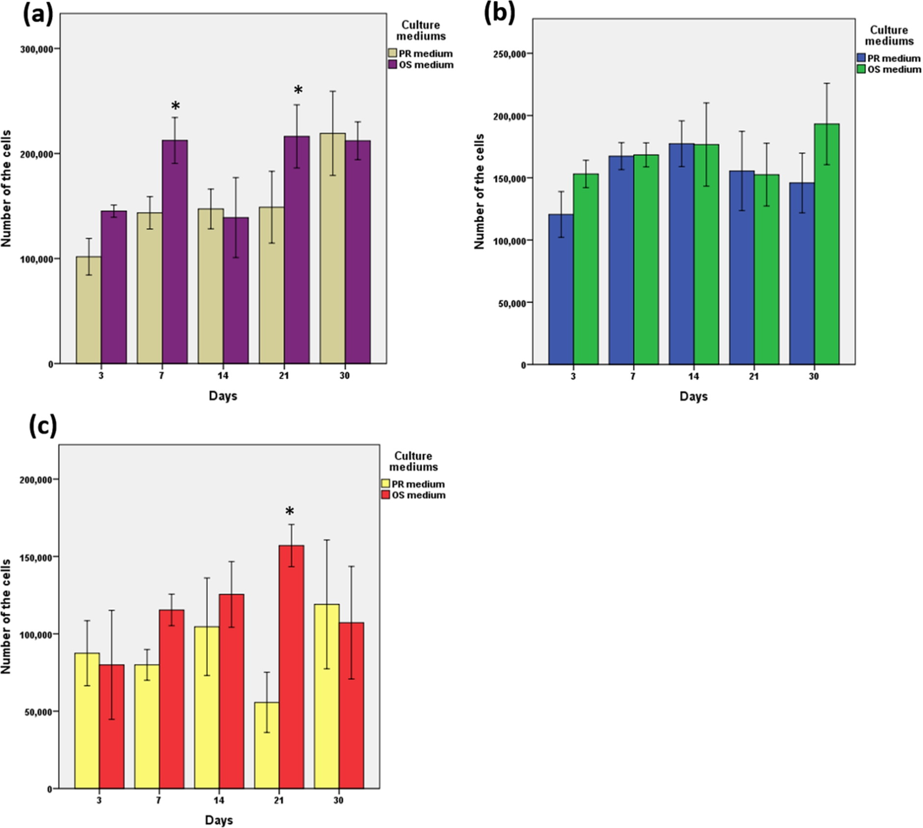
The graphs demonstrated the proliferation of the cells in each group, when cultured in the different mediums; (a) group A, (b) group B and (c) group C. The growth in the OS medium was statistically greater than that in the PR medium on day 7 and 21 of group A (
The ALP activities of the cells in the constructs are demonstrated in Fig. 11. In the PR medium, the highest level of ALP in group A was detected on day 3, then it slightly decreased on day 7. The lowest level of ALP was detected on day 21, but they increased remarkably on day 30. The ALP of group B increased to the maximum level on day 7 and then significantly decreased to the lowest level on day 14 and 21, but remarkably increased on day 30. Low levels of ALP were detected in group C over the observation periods. It was noted that the ALP activities of group A and B were remarkably greater than those of group C over the observation period, and there was no significant difference between group A and B. In the OS medium, the activities of groups A and B increased with time to reach their maximum levels on day 14, and they seemed to be stable thereafter. Similar to the PR medium, the activities of group A and B were obviously higher than those of group C at all time points (
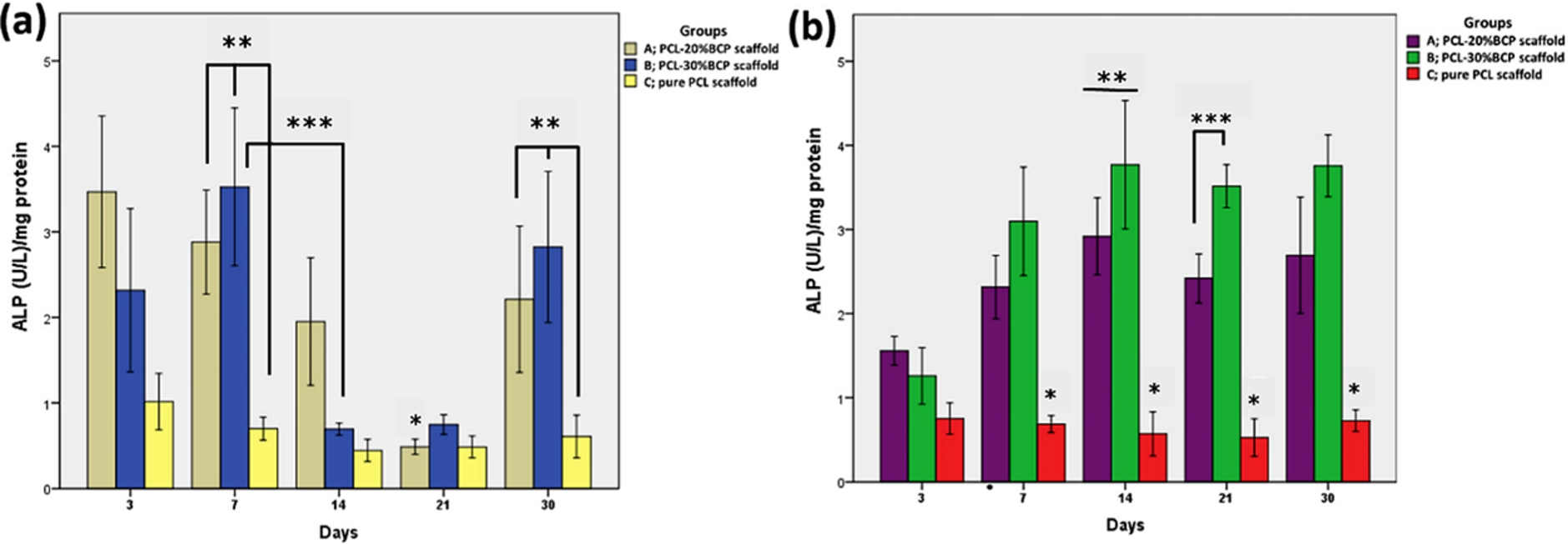
The graphs demonstrate the ALP activities of cells in the constructs over 30 days. In the PR medium (a), the ALP activity of group A on day 21 was significantly lower than other days (
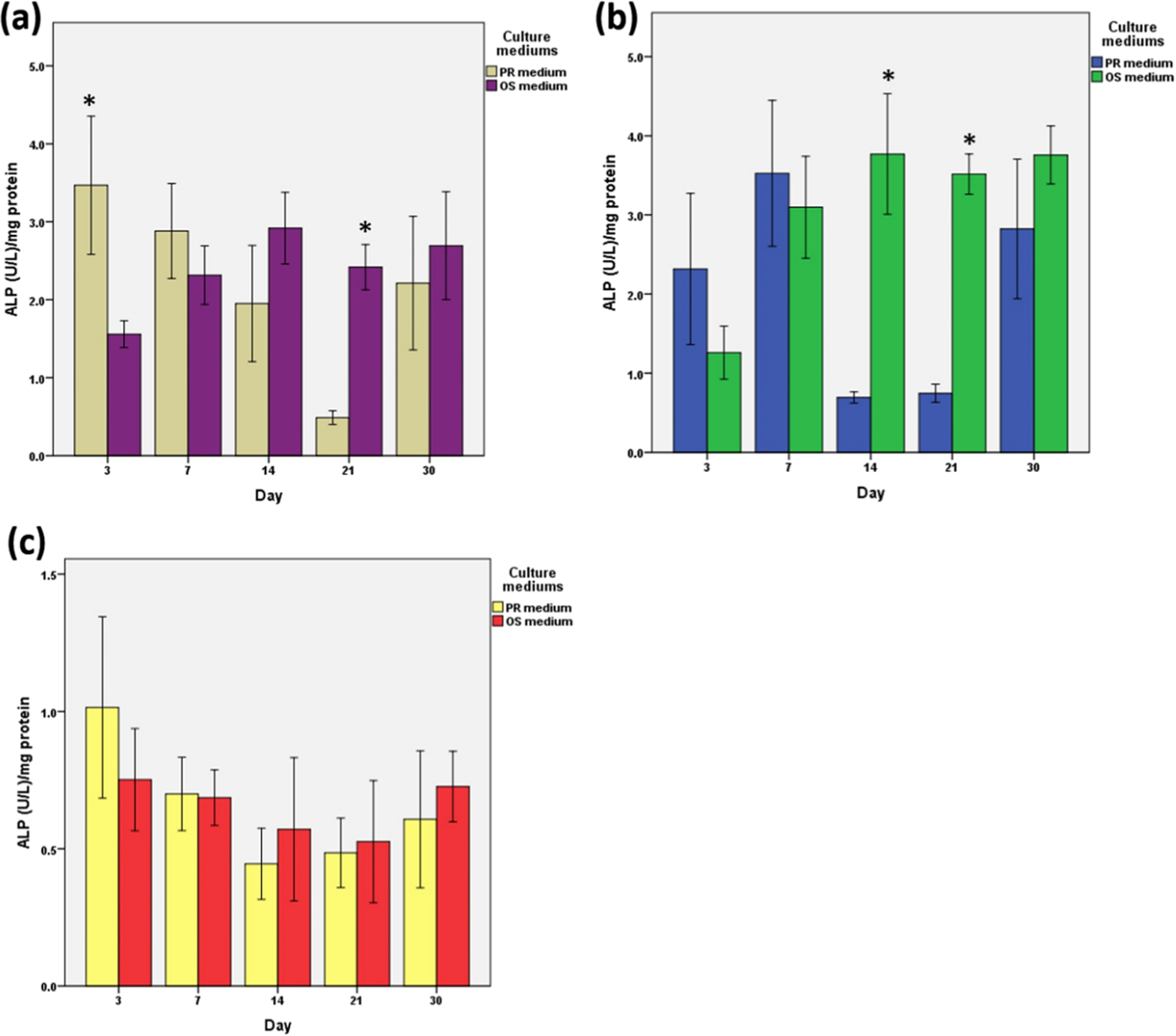
The ALP activities of the cells in each group when cultured in the different mediums are demonstrated: (a) group A, (b) group B and (c) group C. In figure (a), the levels of ALP in PR medium were higher than in OS medium over the first 7 days (
The expression of OCN of the cells in the constructs was demonstrated in Fig. 13. In the PR medium, the OCN levels of groups A and B were stable during the first 21 days, then they significantly increased on day 30 (

Graph (a) demonstrates the OCN levels of the cell-scaffolds constructs cultured in PR medium. The levels of group A and B slightly changed until day 21, and then they increased to the maximum level on day 30. The levels of group A and B were significantly greater than those of group C on day 7, 21 and 30 (
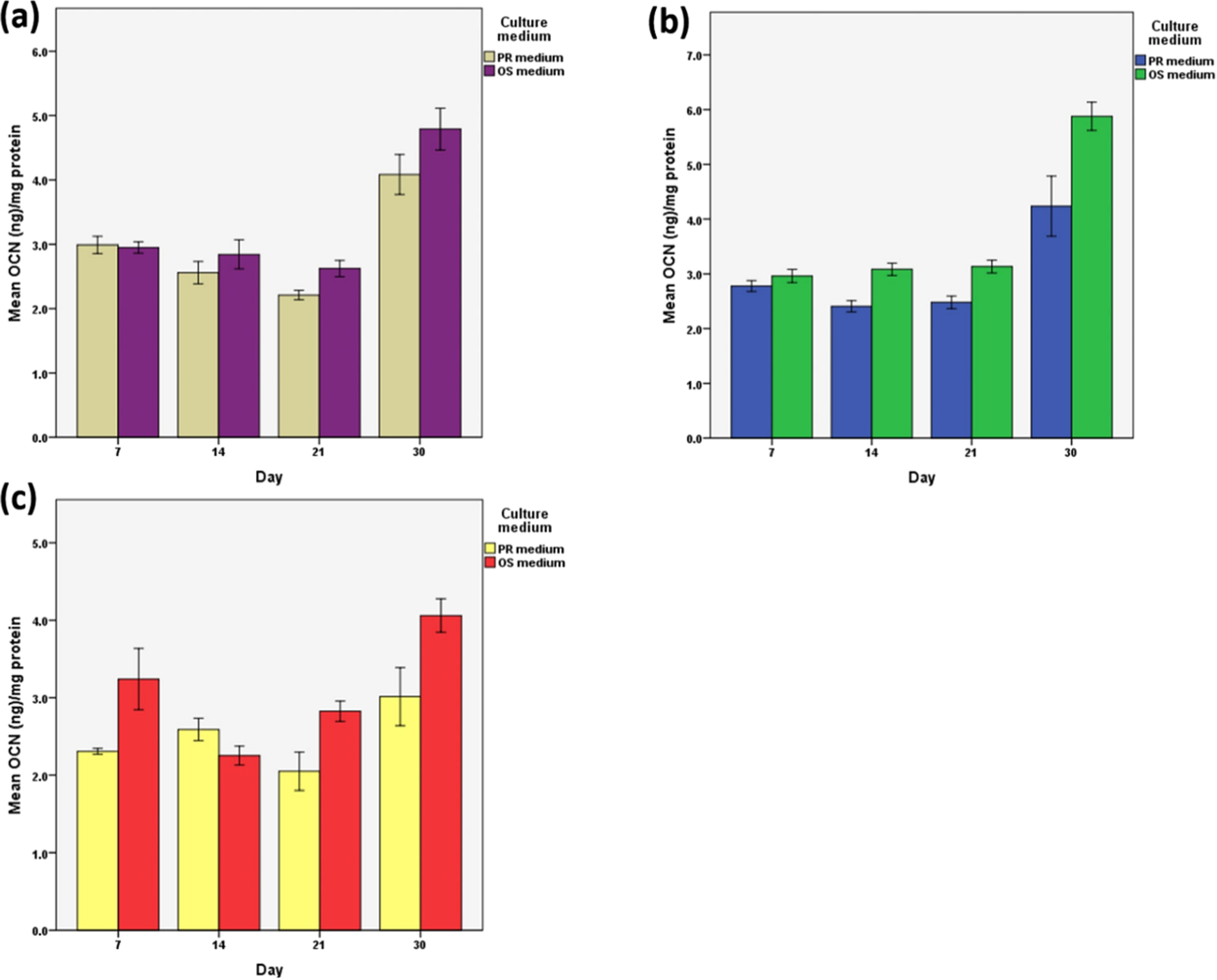
The levels of OCN in the different medium of each group are demonstrated: (a) group A, (b) group B and (c) group C. Similar profiles of OCN in both mediums were detected in groups A and B. In group C, the OCN levels in OS medium were remarkably higher than in PR medium on day 7, 21 and 30 (
This study was performed in order to evaluate the effects of the PCL-BCP MSCM scaffolds on proliferation and differentiation of osteoblasts that directly seeded on their surfaces. Bioactivities of the PCL scaffolds containing BCP filler at the ratios of 20% and 30% were compared with those of the scaffolds without the filler. The ratio of 30% is the maximum amount of the filler, which can be used to fabricate the PCL-BCP monofilaments by the melting-extruding machine [1]. Inconsistency in sizes of the filaments and increase in filament-fracture will occur if blending PCL with fillers greater than 30% by weight. In addition, the ratio of 10% BCP was excluded from the study due to insufficient amounts of the filler to detect its effects. In the experiment, matrix mineralization was not assessed in order to avoid its false positive staining due to its capability to absorb solubilized Alizarin Red S into the scaffolds and stain carbonate apatite layers on the ceramic particles [3] instead of the calcified matrix of the cells.
Regarding the results, the burst release of the calcium and phosphate ions from the PCL-BCP scaffolds on day 7 could increase the entire levels of those ions in the mediums significantly higher than the levels in the PR or OS mediums without the scaffolds. After day 7, higher levels of the calcium ion in the mediums with the scaffolds could be sustained until day 30, whereas, the levels of the phosphate ion were not different. Several studies [24–30] support effects of additional calcium ion in mediums on inducing proliferation, differentiation and extracellular matrix mineralization of osteoblasts. Although phosphate ion is also essential for the cell proliferation and osteogenic differentiation, some studies [25,28] report apoptosis of the cells that induced by additional exogenous inorganic phosphate at high concentrations. Liu et al. [25] revealed that the optimum amounts of the calcium and phosphate ions in culture mediums for supporting growth and osteogenic differentiation of mesenchymal stem cells were 1.8 mM and 0.09 mM, respectively. The authors claimed that the concentration of the calcium ion was more important for the cell differentiation and mineralization than the phosphate ion. The lower or higher calcium concentrations leaded to decreased the cell differentiation, whereas, various concentrations of phosphate had no effects on the cell activities. In addition, the cells could not live over 10 days, when they were cultured in the environment of high phosphate concentration greater than 0.18 mM. Regarding our results, the averages amounts of the calcium and phosphate ions in the mediums after immerged with the scaffolds were lower than those optimum concentrations. However, the mediums were refreshed every 3 days during the culture period, hence, the real time concentrations of the ions that exposing to the cells might be higher than the results. The quantitative measurements of the viable cell numbers in the constructs revealed that the PCL-BCP scaffolds were remarkably superior to the pure PCL scaffolds, whether culturing in PR or OS mediums. Regarding the profiles of the releasing ions and the cell proliferation in Figs 5 and 9, it was clearly that the maximum releasing of the ions on day 7 did not affect the growth of the cells which attaching on the scaffolds. The cells on the PCL-BCP scaffolds rapidly grew on the first 7 days, when cultured in the PR medium as the times of the maximum release of the calcium and phosphate ions. This phenomenon contrasted to the result of our previous study [1], which found that the burst releases of the ions on day 7 retarded the cell proliferation. On the following days 14–30, although the calcium ion sustained release in the lower concentrations, the amounts of the ion still supported the growth of the cells in both groups. The amounts of the calcium ion in groups A and B rapidly decreased from 1.14 mM on day 7 to be 0.84 and 0.96 mM respectively on day 14. In these condition, the concentration of 0.96 mM seemed to be more suitable for the cell proliferation due to the fact that the maximum growth is detected in group B, whilst, a little increase in growth of group A is detected. On day 21, the calcium concentrations of both groups were equal at 0.90 mM, and these conditions made the number of the cells in group A slightly increasing, but, those of group B remarkably decreasing. On day 30, the concentration in group A was still at 0.90 mM, whereas, that in group B slightly decreased to 0.86 mM. That concentration remarkably increased the cell number of group A, but, slightly decreased the number of group B. Therefore, it implies that the amounts of the calcium ion in the medium within the range of 0.90-1.14 mM provide appropriate environment for the growth of the osteoblasts. In our opinion, the concentrations of the releasing ions from the scaffolds had less of an effect on the proliferation of the osteoblasts directly attaching on the scaffolds. In this situation, architecture and surface properties of the scaffolds would be the other major factors that influenced the behaviors of the cells in the constructs. The architectures and the surface morphologies of the MSCM scaffolds are suitable for supporting growth and differentiation of the osteoblasts regardless of the composition of materials. The scaffolds have high porosity of 73.19 ± 3.31% with large pore sizes that allow high oxygen and nutrient transportation throughout the scaffolds. For the PCL-BCP scaffolds, the ceramic filler can produce a calcium-phosphate rich layer or a hydroxyl-carbonate apatite layer on the surfaces of the scaffolds, which serve as a template for hydroxyapatite growth and provide a good environment for bone cells to grow and differentiate to form extracellular matrixes [3,31–35]. The SEM images demonstrated that the osteoblast cells attached and proliferated very well on both PCL-BCP and pure PCL scaffolds. The cells grew in multi-layers, increasing with culture-time, and mineralized matrix formations were generally detected from the second week of culture. By observation, there was no difference in those characters of the cells among the groups of scaffolds. In principle, there are three osteoblast phenotypic stages of rat osteoblasts during the culture period of 4 weeks including proliferation, extracellular matrix (ECM) maturation and mineralization. Dworetzky et al. [36] found that peaks of the cell proliferation occur at days 7, and then decline after day 9, at the time of ECM maturation period is initiated. During the second stage, expression of ALP is peak at day 15, and after day 23, the expression is down-regulated, while levels of OCN expression increase which reflecting the stage the matrix mineralization. Similar to the cell proliferation, our results demonstrated that the cells on the PCL-BCP scaffolds expressed the activity of ALP obviously higher than those on the pure PCL scaffolds when cultured in both mediums. In addition, the maximum levels of ALP of the groups of PCL-20% BCP and PCL-30% BCP in the PR medium were detected earlier at day 3 and 7 respectively. Interestingly, when cultured in the PR medium, the highest levels of ALP of both ratios were detected earlier and their activities were greater than those in the OS medium during the first 7 days. Therefore, it implies that the BCP filler could enhance the bioactivity of the PCL-based scaffold in term of accelerating the proliferation and the early phase of osteogenic differentiation of the cells in the constructs without osteogenic inductive condition from the medium. When comparing between the both ratios, the cells on the PCL-20% BCP scaffolds expressed the maximum activity of ALP when cultured in the PR medium faster than those on the PCL-30% BCP scaffolds, however, there was no significant difference. Regarding late differentiation marker, the profiles of the OCN in PCL-BCP groups slightly changed during the first 21 days and reached the maximum levels on day 30. This sequence is the regular progressive change of the OCN during the osteoblastic differentiation, therefore, it implies that the bioactivities of the PCL-BCP scaffolds have no effect on enhancing the late phase of differentiation of the cells. Interestingly, the levels of OCN in each group were not different, whether culturing the constructs in the PR or OS medium. This contrasts with the results of the pure PCL group where the levels in the OS medium were remarkably higher than those in the PR medium over all the culture periods. It implies that the osteoblasts on the pure PCL scaffold required the osteogenic inductive environment from the culture medium for entering the late phase of osteogenic differentiation, whilst, the PCL-BCP scaffold could support the entire phases of osteogenic differentiation by itself. Similar to the early phase of the cell differentiation, there was no statistically difference for supporting the later phase of matrix mineralization of the osteoblasts between the groups of 20% and 30% of the BCP filler.
Conclusion
The morphologies and the surface properties of the PCL-BCP MSCM scaffolds are suitable to perform functional cell-scaffold constructs used for bone-tissue engineering. The addition of BCP filler in the PCL-based scaffolds obviously increased the bioactivities of the scaffolds in terms of enhancing growth and differentiation of the osteoblasts when compared to the scaffolds without the filler. In addition, the PCL-BCP scaffolds could enhance the early phase of the osteogenic differentiation, even when cultured in non-osteoinduction medium.
Footnotes
Acknowledgements
This research was supported by The Cranio-Maxillofacial Hard Tissue Engineering Center, Department of Oral and Maxillofacial Surgery, Faculty of Dentistry, Prince of Songkla University, Hat Yai, Songkhla, Thailand. The authors would like to thank Mr. Mitchell Allan Bruce Atkins for proofreading.
Conflict of interest
No conflict of interest to disclose.