
Abstract
Background:
Biocompatibility and degradation of poly ε-caprolactone (PCL)-Biphasic Calcium Phosphate (BCP) scaffolds fabricated by the “Melt Stretching and Compression Molding (MSCM)” technique were evaluated in rat models.
Objectives:
Degradation behaviors and histological biocompatibility of the PCL-20% BCP MSCM scaffolds and compare with those of PCL-20% β-tricalcium phosphate (TCP) scaffolds commercially fabricated by Fused Deposition Modeling (FDM) were evaluated.
Methods:
The study groups included Group A: PCL-20% BCP MSCM scaffolds and Group B: PCL-20% TCP FDM scaffolds, which were implanted subcutaneously in twelve male Wistar rats. On day 14, 30, 60 and 90, dimensional changes of the scaffolds and their surrounding histological features were assessed using Micro-Computed Tomography (μ-CT) and histological analysis. Changes of their molecular weight were assessed using Gel Permeation Chromatography (GPC).
Results:
Formation of collagen and new blood vessels throughout the scaffolds of both groups increased with time with low degrees of inflammation. The μ-CT and GPC analysis demonstrated that the scaffolds of both groups degraded with time, but, their molecular weight slightly changed over the observation periods. All results of both groups were not significantly different.
Conclusions:
The PCL-20% BCP MSCM scaffolds were biocompatible and biodegradable in vivo. Their properties were comparable to those of the commercial PCL-20% TCP scaffolds.
Keywords
Introduction
Reconstructing large defects in the maxillofacial region using several different grafting techniques is still very challenging due to the fact that no grafting materials can yet replace the gold standard of autogenous bone grafting. For several years now, using several synthetic materials as bone substitutes has become more popular due to their better osteoconductive and osteoinductive properties. Recently, our research team developed the technique of Melt Stretching and Multilayer Deposition (MSMD) for fabricating three-dimensional (3-D) scaffolds for bone substitution [1–3]. The MSMD scaffolds are specifically designed to have an appropriate interconnecting pore system for enhancing osteogenesis. A microgroove pattern, typically found on the surfaces has proved to support attachment of osteoblasts [1]. In addition, the mechanical properties of the scaffolds are suitable for withstanding forces occurring in real circumstances of reconstruction in the oral and maxillofacial region [3]. To make the fabricating process easier and more practical, the steps of MSMD were simplified and renamed “modified MSMD (mMSMD) or Melt Stretching and Compression Molding (MSCM)” [4]. The 3-D scaffold can be fabricated by only compressing the polymeric monofilament into a glass mold and immersing in warm water. Therefore, the MSCM technique is unique, economical and achieves three major advantages. Firstly, the technique is easier to process; it can instantly be conducted by any surgeon beside his dental chair, during an operation, and thus, the processing time is remarkably reduced. Secondly, fabricating the 3-D scaffold within a glass mold is a closed environment that prevents contamination. Thirdly, using melt blending without any solvents and porogens for fabricating the composite scaffold is considered to be safe for cells and tissue. For the materials, poly ε-caprolactone (PCL) is used as the major component of the scaffold, whilst Biphasic Calcium Phosphate (BCP) which consists of the stable phase of hydroxyapatite (HA) and the more soluble phase of β- tricalcium phosphate (TCP) is used as the filler. Although being a widely used biomaterial and its biocompatibility having been proved, it is known that PCL has an hydrophobic property which takes 2–4 years to totally degrade [5]. Therefore, regarding our previous study [4], addition of BCP which composed of
Prior to clinical trials in humans, this study re-evaluated the biocompatibility and degradation behaviors of the PCL-20% BCP MSCM scaffolds in animal models. Their properties were compared with those of commercial PCL-20% TCP scaffolds fabricated by the Fused Deposition Modeling (FDM) technique for benchmarking. In addition, efficacies of enhancing degradation between the different fillers of BCP and TCP in the PCL-based scaffolds could be compared.
Materials and methods
Scaffold fabrication and study groups
The study groups were divided into 2 groups, Group A: PCL-20% BCP MSCM scaffolds and Group B: PCL-20% TCP FDM scaffolds. The scaffolds of group A were fabricated using the MSCM technique as follows [4]. In brief, PCL pellets (Sigma Aldrich, USA) and BCP particles (
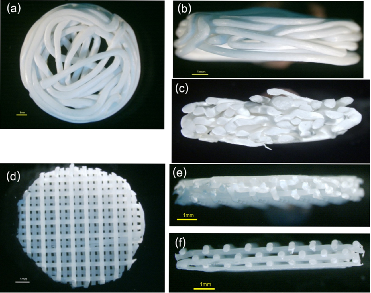
The testing specimens of group A ((a)–(c)) and group B ((d)–(f)): (a, d) superior view, (b, e) lateral view and (c, f) cross-sectional view.
Micro-Computed Tomography (μ-CT) analysis
Prior to the surgical implantation, the scaffolds of both groups were scanned using a μ-CT machine (μ-CT 35, SCANCO Medical AG, Switzerland) at 55 kVp, 145 μA and 4 W (
Gel Permeation Chromatography (GPC) analysis
Prior to the surgical implantation, weight-average molecular weights (Mw) and number- average molecular weights (Mn) of the scaffolds of both groups were measured using GPC processing (Viscotek system, UK) (
Scanning Electron Microscope (SEM)
The scaffolds were coated with Gold-Palladium and their surface morphologies were examined using SEM (JEOL Ltd, Japan) (
The procedure in animal models
Twelve male Wistar rats, weight 200–250 g were used for implanting the scaffolds. The surgical experiment was performed in accordance with the regulations of the university board of ethics committee.
Subcutaneous implantation
For each animal, after it was anesthetized by an intraperitoneal injection of Thiopental 15 mg/kg, six areas on the back (3 sites/each side of the midline, along the spine) were shaved and disinfected with betadine solution and 70% ethanol. Six subcutaneous pockets were created by making 1 cm-long vertical incisions, and then two samples of each group from the pre-operative μ-CT analysis and the second piece of each group from the pre-operative GPC analysis were inserted (Fig. 2). The wounds were then sutured with 4-0 Vicryl. At each time point of 14, 30, 60 and 90 days thereafter, three animals were sacrificed and the specimens including the scaffolds and surrounding soft tissue were retrieved.
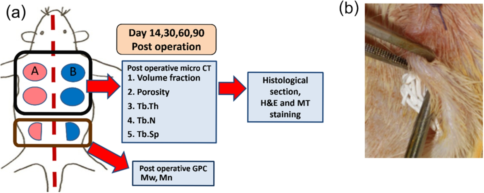
A diagram of the surgical implantation of the scaffolds on the back of each rat (a). On days 14, 30, 60 and 90 after the operation, the animal was sacrificed and the specimens including the scaffolds and surrounding soft tissue were biopsied. The parameters of μ-CT analysis of the first 4 specimens were re-measured, after that they were sent for histological sectioning and staining. The last two scaffolds were retrieved for measuring their Mw and Mn using GPC analysis. The picture (b) demonstrates subcutaneously inserting the MSCM scaffold.
μ-CT analysis
The specimens from days 30, 60 and 90 were fixed in 10% buffered formalin for 48 h prior to scanning (
GPC analysis
For each specimen from days 30, 60 and 90, the tissue surrounding the scaffold filaments was mechanically excluded. The filaments were washed many times with distilled water and some parts of them were collected for GPC analysis using the same protocol as for the pre-operation (
Histological analysis
The specimens from day 14 and days 30–90 (after finishing the μ-CT analysis) were cut along the midline using a cutting-gridding machine (Exakt, Germany), and one side was embedded in paraffin. The serial 5 μm-thick sections were cut at the position of 500 μm from its cutting edge and stained with Hematoxylin and Eosin (H&E), and Masson’s trichrome (MT) (
SEM
After mechanical cleaning, some pieces of the filaments of the scaffolds from days 30, 60 and 90 were collected for coating with Gold-Palladium. Their surface morphologies were re-assessed descriptively using the SEM.
Statistical analysis
The parameters of μ-CT and GPC were analyzed using statistics analysis software (SPSS, version 21, USA). Wilcoxon signed-rank test was applied to compare the differences of those parameters among the healing intervals within each group. Mann-Whitney U-test was applied to compare differences between the two groups at the same time points. The level of statistical significance was set at
Results
All animals tolerated the operation well and were healthy during the observation period. The surgical wounds were intact and healed without complication. The gross specimens from day 30 showed that the scaffolds of both groups were covered with dense fibrous tissue without signs of inflammation and foreign body reaction.
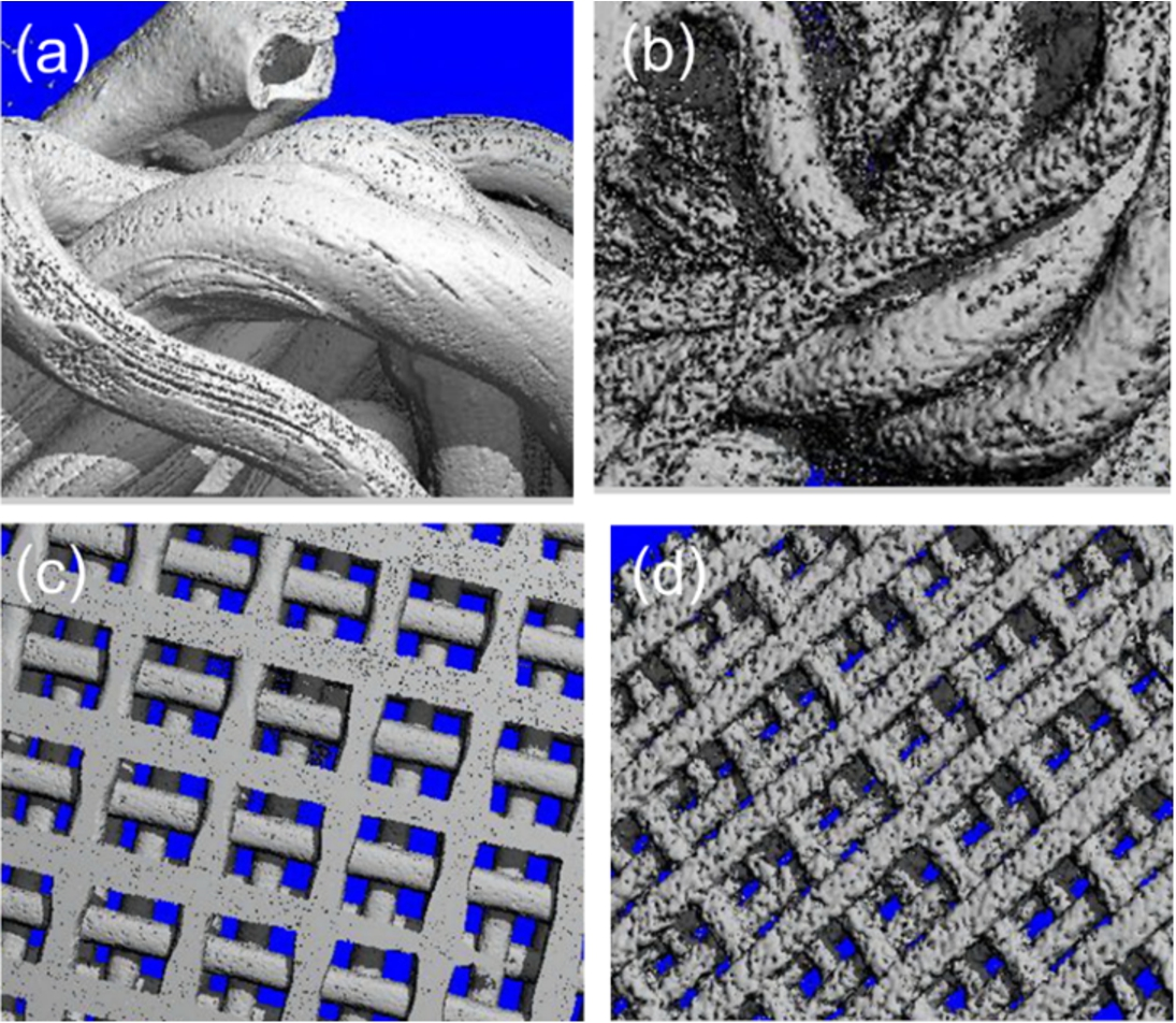
The 3-D reconstructed images of the scaffolds of group A (a, b) and group B (c, d). The pre-operative images (a, c) demonstrate the smooth surfaces of the scaffolds of both groups, whereas, their surface erosion is clearly seen in the post-operative images of day 90 (b, d).
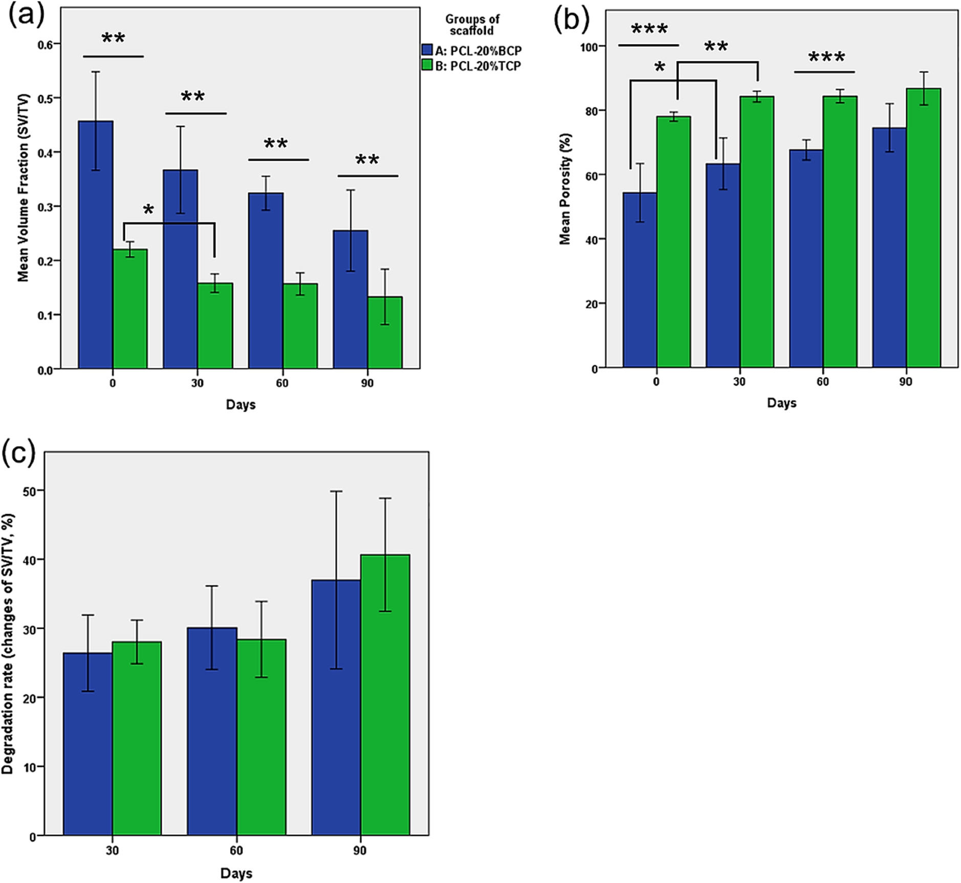
The graph (a) demonstrates that the scaffold volume fractions of both groups decreased with time. The significant difference was detected in group B between day 0 and 30 (
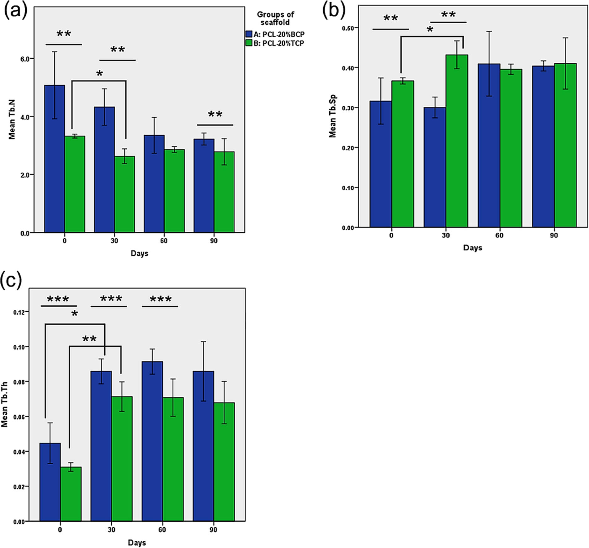
In graph (a), the scaffold trabecular number of both groups remarkably decreased in the first 30 days. The significant difference was detected in group B (
Morphologies of the scaffolds at pre- and post-operation are demonstrated in Fig. 3. The 3-D constructed images showed that surface erosion of the scaffolds was detected in both groups from day 30. The parameters of μ-CT analysis demonstrated the progress of the degradation over the observation period (Figs 4 and 5). The scaffold volume fractions of both groups decreased with time, which corresponding to the increases in the porosities. Differences of the scaffold volume fractions between the pre-operation and the post-operation at each time point can be calculated to be the degradation rates in percentages. It was noted that the degradation rates of the scaffolds of both groups gradually increased in the first 60 days and then remarkably increased on day 90, but no significant difference was detected. In addition, there was no statistical difference of the degradation rates between both groups at every time point. The μ-CT data also demonstrated different characters of the scaffolds. The PCL-BCP MSCM scaffolds had the volume fraction significantly higher (
GPC
Changes of the average molecular weights of the scaffolds are demonstrated in Fig. 6. The data shows that Mw and Mn of both groups slightly changed and no statistical difference was detected. It implies that molecular weight loss was not detected over the observation periods.
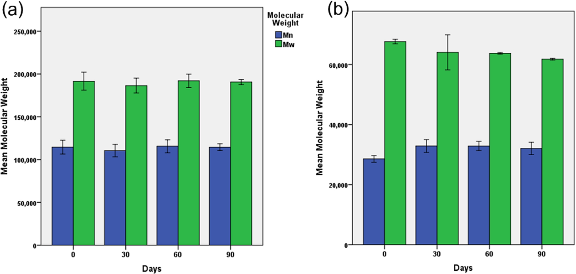
Profiles of the molecular weights of the scaffolds of group A (a) and group B (b). The graphs show that the molecular weights of both groups slightly changed over 90 days. No significant difference was detected at every time point.
After implantation, increase of erosion was clearly seen throughout the surfaces of the scaffolds of both groups (Fig. 7).
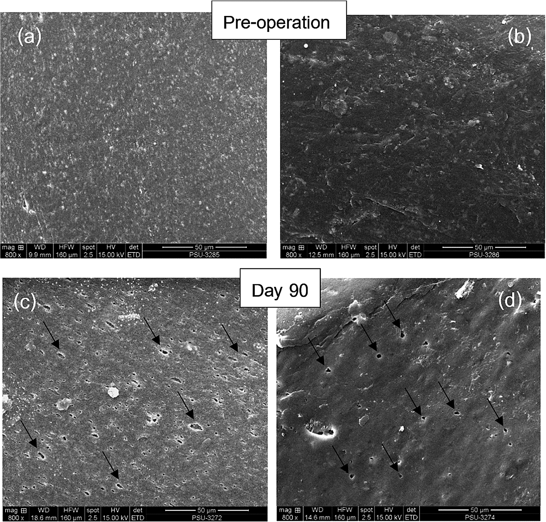
The SEM images of the scaffolds of group A (a, c) and group B (b, d). The pictures demonstrate generalized surface erosion of the scaffolds of both groups on day 90 (arrows).
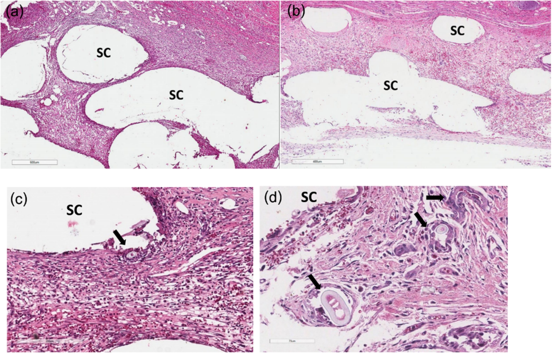
Histological features of the implanted areas are demonstrated in Figs 8–10. During the histological preparation, the scaffold filaments were totally dissolved, hence the scaffold areas of both groups were seen as empty spaces in the histologic images. On day 14 (Fig. 8), the scaffolds of both groups were surrounded by fibrous connective tissue and neo-vascularization was clearly found throughout the inner parts of them. Chronic inflammatory cells and giant cells were occasionally found. On day 30, an increase of neo-vascularization along with denser collagen connective tissue was detected in both groups (Fig. 9). On day 60 and 90, dense collagen fibers with well-arrangements around the scaffold filaments were found, along with well-formed large vascular networks (Fig. 10). By observation, the collagen formation within the scaffolds of group A was denser than those of group B, which had more adipose tissue in their connective tissue stroma. No ectopic bone formation of both groups was detected in the areas of implantation.
The H&E stained sections from day 14: group A (a, c), group B (b, d). In (a) and (b), the scaffolds were surrounded with fibrous connective tissue throughout their interconnecting pores. In (c) and (d), chronic inflammatory cells, which are mainly lymphocytes that were generally found infiltrating in the connective tissue stromal. By observation, more cells were found in group A than group B. Giant cells were occasionally found in both groups and some of them were ingesting the scaffold particles (arrows). Abbreviations:
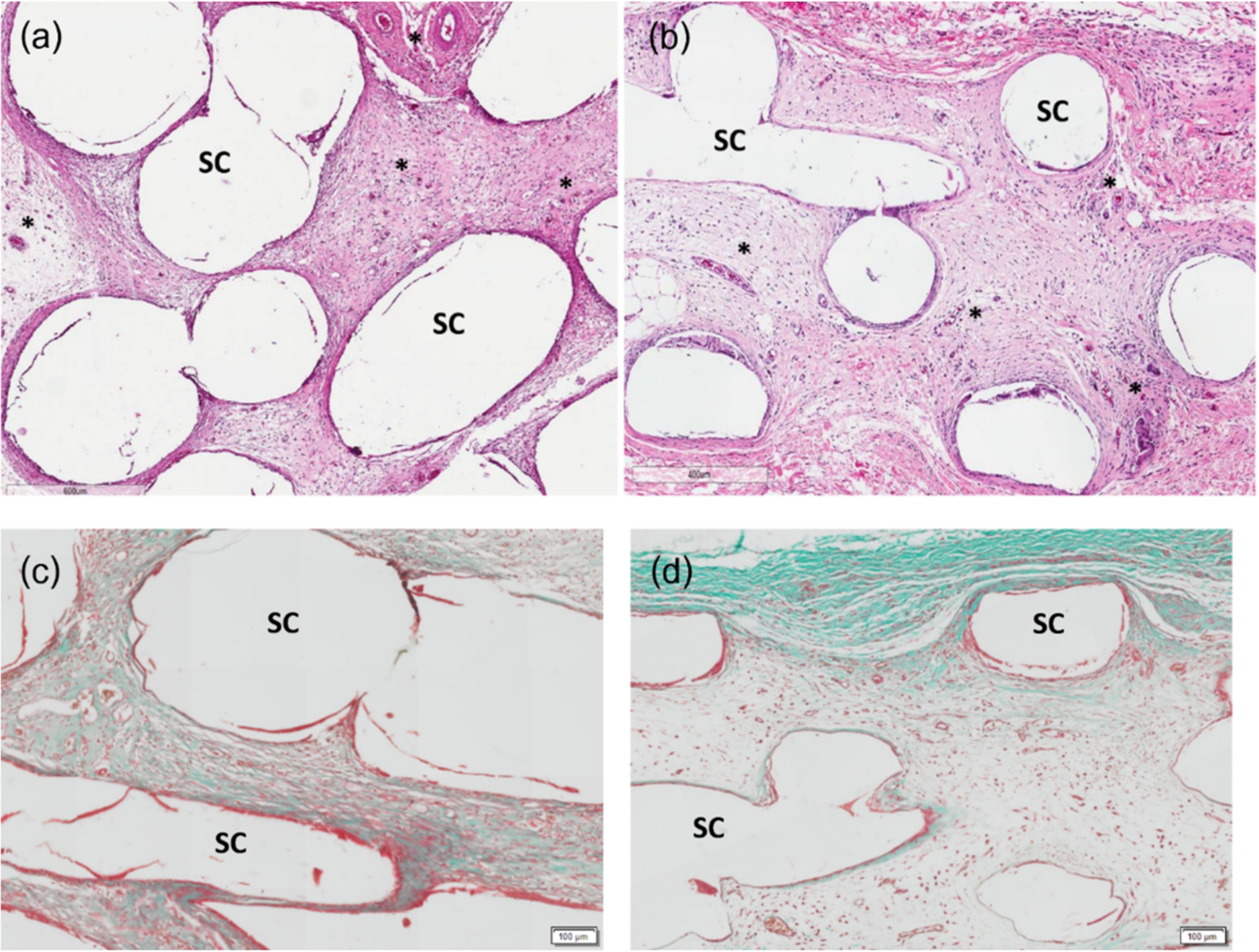
Implanted sites from day 30: group A (a, c), group B (b, d). In (a) and (b), the H&E stained sections show an increase of dense fibrous connective tissue and vascular regeneration (*) without any inflammatory response. In (c) and (d), MT stained sections show the background of dense collagen formation. Abbreviations:

Sections from day 90: group A (a, c), group B (b, d). The sections demonstrate well-formed dense collagen fibers and large vascular networks (*) around the scaffold filaments of both groups. Adipose-like cells were generally found within the connective tissue stroma of the scaffolds of group B (arrows). Abbreviations:
This study revealed two essential properties, biodegradability and biocompatibility, from the two different types of PCL-ceramic scaffolds when implanted in living tissue. Regarding the experiment, combining μ-CT and GPC is an effective method for assessing degradation of polymer-ceramic composite scaffolds due to the fact that changes of the dimension and the volume of the scaffolds as well as their molecular weight can be monitored at the same time. Histological evaluations over 90 days is considered to be commensurate with the bone remodeling process. The degradation behaviors of the scaffolds extending from that period can be predicted by the profiles of their degradation rates using the μ-CT analysis. In principle [6–12], degradation of the PCL-based scaffolds starts through the process of autocatalysis with non-enzymatic hydrolytic chain scission of ester linkages. Afterwards, diffusion of water into inner portions causes swelling and starting bulk hydrolysis throughout the polymer matrix. As degradation advances through the late stage, an onset of weight loss occurs corresponding to a decrease in the rate of chain scission. During those processes, hydrolysis by-produces from carboxyl groups diffuse out to the surrounding environments and form carboxylic acid that would induce acute inflammation and adverse tissue reactions. Therefore, combining some ceramic fillers with PCL is an effective way to not only enhance the degradation of the composite scaffolds, but also to neutralize or buffer the acidic degrading by-products and reduce inflammatory environments [9,13]. The commercial PCL-20% TCP scaffolds used as the benchmark have been proven to support proliferation and differentiation of the osteoprogenitor cells and in vivo bone formation [11,12,14–21]. Regarding the parameters of the μ-CT analysis, the volume fractions, the porosity, the trabecular number and the trabecular thickness demonstrated the different morphologies between the scaffolds of both groups. The parameters of group A, which higher than those of group B at all-time points would imply that the PCL-BCP MSCM scaffolds occupy the areas of interest greater than the PCL-TCP FDM scaffolds. Due to the fact that the MSCM processing has its own limitation of making the less uniform pattern scaffolds when compared with the computerized FDM technique, our scaffolds have the larger filaments and more space occupying. However, both types of the scaffolds consist of the same matrix of PCL and the same ratio of the fillers. Therefore, regardless of the different architectures, their degradation rates in percentages can be compared. In principle, pure TCP is considered more soluble and degrades faster than BCP, which is composed of 70% TCP, however, this study demonstrated that the degradation rates of the PCL-20% BCP and PCL-20% TCP scaffolds were not significantly different. Their degradation rates increased to be greater than 25% in the first 30 days and continued increasing thereafter. It implies that the efficacy of the TCP filler in term of accelerating the degradation rate of the scaffolds was not superior to that of the BCP filler. In contrast, with more composition of HA, BCP has better mechanical strength and it can prolong release of calcium and phosphate ions as well as forming hydroxyl carbonate apatite layers, which are essential for the process of new bone formation [22,23]. The parameters of the volume fractions, the porosities and the trabecular separation correspondingly demonstrated that the degradation of the scaffolds of both groups obviously progressed with time. However, their trabecular thickness significantly increased on day 30 that not corresponding to the other parameters. Regarding the stages of the polymeric degradation, this phenomenon would relate to the stage of massive water diffusion into the inner portions of the PCL matrix. The thickness of both groups seeming stable afterwards would imply their slower diffusion rates. In the living tissue, the scaffold filaments, which are swelling by absorbing the body fluid or plasma, seem to enter the initial stage of bulk hydrolysis. The profiles of the scaffold volume fractions, which continued decreasing during that period, confirms that the process of polymeric degradation continues progressing within the filaments without their outer dimensional change. Correspondingly, the constructed CT images and the SEM images demonstrated generalized surface erosion was found in the scaffolds of both groups over those observation periods. The surface erosion would be not only due to the diffusion of the oligomers and monomers from the hydrolytic chain scission, but also the dissolution and dislodging of the filler materials. Simultaneously, these phenomena would accelerate diffusion of water into the PCL matrix and enhance the mechanism of the polymeric hydrolysis. However, the profiles of unchanged molecular weight of the scaffolds wound imply that they did not yet enter their late stage of molecular weight loss within 90 days. This phenomenon was also corresponding to the previous studies [11] which suggested that major reduction of molecular weight of the PCL-ceramic scaffolds was not found before 24 weeks. Regarding our previous study [4], the in vitro degradation of our PCL-20% BCP scaffolds in SBF was less than 2% over 60 days, being significantly less than that in this in vivo study (
Conclusion
The PCL-20% BCP mMSMD scaffolds have proved to be biocompatible and they can degrade with time when implanted in animal tissue. There is no difference between TCP and BCP when used as the fillers for the PCL-based scaffolds in terms of accelerating the degradation rate of the scaffolds and their histological features.
Ethical approval
This study was approved by the Research Committee of the Animal Care Centre of Prince of Songkla University (MOE 052.11/316).
Footnotes
Acknowledgements
This research was supported by The Cranio-Maxillofacial Hard Tissue Engineering Center, Department of Oral and Maxillofacial Surgery, Faculty of Dentistry, Prince of Songkla University, Hat Yai, Songkhla, Thailand. The authors would like to thank Mr. Mitchell Allan Bruce Atkins for proofreading.
Conflict of interest
The authors have no conflict of interest to report.