
Abstract
BACKGROUND:
The main objective of tissue engineering is to fabricate a tissue construct that mimics native tissue both biologically and mechanically. A recurring problem for tissue-engineered blood vessels (TEBV) is deficient elastogenesis from seeded smooth muscle cells. Elastin is an integral mechanical component in blood vessels, allowing elastic deformation and retraction in response to the shear and pulsatile forces of the cardiac system.
OBJECTIVE:
The goal of this research is to assess the effect of the vitamin A derivative all-trans retinoic acid (RA) and polyphenol pentagalloyl glucose (PGG) on the expression of elastin in human aortic smooth muscle cells (hASMC).
METHODS:
A polycaprolactone (PCL) and the gelatin polymer composite was electrospun and doped with RA and PGG. The scaffolds were subsequently seeded with hASMCs and incubated for five weeks. The resulting tissue-engineered constructs were evaluated using qPCR and Fastin assay for their elastin expression and deposition.
RESULTS:
All treatments showed an increased elastin expression compared to the control, with PGG treatments showing a significant increase in gene expression and elastin deposition.
Background
Despite major advancements in the treatment of cardiovascular disease (CVD), it remains the leading cause of death in the developed world. Replacing blood vessel with diameters larger than 6mm, there are variety of viable vascular graft options, including natural and synthetic grafts [1]; however, for the replacement of small-diameter blood vessels (≤6 mm) such as coronary arteries, autologous blood vessel is the only alternative [2], which in many occasions is not an option due to the lack of available vessels for harvesting or even donor site related morbidity issues. Allografts and xenografts offer another option, but there are immune-related problems related to their use, in which an immune response can reduce the graft patency, requiring life-long treatment to prevent rejection [3]. In addition, the growing geriatric population poses an increasing demand on the already limited availability of common grafts. An alternative to address this unmet need is to focus on the development of tissue-engineered blood vessels (TEBV) and overcome current challenges faced by these constructs. The first TEBV was made by seeding aortic endothelial cells, smooth muscle cells, and adventitial fibroblasts on a collagen-coated thin Dacron mesh scaffold lacking the appropriate mechanical properties [4,5]. The size of the TEBV product market has ignited a strong interest in the private sector for the development of successful FDA-approved products, many of which are currently at different stages of clinical trials. For instance, Humacyte (Humacyte Incorporated, Durham, NC, USA) developed a vascular graft displaying excellent performance in large animal models, while in the Food and Drug Administration (FDA) approval process. Moreover, Artegraft (North Brunswick, NJ, USA) developed an FDA-approved TEBV for arterial bypass applications [2].
As discussed, CVD involving small diameter blood vessels is becoming a leading cause of death. A trend that is expected to continue due to the rise due of an unhealthy aging population [6]. A good tissue-engineering construct aims to mimic the mechanical and biological properties of the native tissue, thus providing the expected functionality. Blood vessels have, two proteins which are major contributors to their mechanical properties; collagen and elastin [7]. These proteins work together to give blood vessels its tensile and burst strength, while simultaneously providing them with their elastic and spring-like behavior. Elastin in blood vessels is organized into elastic lamina sheaths in the medial layer, giving it a resilience critical to withstand the pulsatile flow induced by the beating of the heart [8]. Elastogenesis remains a challenging problem when creating TEBV. A strategy for increasing the elastin content in TEBV is to include the development of elastogenic treatments to induce the production of elastin [9]. The vitamin A derivate, RA and the PGG have been shown to increase elastin production and help fortify it when cultured with smooth muscle cells (SMCs). In vitro studies have shown RA helps modulate vascular SMC phenotype, between synthetic and contractile [10]. PGG is a naturally occurring derivative of tannic acid and can be found in multiple sources including pomegranates, red wine, and mango [11]. Studies have shown that PGG can help restore elastin, while simultaneously preserving any existing elastin [12,13]. In this study it is hypothesized that novel combinations of RA and PGG loaded into an electrospun sheet will provide a suitable scaffold to culture human aortic smooth muscle cells (hASMCs) for TEBV applications.
Materials and methods
Electrospinning polymer solution
A polymer solution of a polycaprolactone (PCL) (average MW 70,000, Scientific Polymer, Ontario, NY, USA) and gelatin from porcine skin (Sigma Aldrich, St. Louis, MO, USA) was prepared for electrospinning. Both the PCL and gelatin were dissolved separately in 2,2,2-triflouroethanol (TFE) (Fisher Scientific, Waltham, MA, USA) at 10 wt%. The polymers were added to the TFE and placed into a shaking incubator heated to 40 °C and allowed to dissolve for four hours. Various PCL:Gelatin ratios were mixed for electrospinning, including 75:25, and 50:50. An additional 2% (v/v) of glacial acetic acid was added to the mixed solutions to improve miscibility between the PCL and gelatin.
Drug loading
RA (Sigma-Aldrich, St. Louis, MO, USA) was dissolved in dimethyl sulfoxide (DMSO) at a concentration of 50 mg/ml to make a stock solution. Similarly, PGG [14] was also dissolved in DMSO at a concentration of 50 mg/ml. Four different drug-loaded polymer solutions were prepared: 0.1 wt% RA, 0.1 wt% PGG, both 0.1 wt% RA-PGG, and a control without RA or PGG. Each batch was made of 10 ml polymer and 0.1% RA or PGG from the stock solution. The solutions were vortexed after the drugs were loaded, to ensure a uniform distribution.
Electrospinning setup
An electrospinning device was setup consisting of a high voltage power supply (Information Unlimited, Inc., Amherst, NH, USA), a syringe pump, and an aluminum collector plate. The entire apparatus was set up within a polycarbonate enclosure with the syringe in a horizontal orientation. A BD plastic 10 mL syringe, with a 22-gauge needle, was loaded with 5 ml of the desired polymer/drug solution. The syringe was then mounted onto the syringe pump, which was programmed to dispense 4.5 mL of solution at a rate of 30 μL/min (1.8 mL/hr). The collector plate was wrapped with a single layr of non-stick aluminum foil. The collector plate was perpendicularly setup 22 cm away from the tip of the needle. The lead from the high voltage power supply was attached to the 22-gauge needle, and the return ground lead was attached to the collector plate. When everything was set, the high voltage power supply was turned on and then the syringe pump was activated. The device was run until all the polymer solution was dispensed, occasionally pausing to clean the needle tip of any dried polymer built-up. After the electrospinning was complete, the scaffold was placed in a bag and stored in a −20 °C until needed.
Scanning electron microscopy (SEM)
Each sample was first sputter-coated with 5 nm of iridium. A Phenom Pro SEM (Thermo Fischer Scientific, Waltham, MA, USA) was used to capture micrographs of the 0.1 wt% RA, 0.1 wt% PGG, 0.1 wt% RA and 0.1 wt% RA-PGG, and the control scaffolds. Three micrographs were taken for each scaffold, and ten fibers from each micrograph were measured. ImageJ software (NIH, Bethesda, MD, USA) was used to measure the average diameter for each scaffold.
Weight loss and release rate
Samples from each scaffold (10 mg) were incubated in 1 ml of Ringer’s solution and placed into a 37 °C incubator. Samples were prepared in triplicate. For weight loss, at each specified time point (i.e., 1, 2, 4, and 6 weeks), the sample was removed from the Ringer’s solution and allowed to dry for 24 hours. After the sample was dry, it was weighed and compared to its original weight. To investigate the release rate, at each specified time point (i.e., 1, 3, 6, 10, and 14 days), the scaffold was removed, and the remaining solution was collected and stored at −20 °C. The concentrations of RA and PGG in the solution were measured through their ultraviolet (UV) absorption peaks at 325 and 570 nm, respectively, using a NanoDrop 2000 spectrophotometer (Thermo Fisher Scientific, Waltham, MA, USA).
Fourier-transform infrared spectroscopy (FTIR)
A Thermo Scientific Nicolet iS 50 FTIR (Thermo Fisher, Waltham, MA, USA) spectrometer with an attached attenuated total reflectance module was used to obtain FT-IR spectra for the scaffolds to verify the composition of the scaffolds.
Cell culture conditions
Human aortic smooth muscle cells (hASMC) were purchase from were purchased from ScienCell Research Laboratories (Carlsbad, CA, USA), and cultured to passage five in smooth muscle cell medium (SMCM) supplemented with FBS (GE Hyclone, Marlborough, MA, USA), smooth muscle cell growth supplement (ScienCell Research Laboratories), and a penicillin-streptomycin solution (10,000 U/mL, Thermo Fisher, Waltham, MA, USA). The cells were cultured in a sterile incubator at 37 °C and 5% CO2. The media was changed every three days inside a sterile biosafety cabinet.
Scaffold preparation for cell seeding
Electrospun scaffolds were prepared for cell seeding by cutting into rectangles measuring 1.6 mm × 3 cm. The sections were sterilized by placing under the UV light of a biosafety cabinet for one hour on each side. Sterile 6-well plates (Corning, New York, NY, USA) were treated with the anti-adherence rinsing solution to encourage cells to attach to the scaffold and prevent cells from sticking to the well bottom. Subsequent washing with DPBS removed any excess anti-adherence solution. After the wells were dry, a single UV sterilized scaffold was added to each well.
Cell seeding
Passage five hASMCs were prepared for seeding at concentrations of 2 × 10⁶ cells/150 μL media. The scaffolds were seeded by pipetting 15 μL of the cell solution uniformly over the top of the scaffold in triplicate for each of the compositions (Control, RA, PGG, “RA+PGG”). The seeded scaffolds were placed into the 37 °C incubator for 4 hours to allow for cell attachment. This was followed by the addition of 4 mL of SMCM to each well. The media was changed every two days for a week.
Viability/cytotoxicity
An Invitrogen (Rockford, IL, USA) viability/cytotoxicity kit for mammalian cells was used to assess the biocompatibility of the scaffolds after 3 days of hASMC culture. The live cells stain with green-fluorescent calcein, while the cell membrane-impermeant nucleic acid dye red-fluorescent ethidium homodimer I stains dead cells by entering through their compromised cell membranes.
Loading cell-loaded scaffolds into the bioreactor
After one week of static culture, the tissue engineering constructs were prepared to be loaded into spinner flasks to expose them to dynamic culture conditions for mechanical stimulation. Silicone tubing with an outer diameter of 4 mm was cut in 3.5 mm length segments. The tubing and spinner flasks were steam sterilized in an autoclave. The seeded scaffolds were sutured over the silicone tubing, to achieve the basic shape of a blood vessel, and were then suspended in spinner flasks filled with enough SMCM to cover the scaffolds. The spinner flasks were placed on magnetic stirrers in the incubator and set to spin at 100 rpm. Half of the media was changed once a week. The scaffolds were cultured for four weeks, taking weekly samples from each flask. The collected scaffolds were placed in cryovials and stored at −80 °C for later qPCR testing.
Gene expression using real-time PCR (QPCR)
Real-Time PCR (qPCR) was used to quantify the expression of elastin in the hASMC seeded electrospun scaffolds. The RNA was extracted by placing the scaffolds in a homogenizer tube with 1 mL of TRIzol (Cat. # 15-596-018, Invitrogen, Carlsbad, CA, USA) and processed for 45 seconds in 15-second intervals. RNA was extracted with the RNeasy plus mini kit (Qiagen, Germantown, MD, USA) following the manufacture’s protocol. The isolated RNA pellet was suspended in 20 μL of DNase/RNase-free distilled water. The concentration of RNA was measured using a NanoDrop 2000 spectrophotometer (Thermo Fisher Scientific, Waltham, MA, USA). The cDNA was synthesized from 300 ng using Applied Biosystems’s high-capacity cDNA reverse transcription kit. The synthesized cDNA added to a PCR plate with Applied Biosystems’s SYBR Green PCR master mix, GAPDH and human elastin forward and reverse primers (Biomol GmbH, Hamburg, Germany). The loaded PCR plates place in Applied Biosystems’s StepOnePlus qPCR machine and programmed according to the SYBR Green PCR mast mix’s protocol (Foster City, CA, USA). Biological triplicate and technical duplicates were used for each sample for each time point. The cycle threshold (C T) value for each gene was extracted from StepOnePlus software to analyze the “fold change” in gene expression using PCR array online data analysis software from Qiagen. We used the ΔΔCT method to calculate the relative expression of the target gene ΔΔCT = (C T target gene − C T endogenous treated) – (C T target gene − C T endogenous control), followed by the fold change calculation using the formula 2ΔΔCt .
Fastin assay for elastin
Total insoluble elastin deposited in the extracellular matrix (ECM) was quantified using the Fastin assay (Accurate Scientific and Chemical Corporation, Westbury, NY, USA). To measure the insoluble elastin within the scaffold, the cell and scaffold were lyophilized and digested in oxalic acid as per manufacturer instruction manual. Since Fastin assay quantifies only soluble 𝛼-elastin, the dried insoluble pellet was subjected to 3 digestion cycles with 0.25 M oxalic acid (100 °C, 1 h in a water bath) and the pooled digests were assayed in the exact same procedure as tropoelastin in media based on manufacturer protocol. The total elastin was normalized to the dry weight which is assumed to be directly proportional to the total cell count.
Statistical analysis
The data was examined using MS Excel (Microsoft Corp., Redmond, WA, USA) and SAS JMP student edition 11 (SAS Institute, Cary, NC, USA). The quantified data from qPCR, fiber diameters from the SEM micrographs, scaffold dissolution, and drug release rate were compared using either unpaired two-tailed t-test or one-way ANOVA with Tukey post-test were performed where appropriate.
Results
Scanning electron microscopy (SEM)
As seen in Fig. 1, low magnification micrographs demonstrate the overall scaffold morphology, including fiber orientation, fiber uniformity, general porosity, and a lack of unwanted inclusions. Fiber orientation was predictably random in all different samples due to the collector plate remaining stationary for the electrospinning duration. The higher magnification micrographs allow for the measurement of individual fiber diameters (Fig. 1). The average fiber diameter for the four different scaffolds ranged between 381 and 516 nm (Table 1). The inclusion of RA and PGG had a statistically significant impact on the average fiber diameter compared to the control scaffold. However, there was no statistically significant difference in the average fiber diameter between the “RA+PGG” scaffold and the control. The fiber diameters could have a potential effect on the dissolution and release rates of the scaffolds due to the different ratios of surface areas to the volume of fibers.
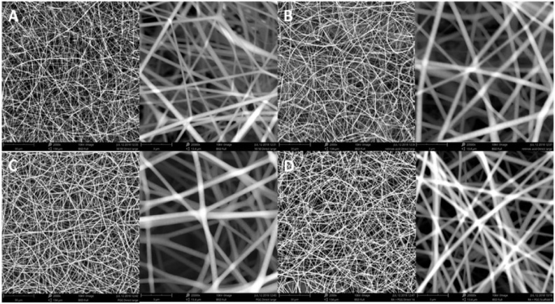
SEM of four scaffolds. A. Control, B. RA, C. PGG and D. “RA+PGG”. Lower magnification (left side) and higher magnification (right side).
Fiber diameter calculated from SEM images
∗ p < 0.05.
Analysis of FTIR spectra confirmed the presence of both PCL and gelatin in the composite scaffold. As seen in Fig. 2A, FTIR measurements were conducted on pure samples of both gelatin and PCL and compared to the 50:50 composite scaffold that was used in this study. The 50:50 composite was chosen because it proved the best for electrospinning and jet formation. Gelatin has characteristic peaks at wave numbers 1630 and 1520 attributed to amide I, amide II, absent in the pure PCL sample. This sample showed the characteristic peaks at wave numbers 1720, 1600, and 1555, which were absent in the pure gelatin sample. The composite scaffolds showed peaks at each of these wavenumbers, confirming both gelatin and PCL were present.
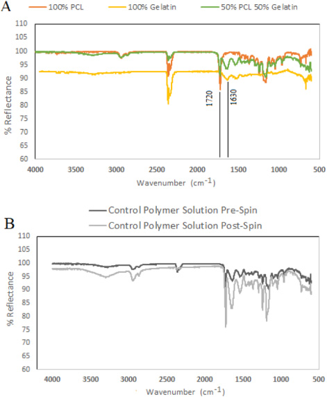
FTIR spectra of polymer. A. 100% PCL, 100% gelatin and PCL/gelatin before electrospinning. B. After and before the electrospinning (n = 3). FTIR spectrum does not change.
Another potential concern when exposing the polymer solution to high voltage is the possible change in molecular structure. FTIR testing was performed on the PCL/gelatin solution before and after electrospinning. As seen in Fig. 2B, the peaks for the pre- and post-spinning were identical, suggesting that the polymer did not degrade during electrospinning.
The scaffolds were submerged in Ringer’s solution and incubated at 37 °C to mimic human tissue fluids. As seen in Fig. 3, most of the weight loss occurred within the first week for all the scaffolds (40–50%). The weight loss in the subsequent weeks slowed dramatically. The initial rapid dissolution can be attributed to a majority of the gelatin being dissolved. Comparing the control with the samples that contained PGG revealed that in week 1, 2, 4 and 6 weight loss was statistically different while RA samples were not. However, there was no significant difference between PGG and “RA+ PGG” samples.
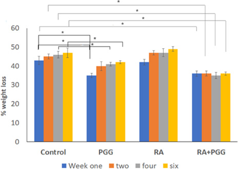
Weight loss of scaffolds. ∗ p < 0.05.
Each scaffold was seeded with 2 × 10⁶ hASMCs and incubated for 3 days prior to viability/cytotoxicity testing. As seen in Fig. 4, all the scaffolds showed a very high ratio of living to dead cells. This suggests that the RA and PGG concentrations are not toxic. The amount of living cells also indicate that despite the fast dissolution of gelatin, many of the cells were still attached to the scaffold. Figure 5 shows the scaffolds in static and dynamic culture.
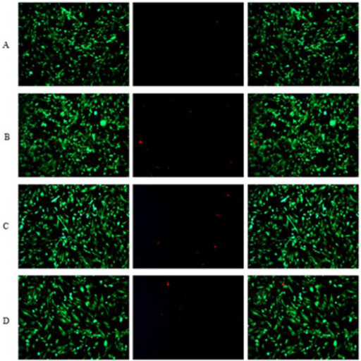
Live/dead cell test. Live-Green/DEAD-Red double-stained hASMCs after 3 days on A. Control, B. RA, C. PGG and D. “RA+ PGG” scaffolds.
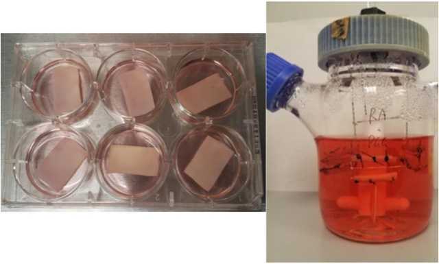
Scaffolds in static and dynamic culture.
To assess if the presence of RA and PGG may have a significant effect on the elastogenesis of hASMCs, these were seeded onto electrospun scaffolds doped with either RA, PGG, “RA+ PGG”, or the undoped control. The scaffolds were incubated for 1 week under static culture conditions, followed by 4 weeks in a spinner flask. Samples were collected from the spinner flasks at the end of each week. As seen in Fig. 6, each of the drug-loaded samples showed at least a 2-fold change compared to the control at week 1. RA showed a 3-fold change after 4 weeks in the spinner flask. PGG showed a significant 4-fold change compared to control after 4 weeks in the spinner flask. The “RA+ PGG” scaffold also showed a 4-fold change compared to the control in week 4. Each of the drug-loaded scaffolds showed an increased fold change statistically compared to the control, with the scaffolds containing PGG showing a greater increase in fold change. Comparing among groups in week 1 showed that both PGG and “RA+ PGG” are expressing more elastin compared to the RA alone. This trend is the same for the rest of the time points.
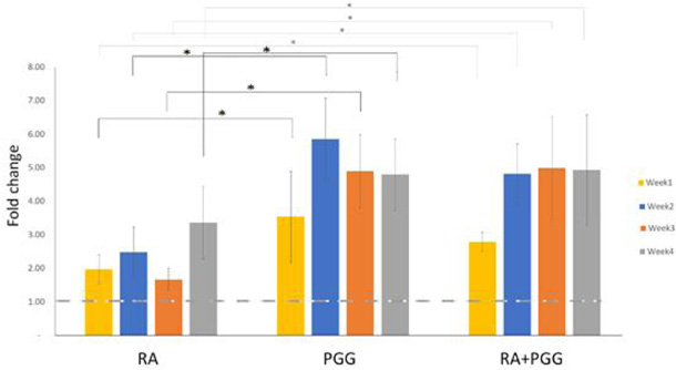
Average relative gene expression (fold change) normalized to control. ∗ p < 0.05.
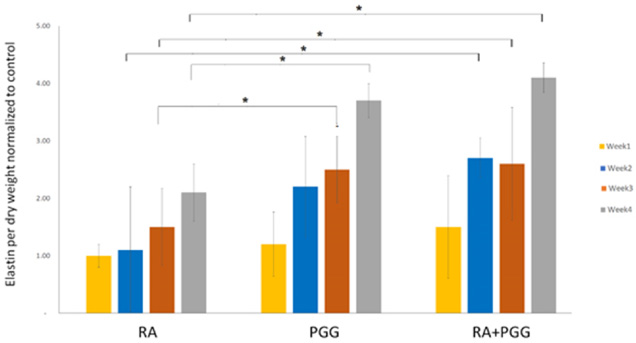
Total insoluble elastin deposited by the cells. ∗ p < 0.05.
Insoluble elastin was at its highest (4-fold change) in the combined PGG and RA samples after 4 weeks, with RA alone having a 2-fold increase. After 1 week, there was no significant difference in the expression of elastin between all experimental groups as shown in Fig. 7. After 2 weeks, there is a spike in mRNA expression in the samples doped with PGG and RA and PGG, that does not seem to translate into protein deposition since we do not see a equivalent spike in Fig. 7.
The current experiment explored the effect of RA and PGG doped scaffolds on hASMCs. Initial testing showed that it is possible to include both RA and PGG into a PCL/gelatin solution for electrospinning. Electrospinning created scaffolds made of randomly oriented fibers of a uniform diameter. During the electrospinning process, small amounts of polymer dried out and accumulated on the tip of the needle. This required periodic pausing and cleaning of the needle tip since these could dislodge and impact the surface of the scaffold affecting its integrity. None of the micrographs show any of these inclusions, indicating that the needle tip was properly maintained for the duration of electrospinning. SEM micrographs revealed the scaffolds were highly porous with a large degree of pore interconnectivity. With this composition and structure we would expect the tissue-engineered construct to degrade at a similar rate to the deposition of new ECM from cultured cells [15]. Each of the electrospun scaffolds consisted of a 50:50 mix of PCL and gelatin. PCL is a well-established biomaterial that degrades over the course of several months to years [16]. In contrast, gelatin readily dissolves when it encounters water. The PCL was intended to provide long term strength while the cells deposit their own ECM. This biomaterial has a burst pressure of 4,000 mm Hg [17], a value very close to natural tissue compared. The gelatin was intended to promote cellular attachment and proliferation [18]. Weight loss studies showed that most of the gelatin in the scaffold was dissolved by the end of the first week, probably due to it not being crosslinked. Therefore, it is likely that the only remaining gelatin would be found at the core of scaffold. Interestingly, both of the scaffolds that contained PGG degraded at a statistically significant slower rate than the two scaffolds without PGG, confirming its crosslinking properties [19]. PGG molecules are able to specifically bind to proline rich hydrophobic regions in elastin and collagen, forming multiple hydrogen bonds with surrounding proteins [20]. The concentrations of RA and PGG in the scaffold were not cytotoxic as seen in live/dead staining. This was an essential consideration as cell survival and proliferation are essential functional components of any tissue-engineering application. qPCR was performed to measure the effect of RA and PGG on elastin gene expression in hASMCs. Cells treated with PGG or RA and PGG showed severalfold increase in elastin expression compared to the control in week 1 and reached a plateau, in the following weeks. The effect of RA on mRNA expression was less pronounced than PGG and reached 3-fold increase in elastin expression after 4 weeks. RA is known to be a regulator of elastin synthesis in lung fibroblasts [21], chick embryonic skin fibroblasts [22] and vascular smooth muscle cells [23]. Recently it has been demonstrated that the expression of LOXL4, a lysyl oxidase (LOX) -like protein, is directly induced by RA at the transcript and protein levels [24]. An increase in the RNA does not guarantee its translation into a protein, however, the Fastin elastin assay results showed that the amount of elastin deposited was proportional to the elastin RNA expression. PGG significantly enhances the deposition of insoluble cross-linked elastin by increasing LOX [12] and binding to soluble elastin, which prevented its degradation [25].
Our results suggest that the addition of PGG along with RA can increase elastin mRNA expression and the resulting deposition of elastin on ECM by hASMCs. The deposition of elastin is crucial for blood vessels as elastic fibers start to form at midgestation and are completed during postnatal development, with no new elastic fiber formation in adults [26]. Typical tissue engineering approaches use a combination of scaffolds and cells that restore tissue function but not any endogenous elastin, and it is often addressed by exogenous elastin substrate. The overall goal of this work was to develop and characterize a RA-PGG loaded scaffold and quantify the endogenous elastin deposition within the matrix. This goal has been successfully met although future studies must look at dynamic environment with a pulsatile flow bioreactor to maximize elastin regeneration.
Conclusions
This research showed the potential of PCL/gelatin doped with “RA+ PGG” as a tissue engineering scaffold for small-diameter vascular grafts. The vascular grafts engineered using hASMCs demonstrated a significant deposition of elastin. Further studies should focus on the mechanical properties of these constructs.
Conflict of interest
None to report.