






Abstract
BACKGROUND:
Understanding the fracture mechanics of bone is very important in both the medical and bioengineering field. Bone is a hierarchical natural composite material of nanoscale collagen fibers and inorganic material.
OBJECTIVE:
This study investigates and presents the fracture toughness of bovine cortical bone by using elastic plastic fracture mechanics.
METHODS:
The J-integral was used as a parameter to calculate the energies utilized in both elastic deformation (J el) and plastic deformation (J pl) of the hipbone fracture. Twenty four different types of specimens, i.e. longitudinal compact tension (CT) specimens, transverse CT specimens, and also rectangular unnotched specimens for tension in longitudinal and transverse orientation, were cut from the bovine hip bone of the middle diaphysis. All CT specimens were prepared according to the American Society for Testing and Materials (ASTM) E1820 standard and were tested at room temperature.
RESULTS:
The results showed that the average total J-integral in transverse CT fracture specimens is 26% greater than that of longitudinal CT fracture specimens. For longitudinal-fractured and transverse-fractured cortical specimens, the energy used in the elastic deformation was found to be 2.8–3 times less than the energy used in the plastic deformation.
CONCLUSION:
The findings indicate that the overall fracture toughness measured using the J-integral is significantly higher than the toughness calculated by the stress intensity factor. Therefore, J-integral should be employ to compute the fracture toughness of cortical bone.
Introduction
Bone is a natural complex hierarchical structure composite material that contains three constituents: water, inorganic (mineral crystal), and organic material 43% by volume (cells, lipids) [1]. Quantitatively, bone primarily contains about 70% minerals, nanoscale hydroxyapatite, 22% proteins (90% collagen fiber) and 8% water by weight [2]. The collagen plays an important role in the toughness of bone, while the minerals content is involved in bone stiffness [3,4]. Hipbone in the human and bovine body is a very important bone as it supports the whole body and is very fragile. In medical science and bioengineering, the understanding of fracture mechanics and fracture behavior of bone is very important. For the evaluations of bone fracture risk, the mechanical behavior of cortical bone is crucial [5–7]. Norman et al. investigated the fracture toughness (K c ) of tibia human bone and compared it with the bovine cortical bone. They cut the longitudinal CT specimens from both bovine and human medial of tibia bone according to the American Society for Testing and Materials (ASTM) E-399. The results indicate that the human cortical bone fracture toughness is 60–68% is considerably weaker than the bovine cortical bone [8]. Libonati et al. investigated experimentally bone fracture toughness and crack propagation. They conducted three pointing bending tests and single edge three point bending tests. They showed that the fracture mode of bone is different from conventional material steel [9]. Tanabe et al. performed quasi-static fracture toughness tests on the bovine hip cortical bone. The fracture toughness (K c ) and strain energy (G c ) are the functions of specimen thickness. The K c and G c were estimated with respect to the crack length. Both transverse and longitudinal specimens showed that the K c and G c are decreased with the increase of the thickness of the specimens [10].
The elastic modulus (E) and shear modulus of bovine cortical bone were experimentally investigated using different span-depth ratios ranging from 6 to 32 by Lefevre et al. [11]. They prepared 29 plates specimens of different dimensions and performed three point bending test using MTS device using crosshead speed of 0.5 mm/min and compute elastic modulus of wide range difference from 6–21 GPa. An et al. studied fracture toughness (K c ) and J-integral using linear elastic fracture mechanics (LEFM) and elastic plastic fracture mechanics (EPFM) of the five different ages range from 25 to 51 years of male human tibia cortical bone and 18 months old of bovine cortical. The K c and J c were computed both experimentally and numerically using ABAQUS software. It has been observed that K c and J c of the bovine cortical are 111% and 108% greater than the human cortical bone [12]. The elastic plastic fracture toughness of human dentin was investigated by Yan et al. using ASTM standard E1820 single edge notched specimens. The J-integral of longitudinal direction is less than transverse direction. The stress intensity factor is 32% less than the fracture toughness K jc calculated by EPFM [13].
In the last three decades, researchers have studied the mechanical properties, such as fracture toughness, stiffness, the density of different bone using theories of linear elastic fracture mechanics (LEFM). The mechanical properties of cortical bone are immensely significant to prevent the fracture of a different bone. The mechanical properties vary with location of bone in a body, the effect of disease, age and also change with region to region [4,14]. Through the parameters of LEFM, such as K c and G c are used to compute the toughness of bone [15,16]. The fundamental assumption of LEFM is based on there is no or slight yielding occurs before fracture. But bones have exhibit some amount of post yield deformation, so the assumption of LEFM could not be applied to bone [17]. For materials, which have crack growth propagation at tip plastically, the theories of LEFM cannot be used to compute their fracture toughness. To accurately investigate the bone fracture, elastic-plastic fracture mechanics is required. The parameter J-integral is used in EPFM to compute the energies used in both elastic and plastic deformations of the hipbone [18,19].
In this paper, the mechanical properties of the male bovine cortical bone in both longitudinal and transverse directions were investigated based on the EPFM. The main goal of this paper is to estimate fracture toughness (K jc ) of the male bovine hip cortical bone using EPFM. The rest of the paper is organized as follows. The experimental setup for fracture toughness tests and tensile tests are illustrated in Section 2. The results of our experimental work are presented in Section 3. Section 4 summarizes the discussions and the conclusion is given in Section 5.
Material and methods
Six fresh male bovine hip bones of different ages (1 year to 3.5 years) and the same area, were collected from the market’s slaughterhouse as shown Fig. 1(a). Twenty four different types of specimens, i.e. longitudinal CT specimens, transverse CT specimens, rectangular (unnotched) longitudinal specimens and transverse specimens for tensile test, were cuts using hacksaw from the mid diaphysis of the bovine hip bone (6 longitudinal CT, 6 transverse CT and 12 for unnotched specimens for both Longitudinal and Transverse tensile test) according to the ASTME-1820 Standard [20]. We supplied a constant spray of clean water during machining; the bone is kept hydrated during processes to retain its moisture contents. For CT tests, 12 CT specimens were prepared in the longitudinal transverse direction to the fiber orientation. The dimensions of CT specimens are 12 × 3 ×14 mm (width × thickness × height) with a crack length (a o ) of 5 mm as shown in Fig. 1(b). In the longitudinal CT specimens, the collagens fibers are parallel to the specimen axis and collagens fiber are perpendicular to the specimen axis in the transverse CT specimens [21,22]. A crack length (a o ) of 5 mm was made in the each specimen using a sharp razor blade. All the machined and unmachined specimens were stored in the freezer until the test. All the CT specimens were tested in the Universal Testing Machine (UTM) as shown in Fig. 2. Unnotched rectangular specimens were prepared to perform the tension test to obtain the elastic modulus. The test was performed through UTM at the same loading rate. The ends of the specimens were covered with scotch tape. The elastic modulus was calculated from the slope of the stress-strain curve [23,24]. All tests were performed at normal room temperature (28 °C). The specimen was fixed in the steel fixture and held in UTM jaws as shown in Fig. 1(d). The speed of the cross head of the UTM is 0.5 mm/min. The tensile load is applied by the machine on the specimens. The applied load on the specimen and its corresponding deformation recorded by a PC connected to the machine during the test. The specimen was loaded until the crack started to grow and the test was stopped at this stage.
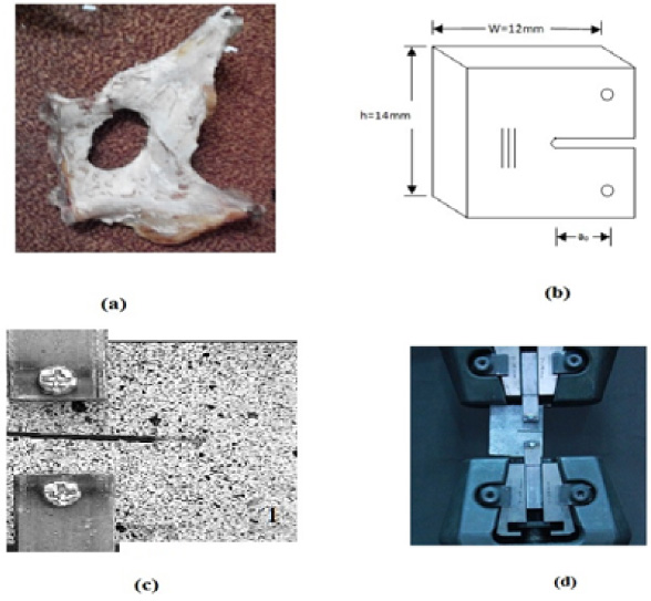
(a) Hip bone of male bovine. (b) The geometry of CT specimens according to ASTM-1820 standard. (c) CT specimen, and (d) The specimen is held with the help of the fixture in UTM.
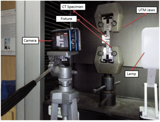
Experiment setup of fracture toughness.
The J-integral indicates the resistance to crack propagation of the material under tensile deformation. J-integral contained two components, i.e. elastic J-integral and plastic J-integral. Figure 3 illustrates these components of J-integral on a load and loading-line displacement diagram for CT specimen. After fracture the J-integral was computed by Eq. (1) for each specimen [20]:
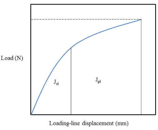
Load (N) vs loading-line displacement (mm) graph of CT specimens for J-integral measurement.
The critical stress intensity factor K
c
is calculated as follows [20]:
From the J-integral, linear elastic equivalent fracture toughness (K
jc
) of the hip bone is also calculated as:
As mentioned earlier, the ex vivo load deform data obtained from the UTM computer control, were used to analyze Young’s modulus, stress intensity factor, and J-integral which show good consensus with the previously published data [26]. The K c was calculated by using Eq. (2) based on the LEFM, as shown in Table 1. It was found that the average K c of the CT specimens in longitudinal and transverse directions is 1.92 MPam1∕2, and 2.5 MPam1∕2, respectively. Furthermore, the average Young’s modulus (E) of unnotched specimens in the longitudinal and transverse directions was found to be 2.11 GPa and 3.03 GPa, respectively.
Fracture toughness in longitudinal and transverse CT fracture specimens
Fracture toughness in longitudinal and transverse CT fracture specimens
After performing fracture toughness tests; the authors used plane stress assumption because the thickness of the CT specimens is less than the standards used in Eq. (3). Despite this, plastic behavior during the CT test was also observed. Therefore, ASTM E-1820 was used to determine J-integral [20]. The average J-integral of an elastic region of longitudinal and transverse specimens were computed 1.51 kPa⋅m and 1.968 kPa⋅m, respectively by Eq. (1). The average plastic deformation component of J-integral is 4.33 kPa⋅m and 5.96 kPa⋅m in longitudinal and transverse directions, respectively. The combining actions of elastic and plastic results of J-integral in both longitudinal and transverse direction are the total J-integral of 5.840 and 7.91 kPa⋅m, respectively. The linear elastic equivalent fracture toughness calculated using Eq. (4) is 3.33 and 4.45 MPa⋅m1∕2 in longitudinal and transverse directions, respectively. Table 2 presents all the average mechanical properties of the hip bone.
Average mechanical properties of the hip bone
N = Number of specimens, (E) = Elastic modulus, (K c ) = fracture toughness, (J el ) = elastic J-integral deformation, (J pl ) = plastic J-integral deformation, (J total) = total J-integral deformation, (K jc ) = linear elastic equivalent fracture toughness (K jc ).
This paper investigates both longitudinal and transverse fractures of the male bovine cortical bone. The stress intensity factor (K c ) and Young’s modulus (E) are computed on the base of the LEFM [27] by the plane stress assumption. In the transverse direction, the average value of K c and E is 1.3, 1.7 times the value of K c and E in the longitudinal direction respectively. In the longitudinal direction, the values of K c and E are smaller than that in the transverse direction [25]. Bone is a complex hierarchical structure, in transverse specimens, the collagen fiber is perpendicular to the crack growth propagation, so more energy is required to overcome the collagen fiber strength in the transverse direction than longitudinal as shown in Fig. 4 [1,28,29]. Hydration of collagen fiber can affect the mechanical properties of cortical bone. Bone consists of about 43% of organics by volume. The organics constitute of bone like bone matrix, have a strong bonding force, and fill the gap between the layers of bone [30].
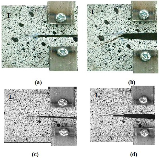
(a, c) The transverse and longitudinal CT specimens before load application and (b, d) Fracture crack propagation in transverse and longitudinal CT specimens.
The J-integral (J total) represents the energy absorption in originally unnotched ligaments. The J-integral determines the bone toughness in both directions (longitudinal and transverse) of collagens fibers of CT specimens of bovine cortical bone. The energy used in the plastic zone is ignored in the LEFM, but the ex vivo experimental results showed that 70–75% of energy was spent in the plastic zone deformation.
To date, the toughness of bone was less investigated by J-integral. Zioupos and Currey computed the J-integral of the 35 years old human femur bone by using a graphical approach and found the J total value is 1.2 kPa⋅m [31]. The G c values reported in plenty of literature are very high than our values [4]. The hoof wall of equine was investigated at different cross head speeds and showed the J-Integral value from 11 to 13 kJ/m2 [32]. In this paper, the elastic deformation J el is calculated by Eq. ((1)) and average values are 1.51 and 1.9 kPa⋅m in a longitudinal and transverse direction, respectively. The average energy in the longitudinal direction is 26% less than the transverse direction. In 2007 Yan et al. studied the single edge notches beam specimens of the bovine femur cortical bone and found that the plastic part of J-integral deformation J pl (5.3 kPa⋅m) in the transverse CT specimens is 4.1 times greater than the average value of elastic J-integral deformation J el (1.3 kPa⋅m) [18]. An et al. prepared CT specimens of bovine tibia cortical bone and their results showed that the plastic J integral deformation J pl (6.635 kPa⋅m) is 2 times larger than the elastic J-integral deformation J el (3.203 kPa⋅m). They found that the J el is smaller than the J pl of the CT specimens of human and cortical bone [12]. In this paper, the average value of J pl is 26% greater than the J el value for CT specimens of bovine cortical bone. The total J-integral of the hip bone is 11–20% less than the bovine femur bone. The average value of linear elastic equivalent fracture toughness (K jc ) in both directions is about 42% greater than the stress intensity factor K c , respectively.
In this paper, elastic-plastic fracture mechanics were applied in both longitudinal and transverse directions to examine the mechanical properties of the bovine cortical bone, since the bone exhibits a slight plastic deformation before fracture. The experimental study of fracture toughness of the bovine cortical bone using ASTM E1820 CT specimens showed that the J-integral is 31% greater in the transverse direction than in the longitudinal direction. The overall fracture toughness (K jc ) computed using EPFM is considerably greater than the stress intensity factor (K c ). Therefore, EPFM theories should be employed to estimate the fracture toughness of human and bovine cortical bone.
Footnotes
Acknowledgement
This work was supported by the Research Program of the Department of Mechanical Engineering, International Islamic University Islamabad.
Conflict of interest
The authors declare that they have no conflict of interest.