






Abstract
OBJECTIVE:
To compare outcomes of unipedicular versus bipedicular approach for percutaneous vertebroplasty for the treatment of thoracolumbar acute osteoporotic vertebral compression fracture (AOVCF).
METHOD:
From November 2014 to September 2015, 382 patients with AOVCF were randomly assigned to the unipedicular and bipedicular groups. Clinical outcomes and complications were compared.
RESULTS:
Both groups were comparable with respect to bone cement leakage and adjacent vertebral fractures (
CONCLUSIONS:
The clinical and radiological outcomes of both procedures were comparable. The unipedicular approach was associated with more nerve root stimulation.
Keywords
Introduction
The prevalence of acute osteoporotic vertebral compression fracture (AOVCF) is increasing with the aging of the population. AOVCF is a frequent cause of persistent back pain, which significantly impairs mobility and quality of life. Since the first report of percutaneous kyphoplasty in 1987 for the treatment of painful hemangioma involving the cervical spine in a young female patient [1], the indications for percutaneous vertebroplasty (PVP) or percutaneous kyphoplasty (PKP) have extended to AOVCF [2, 3]. Many studies have demonstrated that PVP has a high efficacy in immediate pain relief, and the key for successful PVP is to distribute the cement homogeneously within the vertebral body without causing leakage from the bone [3]. Some studies have shown superior long-term outcomes and fewer instances of cement leakage with the bipedicular approach in comparison to the unipedicular approach [4, 5, 6, 7]. However, others appear to favor the unipedicular approach due to shorter surgical time, lower x-ray irradiation and fewer complications [8]. We undertook this single-center, double-blind, randomized controlled trial to compare the clinical outcomes and complications between these two approaches.
Materials and methods
The study protocol was approved by the Ethics Committee at the Honghui Hospital, Xi’an Jiaotong University Health Science Center in China and this clinical trial was registered at the Chinese Clinical Trial Registry (no. ChiCTR-TRC-14004378). Written informed consent was obtained from each participant before the start of the trial.
Random allocation was done using a computer-generated randomization process. The process was double blinded, with neither the patients nor the examiners knowing the test group allocation. Allocation concealment was ensured using serially numbered, sealed envelopes.
CONSORT diagram detailing the study flow from enrollment through analysis. Three hundred and eighty-two patients were randomized to one of two groups: unipedicular group (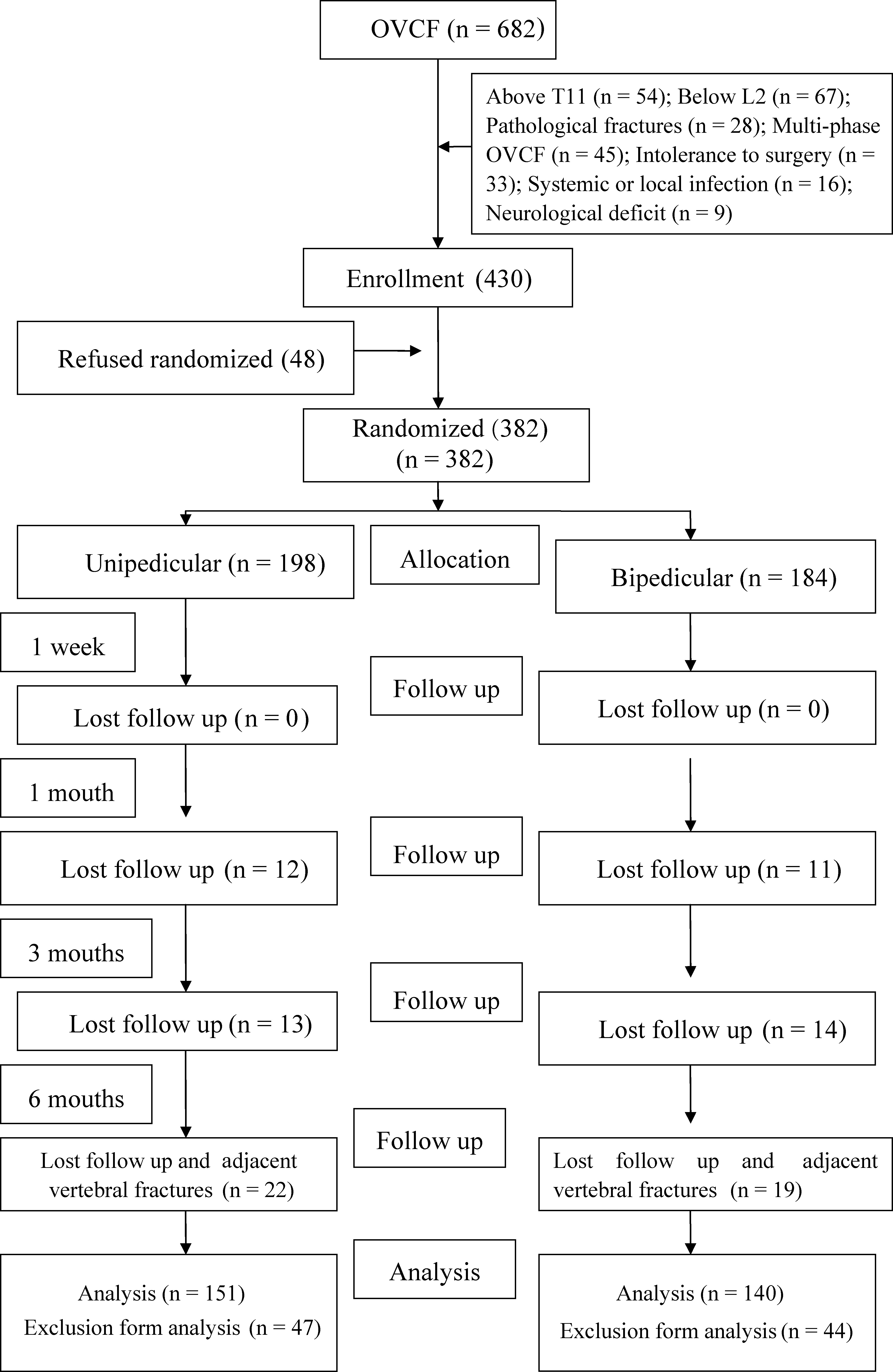
The selection of the study population is shown in Fig. 1. Inclusion criteria were: (1) age between 65 and 75 years; (2) anterior vertebral body compression (15–70% of the vertebral height); (3) pain
The exclusion criteria were: (1) intolerance to open surgery; (2) severe comorbidity (heart, liver, kidney, or lung); (3) systemic or local spine infection; (4) secondary osteoporosis (due to corticosteroid therapy, endocrine disorders or an inflammatory process); (5) uncorrected coagulopathy; (6) spinal metastatic cancer; (7) burst fracture with or without neurological symptoms; and (8) refusal to participate in the study.
All procedures were performed by the same surgeon. The procedure was performed under partial infiltration anesthesia using 1% lidocaine injection.
In the unipedicular group, the trocars were inserted through the lateral margin of the pedicle of the side that was more compressed, at the 3 o’clock position on the right side or at the 9 o’clock position on the left side (about 35–40
CT scan showing the position of inserted trocars, extraversion angle and distribution of bone cement in unipedicular group (A–D) and bipedicular group (E–H).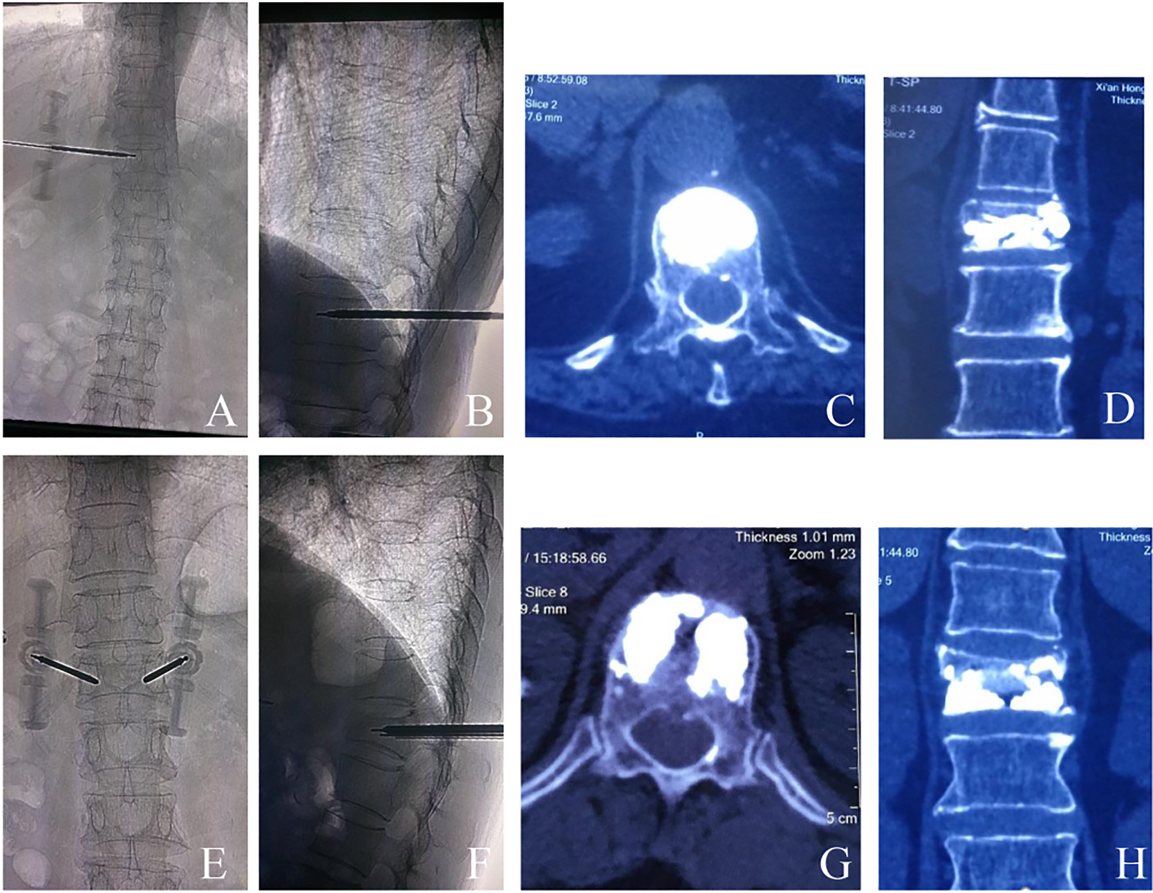
In the bipedicular group, two trocars were simultaneously inserted into both pedicles of the fractured vertebra; the needle entry points were at the 3 o’clock position on the right side and at the 9 o’clock position on the left side, requiring less medial inclination (about 10–15
The injection procedure was carefully controlled under anteroposterior and lateral fluoroscopy. Whenever there was bone cement leakage or the cement reached the dorsal quarter of the vertebral body, the injection was terminated. All patients were prescribed a thoracolumbar hyperextension brace for 1 month and underwent treatment of osteoporosis with 5 mg/day risedronate sodium (for 6 months) to prevent refracture or adjacent vertebral fractures.
Both groups were compared with respect to operation time, volume of PMMA injected, frequency of X-ray fluoroscopy, and perioperative complications, such as nerve root stimulation, bone cement leakage, and adjacent vertebral fractures. CT-3D was carried out in all cases to assess the distribution of bone cement and to look for cement leaks at 1 week post operation. The standing position plain radiographs were obtained at week 1, 1 month and 3 month post-surgery to assess the kyphotic angle. Surgical correction in the angle of kyphosis was assessed by calculating the mean difference between the pre- and postoperative measurements. The postoperative loss of correction was assessed by calculating the mean difference between the measurements made immediately postoperatively and those at the final follow-up examination.
Back pain was assessed with the Visual Analog Scale (VAS) score (11 levels ranging from 0 (no pain) to 10 (the strongest pain having ever been experienced)) and Oswestry low back pain disability index (ODI). The incidence of recurrent and adjacent vertebral fractures was also evaluated. Patients who developed new back pain during the follow-up period were brought back for MR imaging of the thoracic and lumbar spine. The new fractures were determined on MR imaging.
Statistical analyses
All statistical analyses were performed using SPSS software, version 12 (SPSS Inc., Chicago, IL, USA). Between-group differences were assessed using the Student’s
Results
Between November 2014 and September 2015, 682 patients with AOVCF of the thoracolumbar spine were treated at our trauma centre. Of these, 430 met the inclusion criteria. 48 patients refused to participate in the study. All participants were followed up for six months. In the unipedicular group, 28 patients were lost to follow-up and 19 patients had adjacent vertebral fractures. In the bipedicular group, 29 patients were lost to follow-up and 16 patients had adjacent vertebral fractures. Forty-seven (23.7%) of the 198 patients in the unipedicular group and forty-five (24.4%) of the 184 patients in the bipedicular group were not included in the analysis. At the time of follow-up, the preoperative data showed no significant intergroup differences with respect to age, gender and BMD T score (
Baseline characteristics of patients by study group
Baseline characteristics of patients by study group
SD, standard deviation; BMD, bone mineral density.
Clinical and radiological findings by study group
Data are presented as mean
In the unipedicular and bipedicular groups, the mean operation time, frequency of x-ray fluoroscopy and mean volume of cement injected were 35.3
Radiographic findings
The correction angle was higher, and the loss of correction was lower in the bipedicular group as compared to that in the unipedicular group; the differences, however, did not reach statistical significance (
Clinical outcomes
Adjacent vertebral fractures developed in 35 patients (N
VAS score and ODI of patients by study group
VAS score and ODI of patients by study group
Data are presented as mean
Leakage of bone cement outside the vertebral body was noted on postoperative anteroposterior and lateral radiographs and on CT scan in 36 (23.84%) patients in the unipedicular group and in 33 (23.6%) patients in the bipedicular group (
Discussion
AOVCF is associated with pain, deformity, disability and poor quality of life. PVP provides rapid and long lasting pain relief and has gained popularity as an effective treatment modality for AOVCF [6].
In some studies, the bipedicular approach was shown to achieve symmetrical distribution of bone cement in the vertebral body [4]. Others have shown that the bipedicular approach is not required because bone cement is liable to be distributed uniformity in the vertebral body when unipedicular puncture point reaches the median vertebral body [10]. Therefore, which surgical approach should be selected remains controversial. In this study, we sought to compare the two techniques with respect to clinical outcomes and complications in patients with single-level thoracolumbar AOVCF.
The traditional view, as also confirmed in our previous study [11], is that unilateral PKP entails a shorter surgical time and lower x-ray irradiation compared with the bilateral approach [4]. In the present study, the bilateral trocars were simultaneously inserted and PMMA were simultaneously injected in patients of the bipedicular group. The mean duration of operation was longer (31.2
Radiological evidence also indicated the advantage of the bilateral approach in terms of reduction in kyphosis with the loss of reduction being lesser as compared to that in the unilateral group [12, 13]. We found that the correction angle was higher (11.23
Long et al. reported that the distribution of bone cement is critical to the stabilization of fractures of vertebral body and pain relief, and that the stress distribution achieved by unipedicular approach was inferior to that achieved with the bipedicular approach [14]. In our study, the vertebral needle reached anterior third of the vertebral body on the lateral view, but not all cases reached the midline of the vertebral body on the posteroanterior view in the unipedicular group [15]. The bone cement exceeded the midline of vertebral body in all cases, but and exceeded three quarters of the vertebral body in only 113 cases in the unilateral group. To achieve a more uniform distribution of bone cement, intertransverse vertebroplasty may be necessary [11].
Tohmeh et al. found no significant difference between the therapeutic efficacy of unipedicular and bipedicular approaches [10], but Song et al. reported greater improvement in VAS scores in the unipedicular group as compared to that in the bipedicular group (
Previous studies have shown that both bipedicular and unipedicular PVP are relatively safe; however, unipedicular approach results in fewer complications [4]. In the present study, the leakage of the bone cement outside the vertebral body was noted on post-operative anteroposterior and lateral radiographs and CT scan in 36 (23.84%) patients in the unipedicular group and 33 (23.6%) in the bipedicular group (
There are certain limitations of the study which ought to be mentioned. The intervals of follow-up in the study were kept at 1 week, 1 month, 3 month and 6 month post-surgery, which was due to the relatively older age of the patients and consequent inability to comply with more frequent follow-up. Additionally, a multi-centre study with a larger sample size and longer duration of follow-up is needed to confirm our findings.
In conclusion, our study reveals that both unilateral and bilateral puncture during PVP is effective in the treatment of AOVCF. The clinical and radiological outcomes of both treatment procedures were comparable. However, unipedicular approach was associated with greater nerve root stimulation. Unipedicular approach may be used instead of bipedicular percutaneous vertebroplasty for osteoporotic vertebral compression fracture.
Footnotes
Acknowledgments
The Social Development of Science and Technology Research grants of the Department of Science and Technology at Shaanxi Province (grant nos. 2016SF-072 and 2017SF-233) were received in support of this work.
Conflict of interest
None to report.