Optimal seatback angles for automobile drivers’ seats have been investigated based on comfort and back muscle activities; however, radiology supported evidences are scarce.
OBJECTIVE:
The aim of this study was to evaluate optimal range of the seatback reclining regarding torso angles for an automobile driver’s seat to preserve lumbar lordosis.
METHODS:
Thirty-one healthy volunteers were recruited among five body type categories. Lateral lumbar spine X-rays were obtained for the neutral sitting posture without seatback (reference), and with reclining angles of 23 to 33 by 2 intervals. The Cobb angles for the L1-L4, L4-S1, and L1-S1 segments were measured.
RESULTS:
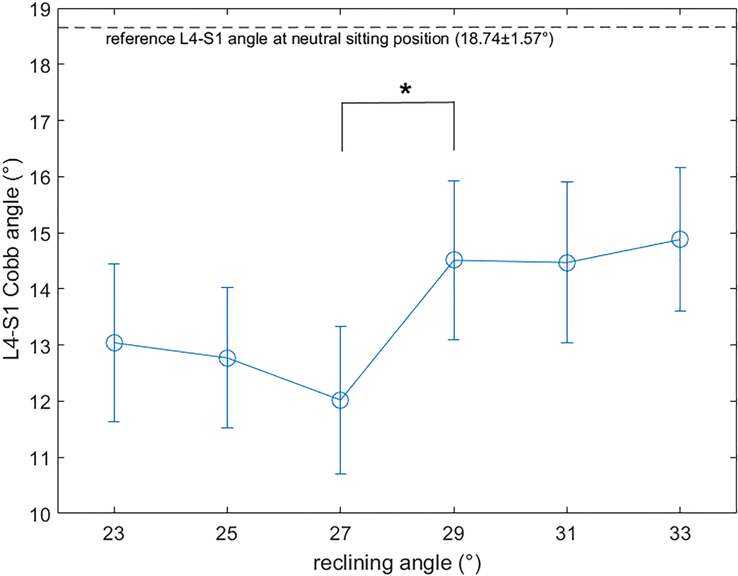
The Cobb angle for L4-S1 was nearest to the reference (18.74 1.57, mean standard error mean) at reclining angles of 29 and 31 (14.51 1.41 and 14.47 1.43, respectively). The Cobb angle at L4-S1 between reclining angles of 27(12.02 1.31) and 29 (14.51 1.41) were significantly different ( 0.001). Tall men showed relatively preserved lordosis angles at all reclining angles. Fat men and short women demonstrated prominent loss of lordosis with excessively kyphotic L1-L4 segment.
CONCLUSIONS:
Reclining angles of 29 to 31 revealed to be optimal for preserving lordosis at the L4-S1 segment. Individualized healthcare-related guideline for driver’s seat adjustment setting is necessary.
Prolonged sitting in driving posture often induces neck and low back pain, which are significant issues, particularly in those individuals who are drivers by occupation [1, 2, 3]. Numerous factors affect pain and discomfort of the driver. Anatomic factors include cervical and lumbar lordosis angle, joint angles such as trunk-thigh angle and knee flexion angle, pelvis rotation, muscular activation, and intervertebral disc pressure [4, 5, 6]. Factors regarding the car seat are seatback reclining angle, height of the seat, tilt angle, sliding position and distance to the handle and brake, headrest position, and lumbar support [5, 6, 7, 8, 9, 10, 11]. Relative hip position and pressure to the seat, sitting posture such as erect, neutral, and slouch postures, hand position on the handle, positioning of the head, and how the drivers place their legs may influence the level of pain and discomfort [12, 13]. In addition, factors such as whole-body vibration and mechanical shocks may affect pain while driving on road [14].
To minimize back pains, optimal seatback angles for automobile drivers’ seats have been investigated over a time based on comfort and paraspinal muscle activities using various seatback settings, such as seatback angle, seat bottom tilting, and lumbar support [5, 7]. Lumbar stabilization has been recently emphasized in relation with mechanical low back pain, and the maintenance of lumbar lordosis angle is known to be essential for structural stabilization [15, 16]. Previous studies showed that spinal length or lumbar lordosis degree was increased when using lumbar support [17, 18]. Lengsfeld et al. [19] showed that the posterior tilt of the seat with a reclining backrest induced an evenly distributed lumbar lordosis, which is preferable from point of view of lumbar spine kinematics. This concept is widely applied in the design of various chair types.
With regard to the seatback angle in the sitting posture, earlier studies investigated the effects on lumbar paraspinal muscle activity and intervertebral disc pressures, and revealed that 10–30 inclination from the upright angle is recommended [5, 7]. However, radiologic investigation on lumbar lordosis angle is limited to date. In 1979, Andersson et al. [20] performed a radiologic study by changing backrest inclination angle from 80 to 110 by 10 interval, and insisted minimal effects were observed on the lumbar vertebral angles. De Carvalho et al. [21, 22] showed that 2–4 cm lumbar support resulted in increased lumbar lordosis, and that lumbosacral lordosis and sacral tilt angle were significantly decreased when changing the standing to sitting position in another study by radiologic investigation. Cho et al. [23] performed a radiological study on healthy volunteers to investigate lumbar lordosis, sacral slope, and pelvic tilt based on various sitting postures, such as sitting with back support, sitting on a 90 angle chair, sitting with anterior support, sitting on a chair, and sitting with legs crossed. They showed that the lumbar lordosis angle was closer to the standing posture while sitting with back support compared with sitting on a 90 angle chair. However, to the best of our knowledge, radiological assessment focusing on specific optimal seatback angles has not been intensively performed to date.
In this study, we aimed to find an optimal seatback angle to preserve lumbar lordosis in the driving posture using radiological assessments. We assumed that the lumbar lordosis in the neutral sitting position without the seatback would be closest to an ideal posture. We investigated how the lumbar lordosis angle changes in relation to the reclining angle of the seatback in an automobile driver’s seat setting, and whether the body type affects the changing pattern.
Methods
Participants
We recruited 31 healthy volunteers from a job recruitment internet website and divided them into five body type groups: tall standard men (group A, 7), middle height skinny men (group B, 6), middle height standard men (group C, 6), middle height fat men (group D, 6), and short standard women (group E, 6). Body types were classified based on the database of human body measurement by governmental investigation organization, Size Korea (http://sizekorea.kats.go.kr). Inclusion criteria for participation were age between 40 and 60 years with driving experience for intermediate-sized car or larger, height and body weight within the range of the five body type groups, and no history of low back pain within 1 year or diagnosed with herniated intervertebral disc in the lumbar spine. Age range was selected to represent age groups with the highest percentage of licensed drivers (www.fhwa.dot.gov) and avoid age groups with high incidence of low back pain and those with higher comorbidities [24]. Subjects with low back pain or intervertebral disc disorder confirmed by medical image, and those who were determined not to be eligible for study participation were excluded. Demographic data of the participants are shown in Table 1.
Demographic data of subjects by body types
Group
No. of subjects
Gender
Height (cm, mean SD)
Weight (kg, mean SD)
BMI (kg/m, mean SD)
A
7
Male
178.2 2.2
79.9 6.6
25.2 2.3
B
6
Male
169.3 2.0
66.2 3.7
23.1 1.0
C
6
Male
170.4 1.9
73.3 2.7
25.3 1.3
D
6
Male
170.6 1.5
83.6 3.5
28.8 1.6
E
6
Female
148.7 2.0
52.7 3.2
23.8 1.3
Total
31
67.8 10.3
71.4 11.8
25.2 2.4
BMI: body mass index; Values are mean standard deviation. Group A: tall height, standard body type; Group B: middle height, skinny body type; Group C: middle height, standard body type; Group D: middle height, fat body type; Group E: short height, standard body type.
Settings
An automobile mock-up driver’s seat was set in a local radiology clinic for X-ray evaluation The mock-up seat resembled a real driver’s seat of a commercial full-sized vehicle with controllable seatback reclining function. For each subject, 7 lateral lumbar spine X-rays were taken in various postures; sitting without seatback in the neutral posture, sitting in the neutral posture with reclining angles of 23, 25, 27, 29, 31, and 33. Reclining angles were set with reference to the torso angles of the dummy model on the car seat. For postures with seatback, the subjects were instructed to place their hip as deep as possible and lie on their back against the seatback.
The Cobb angle at L4-S1 segment (mean angle, error bars indicate standard error mean) at sitting posture according to the seatback reclining angles are shown. Values between 27 and 29 reclining angles were significantly different (* 0.001 by paired test).
Measurements in X-ray image
For each X-ray image, angles of the upper margin of each vertebra (L1, L2, L3, L4, L5, and S1) to the horizontal line was measured with a medical image viewer software, Marosis M-view (Infinitt, Seoul, Korea). The Cobb angle for the L1-S1, L1-L4, and L4-S1 segments were calculated to evaluate the lumbar lordosis angle. The L4-S1 angle was considered the primary segment because the L4-L5 and L5-S1 levels are most related with the pathophysiology in low back pain compared with other segments, accounting up to 95% of all lumbar intervertebral disc herniation [25]. The lordosis angle at the sitting position without seatback in the neutral posture was considered as the reference value. The standardized L4-S1 angle (Cobb angle divided by the reference angle) was also calculated.
Statistical analysis
Descriptive statistical analysis was primarily performed for all parameters in each posture. Paired test for overall analysis and Wilcoxon signed rank test for group analysis were used to compare each parameter between the reference and adjacent reclining angle. A value of less than 0.05 was considered significant. All statistical analyses were performed using SPSS software (SPSS Inc., Chicago, USA).
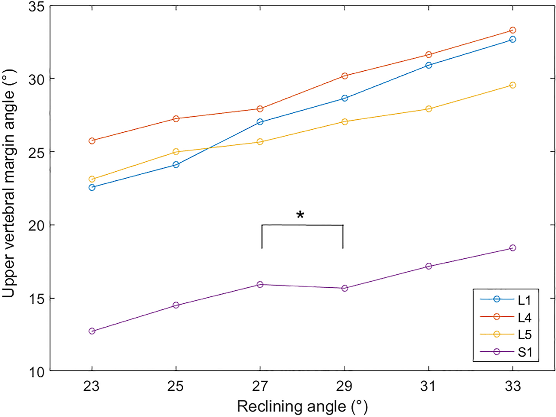
Individual angles of each vertebral upper margin according to the seatback reclining angles are shown. S1 angles at 27 and 29 reclining angles show different pattern compared with other vertebrae. (* 0.001 by paired test).
Results
Effect of reclining angle on lumbar lordosis
In overall analysis, the average reference L4-S1 Cobb angle (L4-S1 angle) was 18.74 1.57 (mean standard error mean (SEM)). Among the investigated seatback reclining angles, the L4-S1 angle was nearest to the reference angle at 29 (14.51 1.41), 31 (14.47 1.43), and 33 (14.88 1.28). However, the L4-S1 angle was significantly different from the reference angle at all reclining angles ( 0.05). Loss of lordosis was the most prominent at the 27 reclining, showing the lowest L4-S1 angle (12.02 1.31), and the angle was significantly different from the L4-S1 angle at 29 reclining (14.51 1.41, 0.001). The changing pattern of the L4-S1 angle at all reclining angles is shown in Fig. 1.
Individual angles of each vertebral upper margin based on the reclining angle are shown in Fig. 2. Although angles of L1-L5 demonstrated increasing pattern with reclining, the angle of S1 upper margin significantly decreased from 27 (15.92 1.71) to 29 (15.67 1.85) reclining ( 0.001).
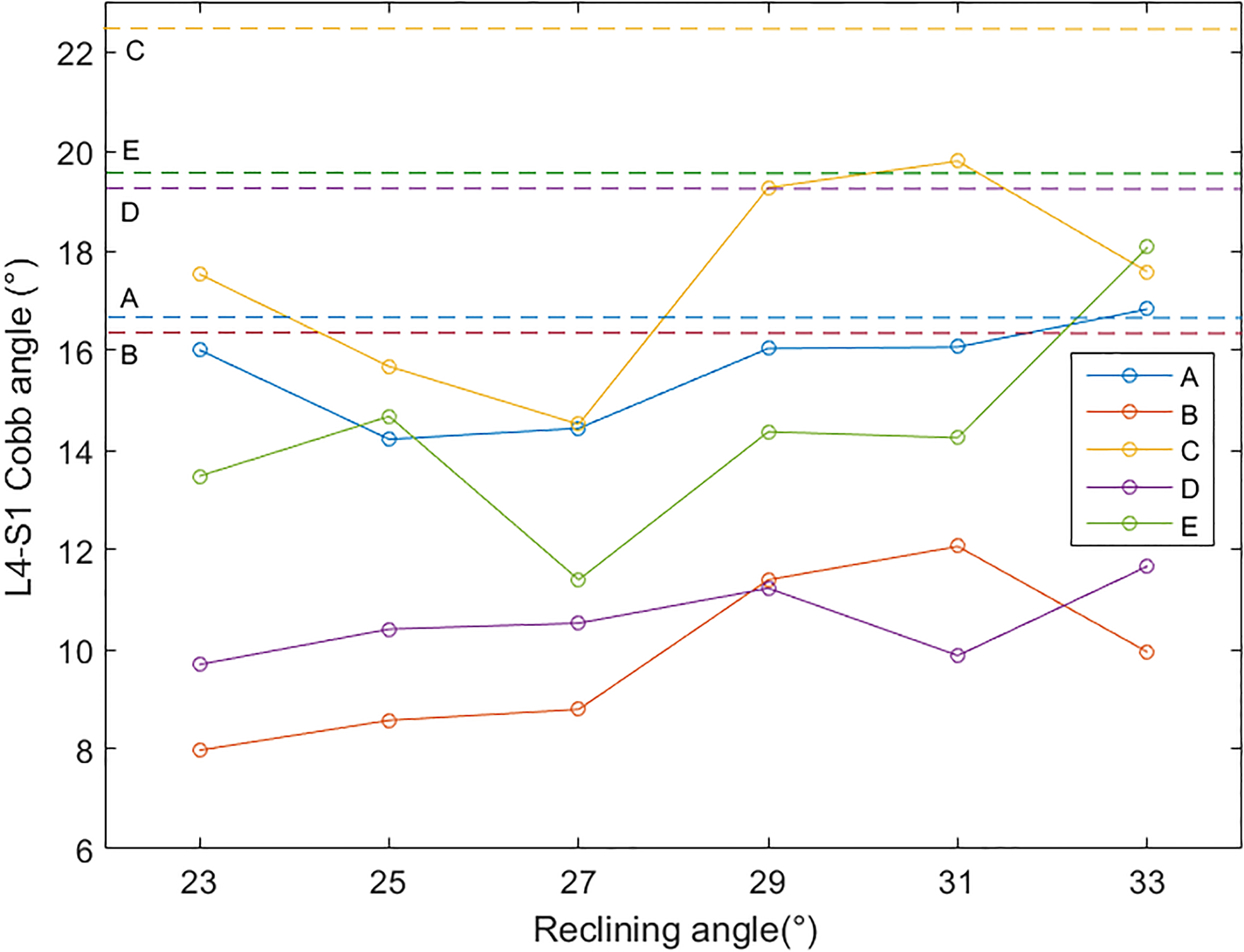
Subgroup analysis results by body types are shown. Groups B and C demonstrated a typical pattern with peak L4-S1 angle at 29–31 reclining range. (Group A: tall height, standard body type; Group B: middle height, skinny body type; Group C: middle height, standard body type; Group D: middle height, fat body type; Group E: short height, standard body type; dashed lines indicate reference angles for each body type group).
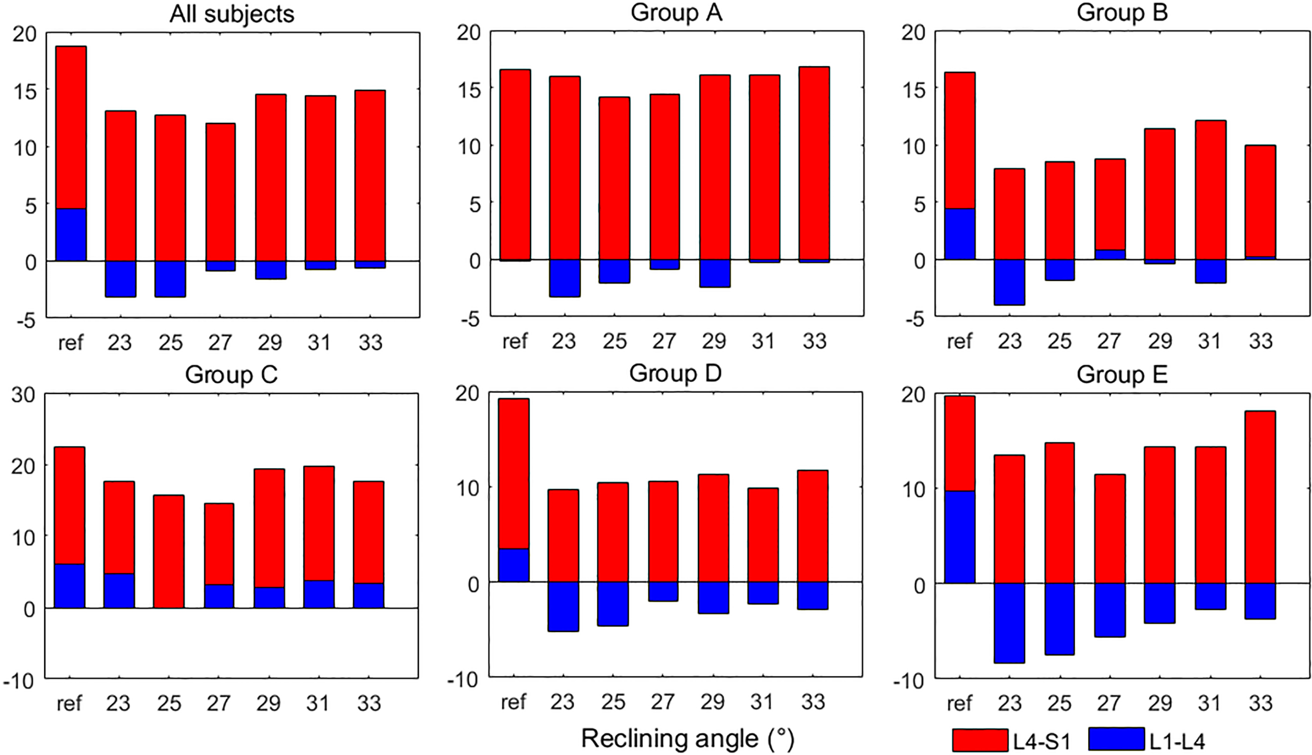
The Cobb angle at L1-L4 and L4-S1 segments for each seatback reclining angle for each body type groups are shown. L1-L4 segments were less kyphotic at higher reclining angles (groups B, C), and groups D and E showed predominantly kyphotic L1-L4 segment at all reclining angles.
Subgroup analysis by body types
Subgroup analysis by body types were performed (Fig. 3). The L4-S1 angles were nearest to the reference angle at 23, 29, and 31 reclining angles (16.01 2.87, 16.04 3.44, and 16.07 2.71, respectively) in tall standard men in group A, whereas a 33 reclining angle resulted in a more kyphotic angle (16.83 2.98) than the reference angle (16.58 3.86), although it was not statistically significant. Groups B and C, middle height skinny and standard men, demonstrated a typical angular pattern showing a peak at a 29 and 31 reclining angle, with the most lordotic angle at the L4-S1. The L4-S1 angle at a 23 reclining angle showed significant loss of lordosis in group B compared with the reference angle (7.98 2.58 vs. 16.31 3.54, 0.028). Groups D and E, middle height fat men and short height standard women, did not show a specific pattern. However, the L4-S1 angle showed a statistically significant difference in group D compared with the reference angle at most of the reclining angles ( 0.05 for 27, 29, and 31, and 0.1 at 23, 25, 33), which demonstrates relatively kyphotic L4-S1 angles compared with other groups.
Analysis by lumbar segments
Segmental analysis, by using the L1-L4 and L4-S1 segments, was performed (Fig. 4). In overall analysis, both the L1-L4 (4.57 2.09) and L4-S1 (18.74 1.57) segments showed a lordotic angle in the reference posture. However, the L1-L4 segment showed kyphotic angles (negative values in degrees) at all reclining angles in the sitting posture with seatback, particularly being more kyphotic at reclining angles of 23 and 25 (3.19 1.45 and 3.15 1.34, respectively). In group A, the characteristics of the segmental angles at reclining angles of 31 and 33 were similar to the reference posture. Groups B and C also demonstrated less kyphotic or lordotic L1-L4 segments at higher reclining angles. However, the L1-L4 segment was predominantly kyphotic at all reclining angles in groups D and E.
Discussion
The degree of lumbar lordosis was shown to be higher at reclining angle of 29 or higher in overall analysis, showing a significantly different lordosis angle from the 27 reclining angle. Middle height men with skinny or standard body type (groups B and C) demonstrated a typical pattern of L4-S1 lordosis angle change based on the reclining angle, suggesting that 29 to 31 may be an optimal range for the reclining angle. Results showed that anterior rotation of the S1 may be a critical factor for preserving lumbar lordosis in a sitting position. Subjects with short height and fat body type tended to show kyphotic upper lumbar segments.
The main finding of this study was that S1 angle was smaller at the 29 reclining (15.67) compared with the 27 reclining angle (15.91), whereas at other reclining angles, the S1 angle and other angles of vertebral bodies increased as the seatback was tilted backwards. This phenomenon was remarkable in all subgroups and in the overall analysis. With regards to this phenomenon, we hypothesized that this may be related with paraspinal muscle activity. Watanabe et al. [26] showed that the core muscle contraction is significantly related with spinal curvature angle, including the sacral angle. Biomechanical analysis of our study data demonstrated decreasing mechanical load on the lower lumbar spine with increasing reclining angle. Therefore, we can infer that lower lumbar paraspinal muscle activity may have decreased with the reclining angles, with the seatback supporting the weight of the lumbar spine. Decreased lumbar paraspinal muscle activity would have led to loosening of the sacral spine from the paraspinal muscle when the reclining angle has exceeded a threshold level, resulting in relative anterior sacral rotation. Because the S1 portion is not fully supported by the backrest, the portion of the passive inclination may have been smaller than that of the lumbar vertebrae. We supposed that the lumbar lordosis was mainly maintained by paraspinal activation at the 27 reclining angle or lower, whereas the passive support of the backrest preserves the lumbar lordosis at 29 or higher. Decrease of lumbar paraspinal activation with preservation of lumbar lordosis may lead to more comfortable and durable posture. O’Sullivan et al. [27] also insisted that modifying the chair or seat type may help reduce lumbar multifidus activation, thus making it potentially advantageous for prolonged sitting.
The subjects with fat body types demonstrated smaller lordosis angles for the L4-S1 segment and relatively kyphotic L1-L4 segment. To the best of our knowledge, the effect of body type or height in the sitting posture has not been specifically evaluated to date. However, obesity or high BMI have been shown to be generally related with kyphotic thoracic curvature or loss of lordosis in lumbar curvature [28, 29]. Fatty degeneration or relatively low proportion of muscle fibers in the paraspinal muscle is related with lumbar kyphosis [30]. However, fat infiltrations in the paraspinal muscles may also cause sway-back posture, which is lumbar hyperlordosis; hence, other factors should be considered [31].
The subjects with short height did not demonstrate a typical pattern for the L4-S1 angle, but the L1-L4 segments were kyphotic in all reclining angles compared with other body type groups. Tall subjects also tended to show a more kyphotic L1-L4 segment than the middle height subjects. No definite previous studies on the relationship between height and the degree of lordosis have been published to our knowledge. We could not conclude the effect of height on lumbar lordosis with this study, but suspected that the contour of the carseat used in this study may have been optimized for people with height nearest to the average, based on the fact that the type of the seat affects the degree of lordosis [25].
This study has several limitations. The number of subjects for each body type group was not sufficient to clarify typical patterns of reclining and lordosis angles. The seat contour may have affected the results, because the commercial car seats have special designs based on ergonomics. The results may not possibly show the same pattern in a different type of seat. In addition, only the angle of the lumbar and sacral vertebrae in the X-ray was evaluated. If electromyography of the paraspinal muscles were acquired simultaneously, we may have derived clinically more significant findings. Finally, this study was performed on the Korean population, which was classified based on their anthropometric data. Further studies on different race or ethnicity groups are necessary to generalize the results.
Conclusion
Reclining of the seatback angle at 29 or more, revealed to be the most optimal angle for the preservation of lordosis in the L4-S1 segment. Fat and short body types tended to show more kyphotic lumbar spine in the sitting position. The results of this study may serve as an evidence for setting healthcare system in an automobile driver’s seat.
Footnotes
Acknowledgments
This study was funded by the Hyundai Motors Group.
Conflict of interest
None to report.
References
1.
MozafariAVahedianMMohebiSNajafiM. Work-Related Musculoskeletal Disorders in Truck Drivers and Official Workers. Acta Medica Iranica.2015; 53(7): 432-8.
2.
LisAMBlackKMKornHNordinM. Association between sitting and occupational LBP. Eur Spine J.2007; 16(2): 283-98.
3.
LyonsJ. Factors contributing to low back pain among professional drivers: a review of current literature and possible ergonomic controls. Work.2002; 19(1): 95-102.
4.
LiCTPengYTTsengYTChenYNTsaiKH. Comparing the effects of different dynamic sitting strategies in wheelchair seating on lumbar-pelvic angle. BMC Musculoskelet Disord.2016; 17(1): 496.
5.
HarrisonDDHarrisonSOCroftACHarrisonDETroyanovichSJ. Sitting biomechanics part I: review of the literature. J Manipulative Physiol Ther.1999; 22(9): 594-609.
6.
MenottiFLabancaLLaudaniLGiombiniAPigozziFMacalusoA. Activation of Neck and Low-Back Muscles Is Reduced with the Use of a Neck Balance System Together with a Lumbar Support in Urban Drivers. PLoS One.2015; 10(10): e0141031.
7.
HarrisonDDHarrisonSOCroftACHarrisonDETroyanovichSJ. Sitting biomechanics, part II: optimal car driver’s seat and optimal driver’s spinal model. J Manipulative Physiol Ther.2000; 23(1): 37-47.
8.
ChenJCDennerleinJTChangCCChangWRChristianiDC. Seat inclination, use of lumbar support and low-back pain of taxi drivers. Scand J Work Environ Health.2005; 31(4): 258-65.
9.
MaradeiFQuintanaLCastellanosJ. Influence of seat tilt motion on discomfort perception during a simulated driving activity. Work.2017; 56(1): 99-110.
10.
PengJWangXDenningerL. Effects of Anthropometric Variables and Seat Height on Automobile Drivers’ Preferred Posture With the Presence of the Clutch. Hum Factors.2018; 60(2): 172-90.
11.
PengJWangXDenningerL. Ranges of the least uncomfortable joint angles for assessing automotive driving posture. Appl Ergon.2017; 61: 12-21.
12.
KilincsoyUWagnerAVinkPBubbH. Application of ideal pressure distribution in development process of automobile seats. Work.2016; 54(4): 895-904.
13.
ParkJChoiYLeeBJungKSahSYouH. A Classification of Sitting Strategies based on Driving Posture Analysis. J Ergon Soc Korea.2014; 33(2): 87-96.
14.
BovenziMSchustMMauroM. An overview of low back pain and occupational exposures to whole-body vibration and mechanical shocks. Med Lav.2017; 108(6): 419-33.
15.
FreemanMDWoodhamMAWoodhamAW. The role of the lumbar multifidus in chronic low back pain: a review. PM R.2010; 2(2): 142-6.
16.
SparreyCJBaileyJFSafaeeMClarkAJLafageVSchwabF, et al. Etiology of lumbar lordosis and its pathophysiology: a review of the evolution of lumbar lordosis, and the mechanics and biology of lumbar degeneration. Neurosurg Focus.2014; 36(5): E1.
17.
EklundJACorlettEN. Shrinkage as a measure of the effect of load on the spine. Spine (Phila Pa 1976).1984; 9(2): 189-94.
18.
MajeskeCBuchananC. Quantitative description of two sitting postures. With and without a lumbar support pillow. Phys Ther.1984; 64(10): 1531-5.
19.
LengsfeldMFrankAvan DeursenDLGrissP. Lumbar spine curvature during office chair sitting. Med Eng Phys.2000; 22(9): 665-9.
20.
AnderssonGBMurphyRWOrtengrenRNachemsonAL. The influence of backrest inclination and lumbar support on lumbar lordosis. Spine (Phila Pa 1976).1979; 4(1): 52-8.
21.
De CarvalhoDECallaghanJP. Influence of automobile seat lumbar support prominence on spine and pelvic postures: a radiological investigation. Appl Ergon.2012; 43(5): 876-82.
22.
De CarvalhoDESoaveDRossKCallaghanJP. Lumbar spine and pelvic posture between standing and sitting: a radiologic investigation including reliability and repeatability of the lumbar lordosis measure. J Manipulative Physiol Ther.2010; 33(1): 48-55.
23.
ChoIYParkSYParkJHKimTKJungTWLeeHM. The Effect of Standing and Different Sitting Positions on Lumbar Lordosis: Radiographic Study of 30 Healthy Volunteers. Asian Spine J.2015; 9(5): 762-9.
24.
WatermanBRBelmontPJ, Jr.SchoenfeldAJ. Low back pain in the United States: incidence and risk factors for presentation in the emergency setting. Spine J.2012; 12(1): 63-70.
25.
PatelN. Surgical disorders of the thoracic and lumbar spine: a guide for neurologists. J Neurol Neurosurg Psychiatry.2002; 73(Suppl 1): i42-8.
26.
WatanabeSKobaraKYoshimuraYOsakaHIshidaH. Influence of trunk muscle co-contraction on spinal curvature during sitting. J Back Musculoskelet Rehabil.2014; 27(1): 55-61.
27.
O’SullivanKMcCarthyRWhiteAO’SullivanLDankaertsW. Can we reduce the effort of maintaining a neutral sitting posture? A pilot study. Man Ther.2012; 17(6): 566-71.
28.
Gonzalez-SanchezMLuoJLeeRCuesta-VargasAI. Spine curvature analysis between participants with obesity and normal weight participants: a biplanar electromagnetic device measurement. Biomed Res Int.2014; 2014: 935151.
29.
Lang-TapiaMEspana-RomeroVAneloJCastilloMJ. Differences on spinal curvature in standing position by gender, age and weight status using a noninvasive method. J Appl Biomech.2011; 27(2): 143-50.
30.
HyunSJBaeCWLeeSHRhimSC. Fatty Degeneration of the Paraspinal Muscle in Patients With Degenerative Lumbar Kyphosis: A New Evaluation Method of Quantitative Digital Analysis Using MRI and CT Scan. Clin Spine Surg.2016; 29(10): 441-7.
31.
PezolatoAde VasconcelosEEDefinoHLNogueira-BarbosaMH. Fat infiltration in the lumbar multifidus and erector spinae muscles in subjects with sway-back posture. Eur Spine J.2012; 21(11): 2158-64.