






Abstract
BACKGROUND:
Multiple sclerosis patients may suffer muscle changes that involve gait disorders of the kinetic and kinematic parameters also their gait may be clinically symmetrical or asymmetrical.
OBJECTIVE:
The aim of this study is to analyze how the muscle change, could affect the biomechanical parameters of foot stability during the gait, by disturb the motor control.
METHODS:
The study group consisted of 13 patients diagnosed with multiple sclerosis, presenting clinically detectable abnormal gait. The biomechanical evaluation included the foot axes and angles -external and internal rotation; the foot angle deviation from the gait direction; the subtalar angle.
RESULTS:
The values of the foot angle were between
CONCLUSIONS:
Biomechanical analysis of the foot angle and of subtalar angle in the patients with multiple sclerosis allows us to objectify the existence of a right-left asymmetry, the behavior ankle-foot during the gait. At the same time this evolution is closely correlated with the contact surface that tends to increase, which means involving the reflex mechanisms that place the foot in the zone of minimum risk and assure the stability of the body.
Background
Current research supports the hypothesis that multiple sclerosis (MS) is an immune-mediated disease that involves muscle change and pain [1]. The incidence of MS is about 50% higher in women than in men. Epidemiology indicates a prevalence of 30–80/100,000 inhabitants in Europe [2, 3].
For ‘normal’ axons, the intern current generated at the nodal level can depolarize the axonal membrane to the next node level, without losing time to depolarize the internode membrane covered by myelin. When the myelin layer is disrupted, the current generated there will be distributed longitudinally and will ‘leak’ across the internode membrane, where there is a low density of Natrium, Na channels [4].
The consequence of this longitudinal redistribution of the current will be either in blocking the transmission of the nerve impulses, because at the nodal level there is an insufficient potential to activate Na channels, or there will be a delay in activating the action potential, because of the time required to depolarize the internode membrane, causing impaired motor control and change of muscle proprieties [5]. In certain axons, which experience chronic demyelination, the action potential is slow but continuous. This suggests that it is possible to increase, in time, the density in the internode region for Na channels, after demyelination.
The ‘normal’, physiological gait requires the integration of the functions of pyramidal, extrapyramidal, cerebellar, vestibular systems, of proprioceptive afferents and the functions of the muscular system. MS patients may suffer from gait disorders of the kinetic and kinematic parameters [6, 7], also their gait may be clinically symmetrical or asymmetrical.
Kinematics and kinetics are directly related to the analysis of the motion and the forces associated with it. In some cases, kinematics is called ‘the geometry of the motion’, describing the fundamental characteristics of the motion in terms of displacement, velocity and acceleration, independent of the forces causing motion [8]. The aim of this study is to present the morpho-functional changes of foot angles, produced by muscle changes, during gait disorders in multiple sclerosis, using biomechanical analyses. This is an observational study that propose to develop a new strategy for evaluation and rehabilitation the MS patients and help, in the future, the clinicians to design the rehabilitation protocols in according with the results of the evaluation of the effects of muscle changes.
Materials and methods
Because this is a pilot study our research included the study group of 13 patients diagnosed with MS, presenting clinically detectable abnormal gait, out of which 7 men and 6 women. The average age of the study group was 40.7 years. The patients were assessed: clinically (evaluation of muscle strength, presence of the pyramidal signs, sensory disturbances and abnormal gait, presence of cerebellar syndrome, assessment of visual and auditory acuity), para-clinically magnetic resonance (MRI) and functionally [Hamilton score, Activities of Daily Living (ADL) scale, Kurtz scale]. From the point of view of the evolutionary stage of the disease, the patients were classified into: for relapsing-remitting MS (RRMS) 7 patients, primary progressive MS (PPMS) 3 patients, secondary progressive MS (SPMS) 2 patients and chronic progressive primary MS (CPPMS) 1 patient. The research was made on Research Center of Human Body Motricity, University of Craiova. This research was carried out in compliance with the principles of ethics covered by the Declaration of Helsinki and the Law No. 206/2004 and it was approved by the Ethics Committee of the University of Craiova – the Research Center for Human Body Motricity (REB-594-16) which has an ethic committee and it is the Institutional Review Board for this research. All participants acknowledged their willingness to take part in the study by signing a written informed consent document.
The biomechanical evaluation of the gait was determined by the analysis of the morpho-functional parameters of the foot using the force plate that can perform static or dynamic measurements (while walking). When the foot touched the contact surface it was automatically detected, and the motion was allowed in both directions on the scanning plate. The initial contact was identified when at least three sensors were activated by a resultant force of approximately 5 N.
We used the platform RSscan International, Olen, Belgium, and were able to carry out measurements at a frequency of 500 Hz in the 2D mode and to record the complete intervention of both plants (Fig. 1).
Recording on the forceplate RSScan.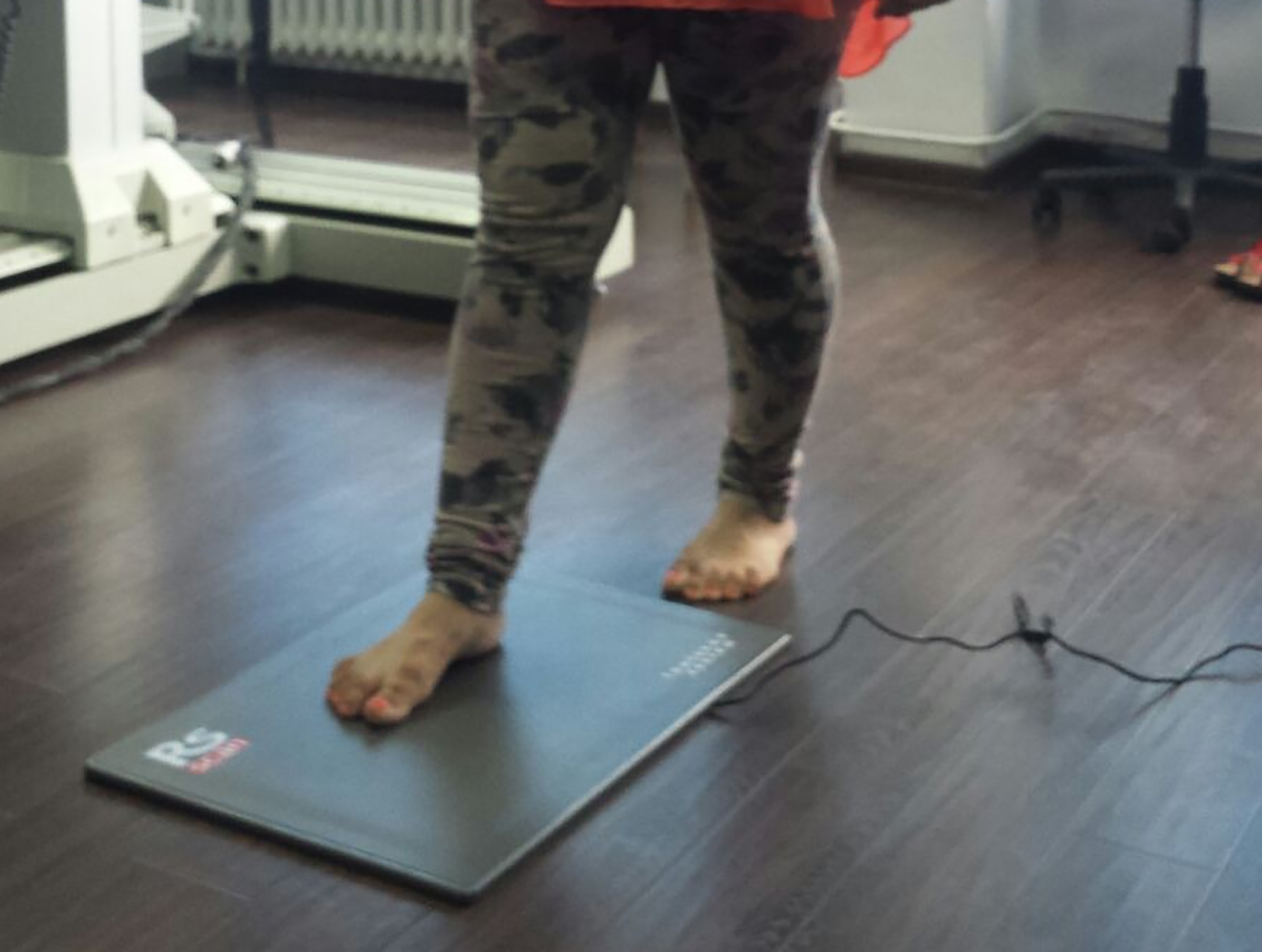
Gait analysis was made by studying the numerical data sorted in the tables or by analyzing the images included in the specific screens with appropriate numerical correspondence (dynamic screen, impulse screen, balance assessment screen, comparison screen, stepping screen). Once the measurements were registered, the software processed the data, analyzing and automatically comparing two sets of measurements. Another important feature was the ability to calculate the average of multiple measurements. The integrated database could save the patients’ personal data, together with their measurements. It was possible to export the results in various formats (Excel) or to transmit the data directly.
In normal gait, the contact time with the platform was 600–800 ms and did not record any significant variations for the two plants.
In our study, we grouped the 8 stages of the gait into 3 stages, rectum: the ground attack stage or the initial contact of the heel to the ground; the semi-support stage, in which the middle region of the plant intervenes; and the propelling stage, in which the load in the metatarsal bones prevailed and that depended on the way in which the tibial-tarsal control was made.
In our study the results of the assessments were expressed in the tables and charts of the evolution and distribution of force and pressure at the plantar level during a gait cycle; the foot balance, the rotation of the heel, the loading of the plantar regions within a gait cycle would be presented later in subsequent graphics, foot axes and angles.
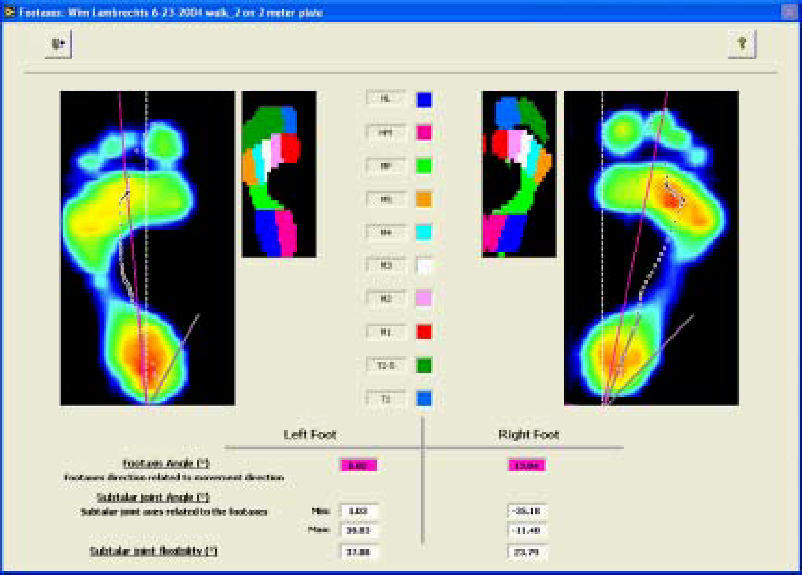
The foot axes and angles provided valuable information about its external and internal rotation (Fig. 2).
Record of the subtalar angle and of the value of the foot axis.
The foot angle expressed the deviation from the gait direction and was quantified by the values expressing the internal rotation (negative values) or external rotation (positive values). The subtalar angle is the angle formed between the axis of the talus and calcaneus and indicates the pronation of the posterior foot during the contact with the ground. During a step it records values that are quantified in minimum and maximum values. The higher the value, the higher the pronation.
In terms of clinical evaluation the patients showed: impaired balance, fatigue while walking, dizziness and numbness in the lower limbs. Muscle weakness (conform to the scale 0–5) was, on average, 3.8 patients in the study group presented pyramidal signs. All the patients showed diplopia, 3 patients had decreased auditory acuity and 5 patients had the cerebellar syndrome.
Para-clinically (MRI), all the patients in the study group showed degenerative focal, infra- and supratentorial lesions of brain damage.
a. Heel rotation evolution-right side; b. Heel rotation evolution-left side.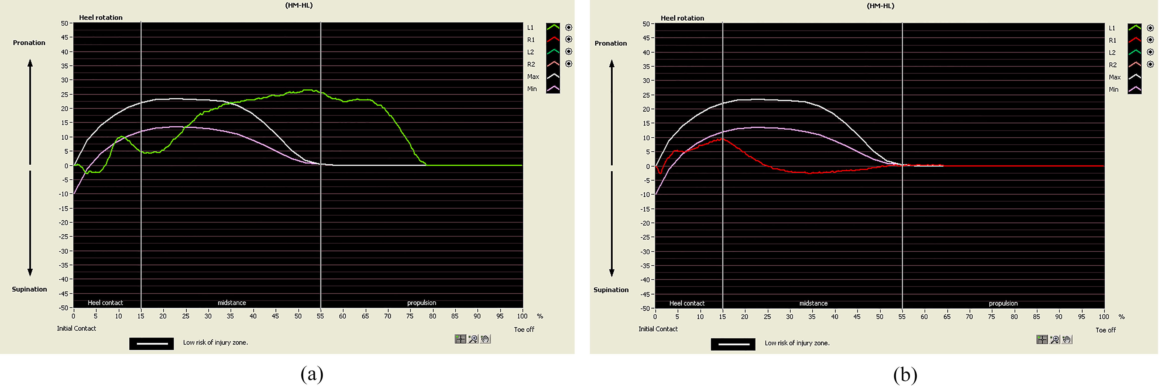
Hamilton score recorded an average of 10-mild depression. ADL score had an average value of 6 points (1 mild dysfunction). Kurtzke score [9] had an average value of 3 (mild disability).
We noted that in most cases there was a relevant, left-right asymmetry, but there were cases in which there was also a right-left symmetry.
Biomechanical analysis showed that the values of the foot angles were between
Values of the foot angle
Values of the foot angle
The values of the subtalar angle of the left and right lower limbs
The values of the subtalar angle of the left and right lower limbs are shown in Table 2.
The foot axis angle is the axis of the foot in relation to the gait direction, and the subtalar angle is in relation to the vertical axis of the foot. The values of the subtalar angle of the left foot indicated an unstable foot, with great instability, in many cases, which enabled the transition from negative values to positive values, that was from the internal rotation to external rotation. In the right foot the values were positive, which meant an external rotation of the foot. We observed, also, there was noticed an excessive abduction of the right foot correlated with the rotation of the heel which recorded negative values (supination).
We noticed a high value of the foot angle, which showed a high pronation, correlated with the disappearance of the contact area of average plantar region.
Analyzing the subtalar angle we gathered information about the rotation of the heel, which gave the asymmetrical character of the gait, with differential evolution from supination to pronation (from negative values to positive values) in phases 1 and 2 of the gait, more frequently in the right foot, while in the left foot there was an evolution in the opposite direction, from pronation to supination in phases 1 and 2 of the gait.
The rotation of the right foot into pronation during the initial contact phase was followed by supination in the single limb support phase, to return to the neutral position during the swing phase, which meant being in free zone of minimal risk (Fig. 3a), which might be correlated with what di Barozzi et al. [10] mentioned, outlining the existing motor control disturbances explained by the proprioceptive disorders [11]. In the left foot the negative values recorded during the contact phase indicated supination and the location of the foot out of the minimum risk zone (Fig. 3b).
In general, it is widely agreed that both feet behave identically during the gait in a normal locomotion (symmetrical gait). Asymmetrical gait is associated with different pathologies. However, recent studies have shown conflicting results and asymmetry even in normal gait [12]. There are different theories that try to explain gait symmetry and asymmetry. Functionally, gait asymmetry is explained by the functional discrepancy of the tasks of the two feet, while working on the propulsion and control of the body. One of the lower limbs is mainly responsible for the control and support of the body weight, while the contra-lateral limb contributes more to the propulsion of the body [12, 13]. Local asymmetry is defined as a statistically significant difference for a single gait parameter, between the left and right foot [12].
The results of the evolution of the heel rotation (as measured by the angle of the foot and by the subtalar angle) show that during the contact phase, when the heel touches the ground, as shown by Houglum [14], the dorsal flexors work more, as they prevent the fall of the foot and tibial anterior muscle intervention in this phase is very important. The action of this muscle group in the subjects with MS mainly contributes to the increased instability of the ankle, as we have previously noted, gastrocnemius muscles exercise less their role as antagonist muscle groups, namely fixing the foot to the ground.
These data indicate a bilateral and complementary disturbance of foot balance, which is supination in one foot and pronation in the contra-lateral foot. These issues are correlated with the results observed when analyzing the heel rotation, namely the existence of an external rotation of the foot which touches the ground and a tendency of the contra-lateral foot from pronation to the neutral position.
These tendencies are probably justified by the existence of certain adaptation mechanisms aimed at maintaining the motor control of the foot and at restoring the balance of the body, as other authors have noted [15], which highlights the importance of developing compensatory motor strategies. Also, the results obtained while analyzing these angles show the stability of the lower limbs during the attack and support phases, emphasizing instability during the swing phase.
We also note that the metatarsal area is within the zone of minimum risk or very close to it, which means that in this area there is a control mechanism of the projection of the centre of gravity, a mechanism that will ensure a dynamic balance; our results are consistent with the results of Castro et al. [16], supporting the importance of making the assessments using the pressure platform in developing a prophylactic program of gait rehabilitation [16], disturbed by the way in which the foot touches the ground during the attack phase, through proprioreceptors. These issues are in accordance with what Kelleher et al. noted, hence the existence of one specific gait pattern in multiple sclerosis [17].
The important negative values of the subtalar angle are present in the patients with normal symmetrical and asymmetrical gait, prevalently the internal rotation. Also, we note significant right-left differences, which reflect the tendency of developing compensatory mechanisms to control the body balance and stability as mentioned by Prosperini et al. [18], through plantar contact and through the control of tibial-tarsal joint that takes the body weight and the ground reaction force [19].
Conclusions
Biomechanical analysis of the foot angle and of subtalar angle in the patients with MS who have clinical gait disorders allows us to objectify the existence of a right-left asymmetry and the behavior of the ankle-foot during the gait. Thus, we note a reverse evolution from the internal to external rotation of the foot (from negative to positive values of the subtalar angle), more frequent in the right foot. In the left foot we note a tendency to evolve from the neutral position to the external rotation or from the external to the internal rotation. At the same time this evolution is closely correlated with the contact surface that tends to increase, which means involving the reflex mechanisms that place the foot in the zone of minimum risk and assure the stability of the body.
Footnotes
Acknowledgments
The authors thank the Research Center of Human Body Motricity and the Research Infrastructure in Applied Science.
Conflict of interest
None to report.
Abbreviations
MS – multiple sclerosis
RRMS – relapsing-remitting multiple sclerosis
PPMS – primary progressive multiple sclerosis
SPMS – secondary progressive multiple sclerosis
CPPMS – chronic progressive primary multiple sclerosis multiple sclerosis