






Abstract
BACKGROUND:
A few studies have noted that paraspinal muscle training is important to reduce pain for patients with lumbar disc herniation (LDH). However, little is known about the exact signs for necessary training of lumbar multifidus muscles (LMM).
OBJECTIVE:
The study aimed to analyze the relationship between the straight leg-raising test (SLR) and the area of fat infiltration in LMM for patients with LDH.
METHOD:
One hundred and fourteen LDH patients were involved in this study. Clinical data were collected from a medical record system. On the MRI images, the cross-sectional areas (CSA) of bilateral fat infiltration in LMM were measured by picture archiving and communication system (PACS).
RESULT:
For 61 patients with inclusive LDH, the positive side of SLR was positively correlated with the side of the larger total CSA of fat infiltration in LMM of L2-S1 (
CONCLUSION:
SLR may be used as an important sign for dysfunction of LMM and can identify the more severe side.
Descriptive statistics of the studied sample (
82 )
Descriptive statistics of the studied sample (
SLR: straight leg-raising test. (+): positive result. CSA: cross-sectional area. LSS: lumbar spinal stenosis. ES: effect size. Data were presented as mean
Despite advances in spinal surgery including endoscopic surgery for lumbar disc herniation (LDH), recent studies on postoperative low back pain have reported high rates of persistent opioid use many years after lumbar surgery [1]. The pain caused by paraspinal muscles and nerves lesion lowers the quality of life in patients [2, 3]. After investigating the relationship between intramuscular pressure or blood flow during posterior lumbar surgery, a study found that paraspinal muscles degeneration after surgery could be explained by direct mechanical damage and increased intramuscular pressure of muscle tissue by the surgical instruments [4]. Several authors have also suggested that stability training involving paraspinal muscles can reduce pain and prevent recurrence after surgery [5, 6, 7, 8, 9, 10, 11]. Among paraspinal muscles, the function of lumbar multifidus muscles (LMM) is to maintain the normal physiological curvature of the lumbar and is considered to be the most important muscle for lumbar segmental stability [12, 13, 14]. In several imaging studies, low back pain has been proven to be related to fat infiltration of LMM [15, 16, 17, 18], while fat infiltration is an important feature of degenerating LMM that predominates in the lumbar spine [19, 20, 21, 22]. It is thought that atrophy of LMM is a result of immobilization and neural compression caused by a herniated disc [23, 24]. Straight leg-raising test (SLR) test is an important sign of spinal nerve root compression for patient with LDH [25, 26].
With the rapid development of evidence-based medicine, evidence should be incorporated into all aspects of clinical care, including rehabilitation of the spine [27]. Nevertheless, we found that only a few exercises are possible after lumbar surgery due to the pain [28, 29]. According to a study by Kim et al., many patients with LDH after lumbar surgery received manipulative rehabilitation (including LMM training), following a protocol only from experience [30]. Thus, studying precise indications for lumbar training is an important consideration and prerequisite to high-quality rehabilitation. To study this issue, Battié et al. [16] found a statistical correlation between the greater side of multifidus signal intensity (fat infiltration) and the side of the herniation at the level below the herniation. Furthermore, due to the direct intervertebral attachments, LMM is potentially better in providing intersegmental stability and its function is a frequent goal of rehabilitation of lumbar [31]. Consequently, clinicians began to investigate the markers of rehabilitation of LMM by imaging technology, such as ultrasound, CT and MRI [29, 31, 32]. However, imaging results and clinical signs are not exactly same. Therefore, the aim of this study was to analyze the relationship between SLR measurements and area of fat infiltration in LMM in patients with LDH and to determine if SLR could be used as an important sign for degenerating LMM to suggest the more severe side. Based on our understanding of the scientific literature in this area, we hypothesized that sciatica on one side caused by LDH leads to altered neuro-muscular functioning, which in turn results in atrophy of LMM on the same side, which is seen as fat infiltration. Moreover, no similar studies had mentioned the exact association between SLR and area of fat infiltration in LMM for patients with LDH before.
Bilateral CSA of fat infiltration was obtained in lumbar multifidus at each level.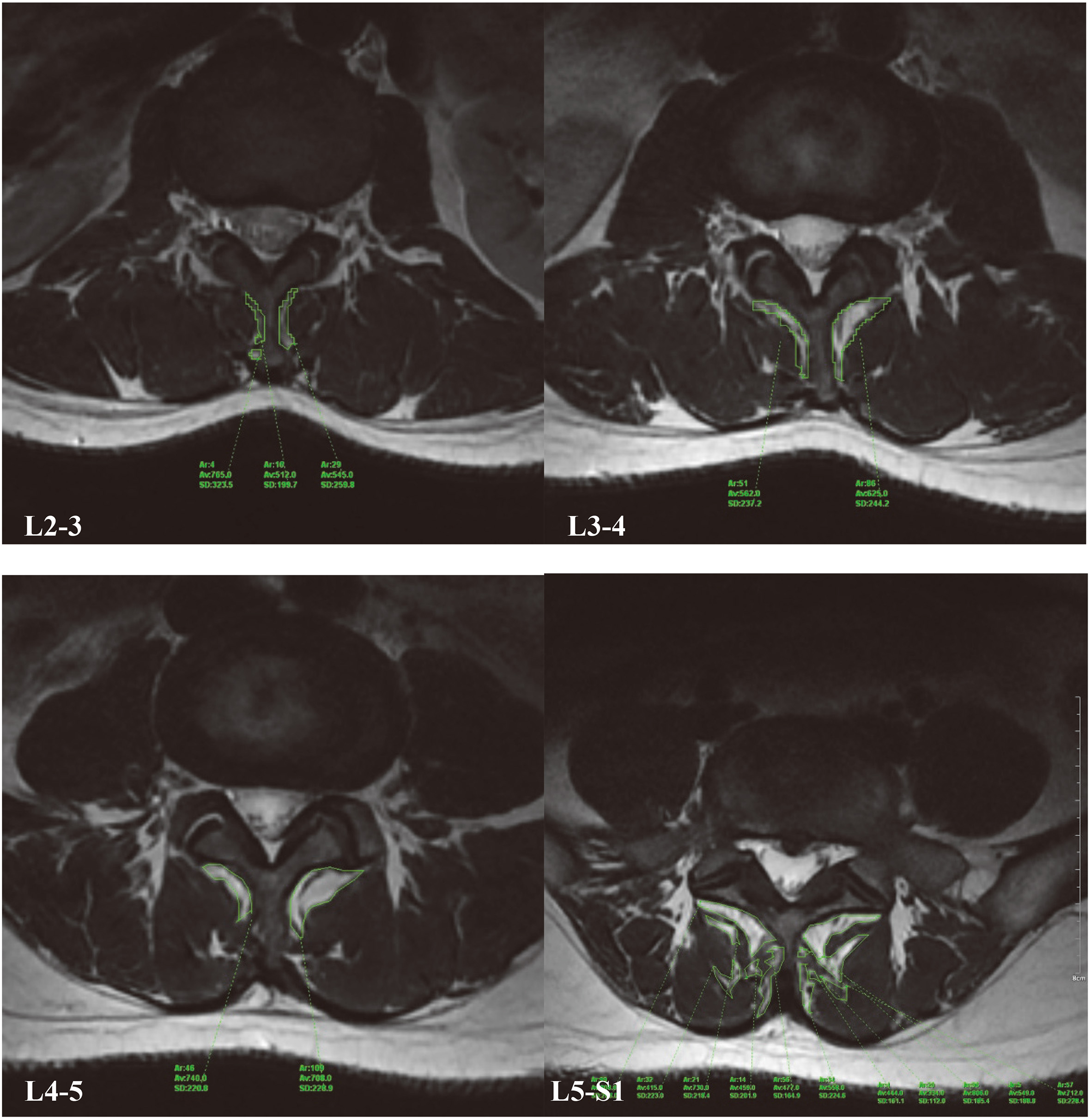
Participants
This study retrospectively followed 114 patients with LDH who initially came to our clinic for percutaneous transforaminal endoscopic discectomy (PTED) between March 2017 and November 2017 in the Pain and Rehabilitation Medicine Centre of Navy General Hospital. Demographic details, duration of symptoms and clinical data including angular degree of SLR were recorded from patients’ medical records. In all, 32 cases were excluded for the following reasons: 10 had no records of SLR, 14 had no records of Magnetic Resonance Imaging (MRI), or only computed tomography or X-ray films, 3 had a history of severe spinal trauma, 2 had a space occupying the lesion of the spine, and 2 had diagnoses were not in accordance with the description of medical records. Inclusion criteria were as follows: (1) Patients met the diagnostic criteria of LDH according to the medical records and final diagnosis; (2) Posterolateral disc herniation at the following disc levels: L4-5 and L5-S1. Primary exclusion criteria were as follows: (1) Diseases of the spinal canal caused by metastatic tumor or spine infection; (2) History of spinal trauma or spinal deformity; (3) Syndrome of cauda equina; (4) Permanent disability of the lower limbs; (5) Incomplete medical records. Finally, 82 patients (42 females and 40 males) were included, of which 21 had combined lumbar spinal stenosis (LSS). The mean age of the subjects was 56.15
SLR test methods
With the patient lying down on his or her back on an examination couch, the examiner lifts the patient’s leg while the knee is straight. If the patient experiences sciatic pain when the straight leg is at an angle less than 70 degrees, then the test is positive. Otherwise it is negative.
Comparison of CSA of fat infiltration in mm
for two sides with SLR(+) and SLR(-)
Comparison of CSA of fat infiltration in mm
SLR: straight leg-raising test. (+): postive result. (-): negative result. CSA: cross-sectional area. LDH: lumbar disc herniation. ES: effect size. Data were presented as mean
Three spinal doctors, who had been trained professionally and had been qualified, were responsible for collecting MRI data and for data processing. Lumbar MRI was performed used Siemens 3.0 T Magnetom vision and patients were placed in the prone position in the MRI device. The picture archiving and communication system (PACS) was applied to analyze the T2-weighted axial images at the inferior endplate of L2, L3, L4 and L5 bilaterally. On the endplate levels the contour line was constructed by polygon points around the outer edge of the fat infiltration in LMM. For details see Fig. 1. The measurements were performed separately for the right and left sides. To reduce the error of measuring, all the mean values were calculated from the three physicians mentioned above, including L2-S1 in total. Total CSA
Statistical evaluation
Statistical analyses of the findings were performed with the SPSS for Windows v.10.0 software program. Data were presented as mean
where:
Results
In accordance with a prior study [33], we also found that the mean CSA of female was larger than that of males at L2-3, L3-4 and L5-S1 levels (
Although not quite the same as the findings of Hides et al. [40], we found that the CSA of fat infiltration on the side of SLR(+) was significantly larger than that on the side of SLR(-) , but only at L3-4 and L4-5 levels for 61 patients with inclusive LDH (
In contrast to our hypothesis, we found no correlation between the side of the larger CSA of fat infiltration and the side of straight leg raising with a positive result at any single level for 82 patients, including L2-3/L3-4/L4-5/L5-S1 (all
Correlations between the sides of the larger CSA of fat infiltration in LMM and the sides of SLR with a positive result
Correlations between the sides of the larger CSA of fat infiltration in LMM and the sides of SLR with a positive result
SLR: straight leg-raising test. (+): positive result. CSA: cross-sectional area. LDH: lumbar disc herniation.
We also found that there were no significant correlations between the sizes of the larger CSA of fat infiltration and the angular degree of SLR with a positive result for patients with LDH (all
Correlations between the area sizes of the larger CSA of fat infiltration and the angular degree of SLR with a positive result
LR: straight leg-raising test. (+): positive result. CSA: cross-sectional area. LDH: lumbar disc herniation.
Our study included lumbar levels from L2-3 to L5-S1, and not only L4-5 and L5-S1 as Battié’s study [16] included, mainly considering that LMM is a group of muscles which stretches across several lumbar vertebral segments. In addition, given that statistical significance does not offer an indication of how important the result of the study is [41], ES was included in our study. As is apparent from Table 1, the CSA in LMM of each segment was significantly larger than that of the last segment in L2-S1, and the sexual differences of CSA were considered clinically significant in L2-3 and L3-4 (all
The difference between the side of SLR(+) and SLR(-) was found in present study, and it indicated that the CSA of fat infiltration in LMM on the side of SLR(+) is larger than the other side at L4-5 levels for 61 patients with inclusive LDH (Table 2). Note that this difference was considered both statistically significant and clinically significant. However, for L3-4, although
For inclusive LDH in our study, it is obvious from Tables 2 and 3 that we may determine the side of dysfunctional LMM by the side of SLR with a positive result, which can be confirmed further by the CSA of fat infiltration at L3-4 and L4-5 levels. However, according to Table 4, the degree of degeneration of LMM cannot be evaluated by the degree of SLR, since there were no significant correlations between the sizes of the larger CSA of fat infiltration in LMM and the angular degree of SLR with a positive result for patients with LDH.
There were several limitations in this study. The first problem is the measure of fat infiltration in LMM. There were many tiny and scattered fat gaps in LMM, and so we could not construct the contour line accurately around the outer edge of the tiny fat infiltration on MRI. Thus, the measure of CSA of partial fat infiltration was not very precise. In addition, it was difficult to exclude subjective factors completely although we had calculated a mean value of MRI data from three different professional physicians. Moreover, the approach may need to be different for males and females in future studies because there are gender differences on the CSA of LMM, according to Hides et al. [40]. Similarly, the present study also showed the significant difference between males and females (Table 1). The third problem is the low number of enrolled patients. If a larger number of subjects were included, the results may be more reliable.
In conclusion, the study findings suggest that SLR may be used as an important sign for dysfunction of LMM and can suggest the more severe side for inclusive LDH. However, the degree of degeneration of LMM can not be evaluated by the degree of SLR. We hope that this study may remind lumbar surgeons to protect the LMM during the operation.
Footnotes
Acknowledgments
This study was supported by the innovation fund of the Naval General Hospital (CXPY201721).
Conflict of interest
None to report.