






Abstract
Keywords
Introduction
Breast cancer has remained the leading cause of tumor mortality in women older than 40 years of age [1]. DM has been widely applied in breast cancer screening and has been demonstrated its efficiency in the detection of early breast cancer. DM has a higher sensitivity and specificity (80%–90%) in fatty breast, but the diagnostic performance of DM is poor in extremely dense breast due to overlapping dense fibroglandular tissue that may obscure or completely conceal a lesion [2–4]. It is well known that breast MRI is considered to be the most sensitive tool and the best supplementary method to conventional imaging in breast diseases [5]. Furthermore, the sensitivity of breast MRI after enhancement in the detection of breast cancers could reach 90–100% and avoided the limitations in dense breast tissue [6, 7]. Nevertheless, breast MRI has some shortcomings. The specificity is relatively low and ranges from 60 to 70%, which leads to a high rate of overtreatment [8]. Moreover, there are some other restrictions, such as higher cost, lower efficiency and availability also existed.
DBT is a rising imaging modality that acquired the images of a breast at numerous angles within a short scan. All images will be reconstructed into a series of thin, high-resolution slices that can be showed individually or in a dynamic movie mode [9, 10]. Through this technology, DBT may overcome the limitations of DM mentioned above; consequently, improving the sensitivity and specificity compared with that of conventional mammography. Until now, most scholars have focused on comparisons of the diagnostic ability of DM with DBT and have reached a consensus that DBT is superior to DM [11–13]. However, a direct comparison between DBT and MRI in symptomatic women was seldom reported [14]. We consider that a direct comparison of the two methods in the field of sensitivity, specificity, overall accuracy, PPV and NPV will potentially improve our understanding of DBT as a new diagnostic modality.
Therefore, the purpose of the present study was to compare the diagnostic efficiency of DBT plus DM and MRI plus DM and to seek an optimal imaging combination for patients with suspected breast lesions.
Materials and methods
This study was a prospective study that was funded by a Shanghai Cancer Center grant (HX-1317). This study was approved by our institutional review board (No.: IRB1405135-6), and written informed consent was obtained from all patients. The inclusion criteria was listed as follows: 1. No breast surgery within 5 years 2. Aware of the research content and sign the informed consent 3.DM, DBT and MRI images were available in all patients prior to surgery or biopsy 4. All patients are proven by pathology. The exclusion criteria was listed as below: 1. Pregnant women and nursing mothers 2. Patients were lost to follow-up 3. Patients were treated with neoadjuvant chemotherapy prior to image acquisitions 4. Image quality does not meet the diagnostic requirements 5. Patients with claustrophobic or chronic renal failure.
Patient data
The inclusion and exclusion criteria are summarized in Fig. 1. Between June and December 2014, 227 consecutive patients with suspected lesions (by clinical palpation or ultrasound) and who satisfied the inclusion criteria all underwent DM, DBT and MRI at our hospital. Among these patients, 30 were ineligible for this study for the following reasons: sixteen underwent needle biopsy at other hospitals before imaging examination, eight had received neoadjuvant chemotherapy, three had breast implants and three lost follow-up. Therefore, a total of 197 patients with a mean age of 54.3 years (range, 28–66 years) were finally enrolled. Among the 197 patients, 139 women (70.5%) had palpable masses, 42 exhibited symptoms of pain (21.3%), 7 had nipple discharge (3.6%), 6 had nipple retraction (3.1%) and 3 (1.5%) experienced skin thickening (Table 1).
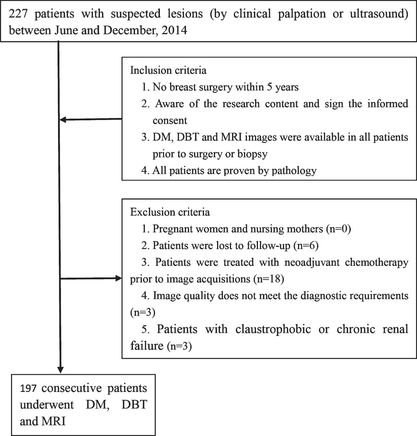
Flowchart of study population.
Characteristics of patients and lesions
IDC = indicates invasive ductal carcinoma; DCIS = ductal carcinoma in situ.
DM and DBT were performed using the same mammography unit (Selenia Dimensions; Hologic, Bedford, MA, USA). Craniocaudal (CC) and mediolateral oblique (MLO) views were performed in all patients. DBT and DM images were acquired with one compression for each projection. During tomosynthesis imaging, x-ray tube rotation was performed through the angular range of 15° (–7.5° to +7.5°), and a series of images were acquired; these images were then reconstruct as tomographic images with a slice thickness of 1 mm. In the light of radiation dose, supposing a breast was compressed into a thickness of 4.0 cm and a 60% fibroglandular fraction, DBT (MLO&CC) acquisitions leaded to a 13% higher mean glandular dose (MGD) per view than a DM image (1.70 and 1.50 mGy, respectively); both doses were lower than the Mammography Quality Standards Act (MQSA) dose limit.
All MRI examinations were performed on patients in a prone position using a 3.0-T MRI (Signa HDxt; GE Healthcare, Milwaukee, WI, USA) with a dedicated breast coil (HD 8-Channel Volume Imaging For Breast Assessment [Vibrant] Breast Array). The patients received the exam on days 7–14 of the menstrual cycle if they were premenopausal. A transaxial T1 fast spin echo (FSE) sequence [repetition time (TR)/echo time (TE) of 600–800 milliseconds (ms)/8.9–10.5 ms] and a short time inversion recovery (STIR) sequence TR/TE of 3600–4200 ms/100–120 ms] were performed before contrast. Both sequence had 35 excitations, a matrix of 256×256, field of view of 25×25 cm, thickness of 2 mm, and gap of 0.5 mm. Contrast agents (Magnevist; Bayer Schering Pharma, Berlin, Germany) were injected intravenously (total dose, 0.2 mmol/kg of body weight and flow rate of 2 ml/s,) with a high-pressure syringe (Mallinckrodt, Liebel-Flarsheim, Cincinnati, OH, USA) and was followed by a 20-ml saline flush. A series of seven 1-minute axial T1 fat-suppressed enhanced pictures were obtained with a Vibrant sequence. Besides delayed high resolution sagittal T1 fat-suppressed enhanced images were also acquired with a sagittal Vibrant sequence. The scan parameters were as follows: TR/TE of 6.5–9.0 ms/3.3–4.5 ms, 2 excitations, and a matrix of 256×256, a field of view of 30×30 cm, a thickness of 3 mm, and a gap of 0.5 mm. Three-dimensional Maximum Intensity Projection (3D-MIP) reconstructions with dynamic enhanced curve were generated when necessary.
The Hospital Information System (HIS) and the Picture Archiving and Communication System (PACS) were available to radiologists for the interpretation of images from all three modalities.
Study design and image analysis
All images from DM alone, DBT plus DM and MRI plus DM were interpreted independently by two experienced radiologists (radiologist 1 with experience of 12, 10, and 2 years in DM, MRI and DBT, respectively; radiologist 2 with 20, 15 and 3 years, respectively). All images were divided into four sessions, and every reading session included one-quarter of all the images of DM alone, DBT plus DM and MRI plus DM, which were randomized and displayed in alternate sequence. Each reading session was separated by at least 3 weeks to minimize the memory effects of the interpreters. The radiologists were blinded to the clinical issue including the results of previous examinations, and they were required to document all visible lesions using a graphical interface and write down the number and location of all lesions with a standardized template. Each identified lesion was evaluated in terms of the Breast Imaging Reporting and Data System (BI-RADS, 5th edition for mammography and 2nd edition for MRI, 2013, American College of Radiology) categories [15, 16]. We defined the BI-RADS categories of 1 to 3 as normal or benign, while cases ≥4 were considered abnormal or malignant. The BI-RADS category of each lesion detected by all imaging modalities was compared with the histopathology results of the core biopsy, vacuum-assisted stereotaxic biopsy or surgical excision specimen.
The lesions detected by each imaging modality, which were documented in a standardized template by the interpreters, were considered concordant with histopathology if they were located within a distance of 2 cm. Additionally, the location of the lesions was supposed as accurate if the lesions were located less than 2 cm away from the location identified in surgery, which were available in the HIS. All the pathology specimens were reviewed by a pathologist with rich experience in breast diseases.
Statistical analysis
We used SPSS Statistics 21.0 (IBM Corp., Armonk, NY, USA) and Medcalc 12.7.2 (Medcalc software, Ostend, Belgium) for statistical data analysis. A ROC curve in accordance with the BI-RADS classification was generated for DM alone, DBT plus DM and MRI plus DM. We calculated the area under the curve (AUC) of all three modalities and compared the presentation of the ROC curves as described in the article by Park et al. [17]. The sensitivity, specificity, accuracy, PPV and NPV of each diagnostic modality were also calculated. Comparisons were performed using McNemar’s test (sensitivity, specificity, accuracy) and Fisher’s exact test (PPV, NPV). A P < 0.05 by two-tailed t-test was considered significant.
Interobserver agreement in the classification of lesions according to the BI-RADS lexicon was assessed using Cohen’s Kappa analysis (0–0.20, poor agreement; 0.21–0.40, fair agreement; 0.41–0.60, moderate agreement; 0.61–0.80, good agreement; 0.81–1.00, excellent agreement).
Results
Patients and lesion characteristics
In this study, 197 patients were enrolled, and 238 lesions were identified (based on any imaging test) and confirmed by histopathology (153 were malignant, whereas 85 were benign). The specific histologic distributions of all the lesions are described in Table 1. Lesions that were benign according to biopsy but that were not excised were followed-up with conventional breast imaging tools for at least 12 months (average follow-up duration, 16.8 months; range, 10.4–22.6 months). Among 238 lesions, 170 (71.4%) were unifocal, 38 (16.0%) were multifocal or multicentric in 12 women, and 30 (12.6%) were bilateral in 15 women. The breast fibroglandular composition in the patients was classified in terms of the BI-RADS lexicon: 10 cases of nearly entirely fatty breasts (composition a), 38 scattered fibroglandular densities (composition b), 128 cases of heterogeneously dense breasts (composition c) and 21 cases of extremely dense breasts (composition d).
Diagnostic performance
The ROC curves for DM alone, DBT combined with DM and MRI combined with DM are shown in Fig. 2. We listed and compared the AUC for all three imaging methods (Table 2). The AUC values of DM alone, DBT plus DM and MRI plus mammography for radiologist1 (R1) were 0.849, 0.9073 and 0.939, respectively; for radiologist2 (R2), the AUC values of each modality were 0.850, 0.900 and 0.935, respectively. The AUC of DM alone was obviously lower than that of DBT combined with DM (R1, P = 0.0204; R2, P = 0.0239) and that of MRI plus DM (R1, P = 0.0006; R2, P = 0.0009), while no significant difference was found between DBT plus DM and MRI plus DM (R1, P = 0.1262; R2, P = 0.0843) (Fig. 2). We also calculated the accuracy, sensitivity, specificity, PPV and NPV values for the three diagnostic methods for each radiologist, which are shown in Table 3. The accuracy (R1, 71.8%; R2, 71.4%), sensitivity (R1, 71.9%; R2, 71.2%) and NPV (R1, 58.7%; R2, 58.1%) of DM alone were lower than those of DBT plus DM (accuracy: R1, 85.3%, P = 0.001; R2, 83.6%, P < 0.001; sensitivity: R1, 92.1%, P < 0.001; R2, 90.8%, P < 0.001; NPV: R1, 83.8%, P < 0.001; R2, 82.8%, P = 0.001) and those of MRI plus DM (accuracy: R1, 90.3%, P = 0.001; R2, 90.7%, P < 0.001; sensitivity: R1, 94.7%, P < 0.001; R2, 95.4%, P < 0.001; NPV: R1, 89.7%, P < 0.001; R2, 90.9%, P < 0.001), whereas no significant difference was found between DBT plus DM and MRI plus DM (accuracy: R1, P0.644; R2, P = 0.360; sensitivity: R1, P = 0.502; R2, P = 0.359; NPV: R1, P = 0.340; R2, P = 0.218) (Fig. 3).
ROC analysis of mammography alone, tomosynthesis plus mammography and MRI plus mammography
ROC analysis of mammography alone, tomosynthesis plus mammography and MRI plus mammography
P valuesa between mammography alone versus tomosynthesis plus mammography. P valuesb between mammography alone versus MRI plus mammography. P valuesc between tomosynthesis plus mammography versus MRI plus mammography. *Data in parentheses are 95% CI.
The diagnostic sensitivity, specificity, PPV, NPV and accuracy of DM alone, DBT plus DM and MRI plus DM
P valuesa between mammography alone versus tomosynthesis plus mammography P valuesb between mammography alone versus MRI plus mammography. P valuesc between tomosynthesis plus mammography versus MRI plus mammography.
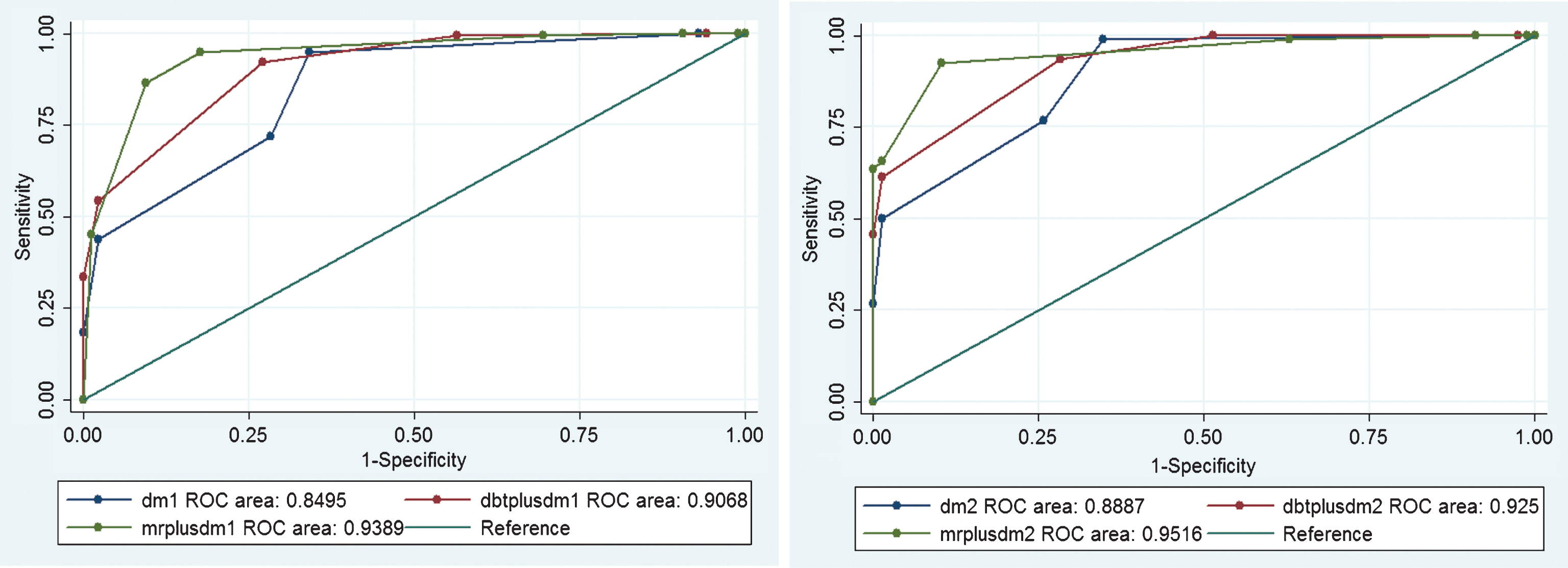
(a) ROC for radiologist 1; (b) ROC for radiologist 2.
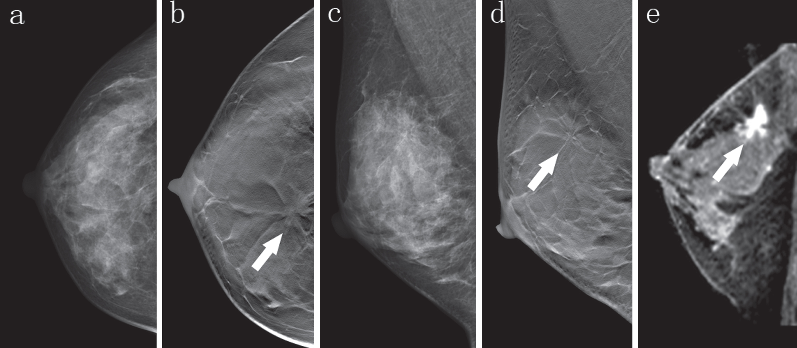
A 47-year-old woman with palpable mass in her right breast. (a, c), Digital mammography CC and MLO projections show no significant abnormality, classified as BI-RADS 1. (b, d), CC and MLO projections of DBT show an obvious architectural distortion with a mass in right breast (arrow), classified as BI-RADS 5. (e). Sagittal T1-weighted post-contrast enhanced subtraction MR image shows an irregular mass with enhancement (arrow), which is consistent with DBT. Hispathology: Invasive ductal carcinoma.
The interobserver agreement of DM alone, DBT plus DM and MRI plus DM was excellent (k = 0.894, 0.919 and 0.882, respectively).
Discussion
In our study, the diagnostic performance of DM alone, DBT combined with DM and MRI combined with DM was compared in women who had suspected breast lesions. We found that the AUC, accuracy, sensitivity, and NPV of DM alone were all significantly lower than those of DBT plus DM and MRI plus DM for the two radiologists. Although the AUC, diagnostic accuracy, sensitivity, and specificity of MRI plus DM were slightly higher than those of DBT plus DM for the two radiologists, the differences were not statistically significant.
The inferiority of DM alone in terms of diagnostic performance may be specific to dense breasts. DM is a conventional two-dimensional (2D) imaging modality, which leads to the loss of ‘depth vision’, and thus a lesion may be obscured or hidden by the overlapping fibroglandular tissue; alternatively, a lesion with the same attenuation as the surrounding tissue may also obscure the lesion, and either case will result in a false-negative interpretation. On the contrary, the superposition of normal tissue may also obscure a tumor, which would result in false-positive identification; this in turn may diminish the PPV and the specificity of DM. DBT is a novel imaging tool that could export tomographic images and volumetric three-dimensional (3D) images, which will reduce tissue overlapping. In addition, the shape and edge of the lesions will be shown more clearly, and thus, this method increases the sensitivity and accuracy of the detection of breast lesions. Most previously published articles have focused on the comparison of the diagnostic performance between DM and DBT or DBT plus DM in the screening or diagnosis of women. A consensus has been reached that DBT plus DM clearly has a higher diagnostic performance than DM alone [18–20]. The findings of our study are consistent with those of previous studies and ensure the advantage of DBT plus DM over DM alone in terms of sensitivity, NPV and accuracy [21–24] (Fig. 4). Some initial studies found that the performance of DBT in the detection of calcifications was inferior to that of DM [25], while Wallis MG et al. found that DBT was better than DM in this regard [26]. Our study also confirms that DBT allowed for the detection of calcifications and was superior to DM (Fig. 5).
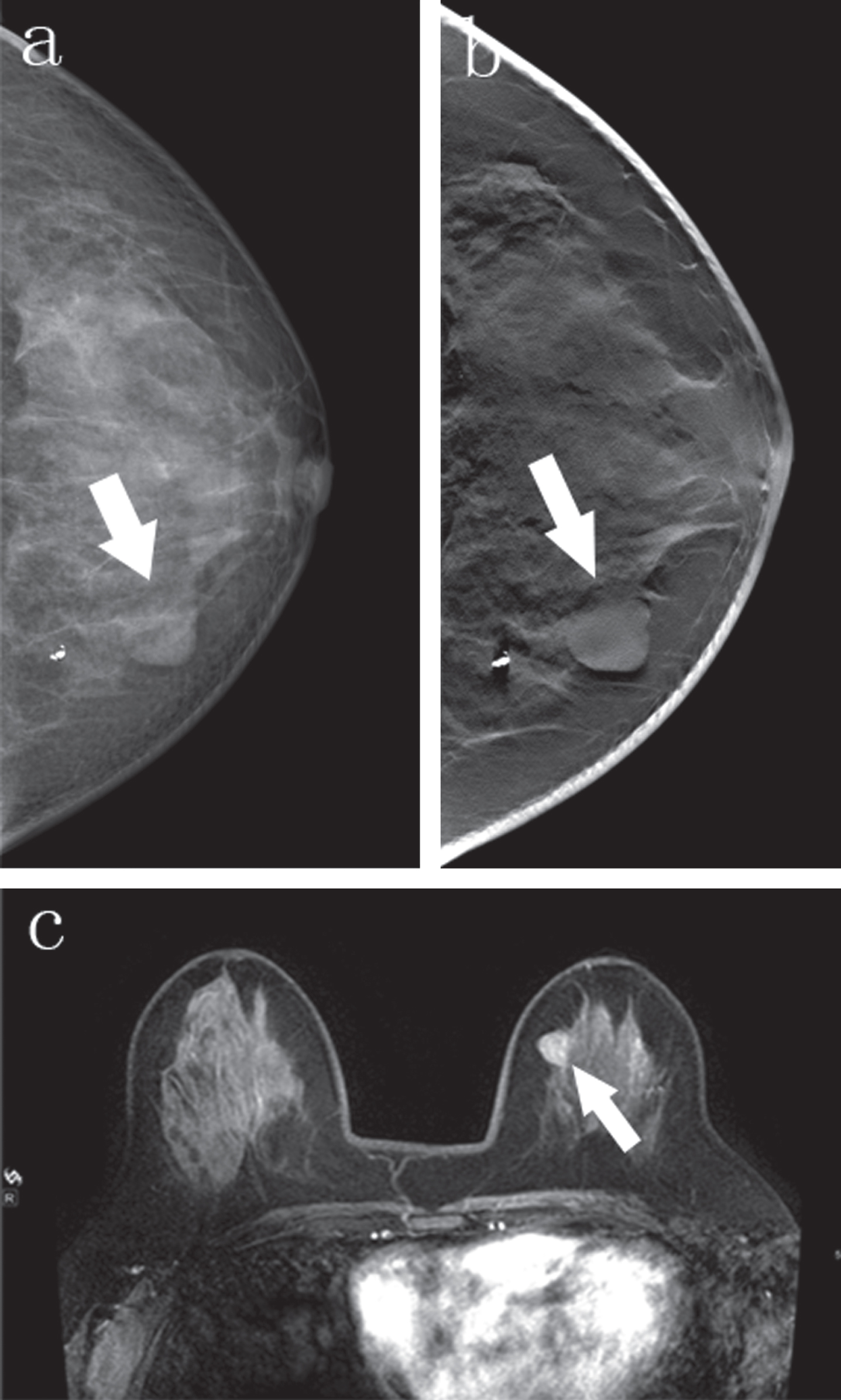
A 40-year-old woman with palpable mass in her left breast. (a), Digital mammography CC projection shows a mass, part of the margin is obscure, classified as BI-RADS 4A for radiologist1, 3 for radiologist 2. (b), CC projection of DBT shows margin of the mass clearly, typical benign lesion, classified as BI-RADS 3 for both radiologists. (c). Axial T1-weighted post-contrast enhanced MR image shows an regular mass with homogenous enhancement (arrow), which is very accordant to DBT. Hispathology: Fibroadenoma.
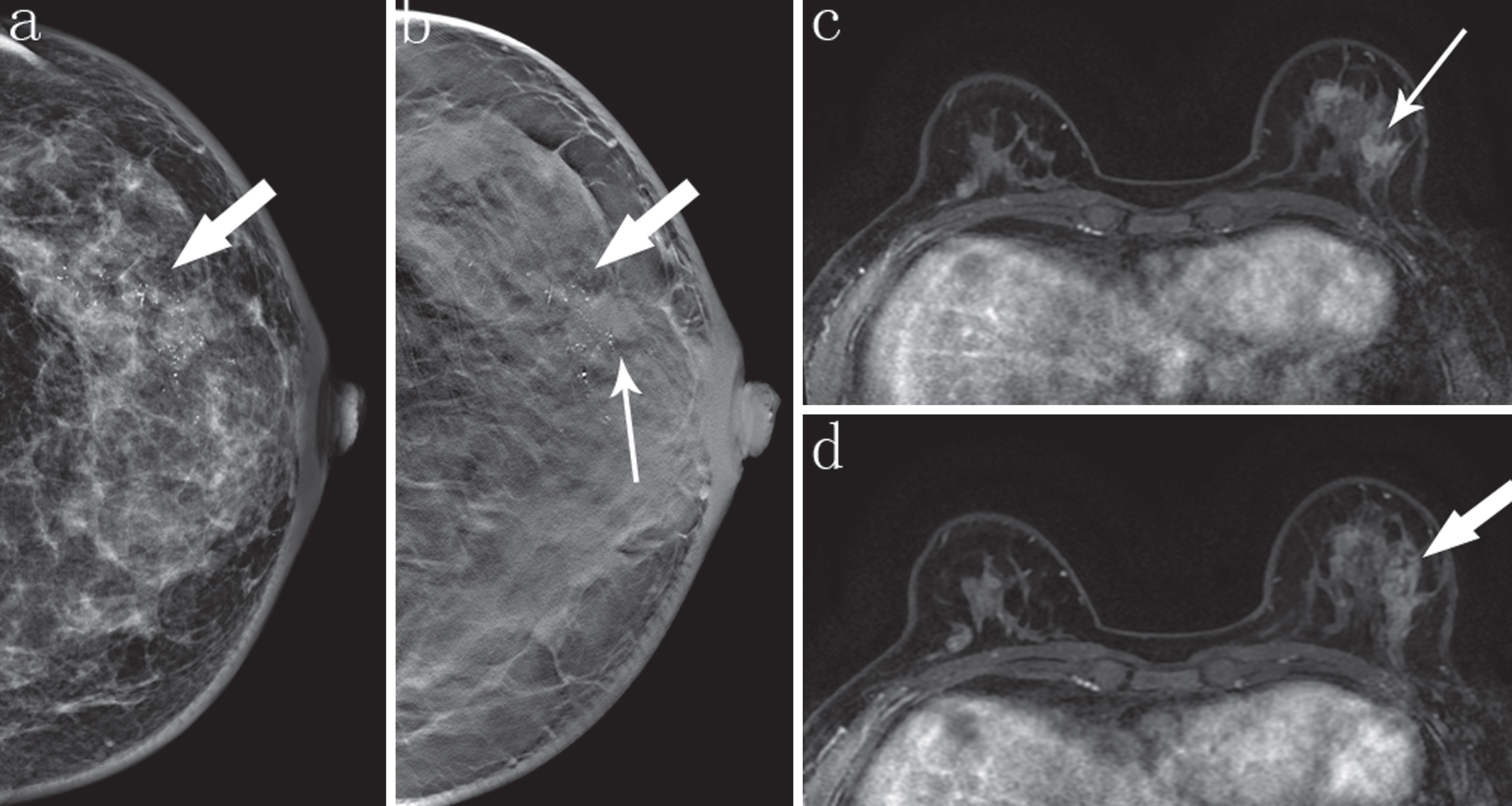
A 51-year-old woman with palpable mass and pain in her left breast. (a), Digital mammography CC projection shows fine pleomorphic calcifications in segmental distribution, classified as BI-RADS 4B for radiologist1, 4C for radiologist 2. (b), CC projection of DBT shows not only calcifications in segmental distribution (thick arrow), but an irregular mass (thin arrow), typical malignant lesion, classified as BI-RADS 5 for both radiologists. (c). Axial T1-weighted post-contrast enhanced MR image shows a regular mass (thin arrow) and segmental non-mass enhancement (thick arrow), which is highly consistent with DBT. Hispathology: IDC with DCIS.
To date, MRI is regarded as the best imaging tool for the preoperative assessment of women with breast lesions. Indeed, in clinical practice, breast MRI is widely used as the most important supplementary imaging modality to conventional imaging tools (DM and ultrasound) not only in preoperative assessment but also in the observation of the response of breast cancer patients to neoadjuvant chemotherapy patients. The superiority of breast MRI is apparent for several reasons. First, unlike DM and DBT, the diagnostic performance of MRI is not dependent on breast tissue density [27]. Second, besides well-demarcated morphological and marginal characteristics of breast lesions, dynamic contrast-enhanced MRI can further reflects the enhancement characteristic of breast lesions. The post-contrast enhancement kinetic curve is available to assist evaluating the characteristics of breast lesions. In this study, the accuracy, sensitivity, and NPV of MRI plus DM were the highest among the three modalities, which is in accordance with previous studies [20, 28]. However, the merits of MRI mentioned above should be considered with its defects such as that MRI is time-consuming, costly, unsuitable for claustrophobic patients or those with renal failure and has high false-positive rates. Its shortcomings in terms of inefficiency and high cost are particularly apparent in China because of it has the largest population in the world. Even in some small cities, MRI machines are unavailable. Moreover, false-positive results may lead to an unnecessary biopsy and mastectomy [29]. In terms of PPV, our study showed that the PPV of MRI plus FFDM was higher than that of DBT plus FFDM and DM alone, which is different from the result described in the previous articles [30, 31]. The reason for this discrepancy may be that the previous articles both focused on screening MRI, whereas our study is a diagnostic study, in which the enrolled patients were all symptomatic women; most of the lesions are unifocal (170/238), and therefore, the radiologists can detect more true positive lesions and fewer false-positive lesions, which results in a higher PPV of MRI plus DM.
To our knowledge, very few studies have focused on the direct comparison of the diagnostic ability of DM alone, DBT plus FFDM and MRI plus DM. In one published article, the authors compared the diagnostic performance of three imaging tools; however, in that study, only a patient population with cancer was enrolled, and the observers were conscious that all patients were possible for having had at least one incidence of breast cancer before image analysis, which may have imported a bias [14]. In another previous article that compared DBT and MRI, only a population with cancer was enrolled. The authors found that the addition of MRI to the combination of mammography, DBT and ultrasound makes no difference to the diagnostic performance. However, in this study, besides DBT, ultrasound results were also introduced that make the direct comparison between DBT and MRI impossible [27]. Some other studies assessed the value of a second-look DBT after MRI was used to detect additional lesions [32, 33]. According to those studies, DBT increased the detection and characterization of additional lesions identified by MRI, which indicates that a combination of either imaging modality might improve the overall diagnostic value for breast lesion evaluation. Ultrasound has been widely used in the breast examination, especially in the dense breasts, yielding good results. As reported in the paper written by Chen YP [34], the diagnostic performance of Virtual Touch Tissue Quantification (VTQ) or Virtual Touch Tissue Imaging Quantification (VTIQ) was moderate to good for solid breast tumors. Although both methods have higher sensitivities in tumors ≥10 mm, their overall diagnostic performance was similar for all sizes. For the advantages of ultrasound, we will compare the diagnostic efficiency of DBT with ultrasound in the subsequent study.
Our study is a direct comparison of the diagnostic ability of DM alone, DBT plus FFDM, and MRI plus DM. Our study enrolled patients who were symptomatic women rather than a cancer-only population. Furthermore, the radiologists were unaware of the clinical information and results of previous imaging examinations, which may have reduced the bias. The interobserver agreement between the two radiologists was excellent in our study, which means that the difference was not caused by the readers’ experience.
Nevertheless, our study has several limitations. First, only symptomatic women were included, which may have brought in a bias. Thus, our finding may not be suitable for a screening population. Second, not all lesions were confirmed by surgical pathologists, and less than half of the benign lesions were proved by core biopsy or vacuum-assisted stereotaxic biopsy, though all benign lesions remained stable during the follow-up circle. This may have resulted in an overestimation to the actual sensitivity of each imaging modality by ignoring some of the false-negative lesions. Third, non-dense lesions and calcified lesions were uncommon in this study, and thus a further comparative study needs to be performed according to the different types of lesions. Fourth, most of the lesions in our study were unifocal, and the results may be different in multifocal or multicentric lesions, which require additional research in a larger population.
In conclusion, DBT and MRI are both good methods to supplement DM. The diagnostic performance of DBT plus mammography is similar to that of MRI plus mammography. If our results are validated in larger studies, our findings may widen the understanding of DBT and provide a new, economic and convenient imaging tool, besides MRI, for symptomatic women in future clinical practice.