






Abstract
BACKGROUND:
Duplex ultrasound is the first choice in diagnostics and surveillance of stenoses of the internal carotid arteries before and even after surgery. Therefore, the quality of duplex ultrasound is crucial to investigate these vascular pathologies.
OBJECTIVE:
Aim of this study was the evaluation whether different surgical techniques affect the postoperative quality of duplex ultrasound.
METHODS:
In a time period from January to May 2015 duplex ultrasound of the cervical vessels was performed in 75 patients after unilateral endarterectomy of the internal carotid artery at our department between 2006 and 2012. Thereby, the non-operated contralateral side served as a control. Study groups were defined by the surgical techniques of eversion- or thrombendarterectomy with patch plasty using different patch materials and/or a haemostatic sealant. Duplex ultrasound analysis included acoustic impedance, extinction of ultrasound, thickness of skin and individual anatomic aspects of the patients.
RESULTS:
Carotid endarterectomy itself reduced intravascular grey levels, skin thickness and increased extinction of duplex ultrasound when compared to the non-operated side of the neck. In contrast, neither the kind of chosen operative technique nor the use of different patch materials or the application of a haemostatic sealant showed an effect in this regards.
CONCLUSIONS:
Whereas carotid endarterectomy per se worsens the quality of postoperative duplex ultrasound, the different analysed surgical techniques as well as used patches and the application of a haemostatic sealant can be assumed to be equal regarding the quality of postoperative ultrasound.
Introduction
Arteriosclerotic stenoses of the internal carotid arteries (ICAs) are a pathologic process with high prevalence [1] caused by different risk factors like age, sex, nicotine abuse, arterial hypertension and hypercholesterinaemia [2]. Although arteriosclerotic plaques can remain stable for years, there is a risk for plaque rupture leading to local thrombus formation and subsequent embolisation or complete occlusion of the ICA resulting in amaurosis fugax, retinal infarction, transient ischemic attack or even stroke. Therefore, anti-platelet medication and treatment with statins as well as local therapy in means of carotid endarterectomy (CEA) or carotid artery stenting (CAS) are recommended in both symptomatic and asymptomatic patients depending on the grade of stenosis [3, 4]. CEA can be performed either as an eversion endarterectomy (EEA) or a thrombendarterectomy (TEA) with mandatory patch plasty. Nowadays, CEA is routinely performed under anti-platelet medication [5], therefore local haemostatics might be applied to achieve sufficient haemostasis at the suture line.
Following CEA or CAS a regular surveillance primarily by means of duplex ultrasound (DUS) is recommended, providing a safe and accurate method for follow-up including detection and quantification of recurrent stenosis [6, 7]. An additional focus of DUS surveillance is to detect a possible stenosis of the contralateral ICA. However, the quality and significance of this DUS depends on the user’s experience and the patient’s individual morphological characteristics. Thereby, postoperative fibrosis and scar formation might attenuate ultrasound waves and alter the quality of DUS. Additionally, it is known that glutaraldehyde-fixed bovine pericard patches used for patch plasty in TEA might lead to a calcific perivascular degeneration and local cytotoxicity [8, 9] that might interfere with DUS during postoperative surveillance. Application of the haemostatic sealant TachoSil® is known to induce fibrovascular granulation tissue [10] and might influence postoperative scar formation and subsequently reduce the quality of DUS.
Aim of this study was to analyze whether the performed different surgical techniques including the use of different patch materials or the application of TachoSil® have an influence on the postoperative quality of DUS.
Methods
Written informed consent of all volunteers was obtained before recruitment into the study in accordance with the votum of the local ethics committee (A 2015-0020). The study was performed in accordance with the ethical guidelines of Clinical Hemorheology and Microcirculation [11]. The study population consisted of 75 patients who underwent unilateral CEA due to symptomatic or asymptomatic stenoses of the ICAs between March 2006 and June 2012 at our department. An overview on the epidemiologic patient criteria is given in Table 1. The surgical techniques included EEAs (n = 20) and TEAs (n = 55). Following TEA the ACI was patched either with Dacron (Hemagard Carotid Patch, Maquet Getinge Group, Intervascular, La Ciotat Cedex, France) or bovine pericardium (XenoSure, LeMaitre Vascular GmbH, Sulzbach/Ts., Germany). Since 2008 the haemostatic sealant TachoSil® (Takeda Austria GmbH, Linz, Austria) was frequently applied to cover the suture lines and to reduce the risk of postoperative bleeding/hematoma. Thus, four subgroups were defined: a) TEA with Dacron patch plasty and TachoSil® (n = 20); b) TEA with Dacron patch plasty without TachoSil® (n = 20); c) TEA with bovine pericardium patch plasty and TachoSil® (n = 15); d) EEA with TachoSil® (n = 20).
Demographics and clinical characteristics of the 75 patients (55 TEAs and 20 EEAs)
Demographics and clinical characteristics of the 75 patients (55 TEAs and 20 EEAs)
The ultrasonographic examination of the patients was carried out by a single angiologist (T. Heller) using a Toshiba AplioXG SSA 770A/80 color duplex system (Toshiba, Minato, Japan) with a linear transducer (PLT-604AT) in THI mode with a frequence range of 4.0–9.2 MHz (mean frequence 7.6 MHz), a dynamic range of 65, an edge enhancement of 2 and a mechanical index of 0.7. DUS included examination of the common carotid arteries, carotid bifurcations, internal and external carotid arteries. As mentioned before, only patients after unilateral surgery of the neck were included into the study with the non-operated side serving as control. DUS analysis included the documentation of extinctions of the ultrasound signal and impedance changes, which were quantified by subcutaneous and intravascular grey scales using the Java-based image-processing program ImageJ (release 1.48v, National Institutes of Health, Bethesda, USA). These variables were assessed as parameters for tissue remodelling that might interfere with DUS. Acoustic impedance assessment is based on phase contrasts between the assessed signal and a reference. Thereby, the phase contrasts are influenced by the cellularity within the layers of the skin and the acoustic impedance that might be influenced also by connective tissue, scars and the extracellular water content. For a valid and reproducible analysis, the regions of interest were defined at the carotid bifurcation and between the skin and carotid vessel wall in a right angle to the surface of the skin. Mean grey values were assessed between 0 (black) and 255 (white). Skin thickness was defined as distance between the skin and the carotid bifurcation.
Regarding individual anatomic characteristics of the patients, neck length and circumference as well as the body mass index (BMI) of the patients were ascertained. Thereby, neck length was defined as the distance between the chin and the sternum. The neck circumference was measured at the height of the thyroid cartilage.
All statistical calculations were performed with the SAS software (release 9.3, SAS Institute Inc., Cary, NC, USA). Quantitative variables are presented by mean value and standard deviations. For qualitative parameters, absolute and relative frequencies are given. For comparison of all four groups, Chi2 test or Fisher’s exact test were used for qualitative factors. The mean values of the the four groups regarding the variables age, BMI, neck circumference and neck length were compared by a one way ANOVA, as all these variables were approximately normally distributed. For non normally distributed variables Kruskal-Wallis-test has been used instead. Two paired samples (operated and not-operated side) were compared using McNemar’s test (in the case of a binary factor) or t-test for paired samples. Results are considered significant for p values below 0.05.
The timespan between CEA and the performance of DUS within this study raged from three to nine years in our study population. Analysis of patients’ individual characteristics that might influence the quality of DUS, including neck length, perimeter of the neck, BMI and gender, revealed no significant differences between the four subgroups of different performed surgical techniques and chosen patch materials. Details are depicted in Table 2. In 6 patients (8.0%) a recurrent stenosis of the ICA was seen, and in 17 patients (22.7%) an ICA high grade stenosis on the contralateral side was detected.
Non-susceptible parameters that might influence duplex ultrasound quality in 75 patients (55 TEAs and 20 EEAs)
Non-susceptible parameters that might influence duplex ultrasound quality in 75 patients (55 TEAs and 20 EEAs)
Values are means±SD. Statistical analysis of neck perimeter (p = 0.2852), neck length (p = 0.6486) and body mass index (p = 0.9606) revealed no significant differences between the subgroups.
Comparing the effect of the endarteriectomy itself, the distance between the surface of the skin and the internal carotid artery was significantly reduced on the operated side (1.34±0.39 cm) in comparison to the non-operated side of the neck (1.76±1.68 cm, p = 0.0344, Fig. 1A). Areas showing a loss of signal were found significantly more frequent on the operated side (23.3%) than on the non-operated side (4.1%, p = 0.0005, Fig. 1B). While subjective assessment of impedance changes in DUS revealed no different frequency of occurrence between the operated and the non-operated side of the neck (Fig. 1C), grey scale analysis showed markedly reduced grey levels in the subcutaneous tissue on the side of operation (91.71±19.42) and significantly reduced intravascular grey levels (6.58±8.44) compared to the non-operated side (subcutaneously: 95.38±16.60, p = 0.0918, Fig. 1D; intravascular: 15.64±13.85, p < 0.0001, Fig. 1E).
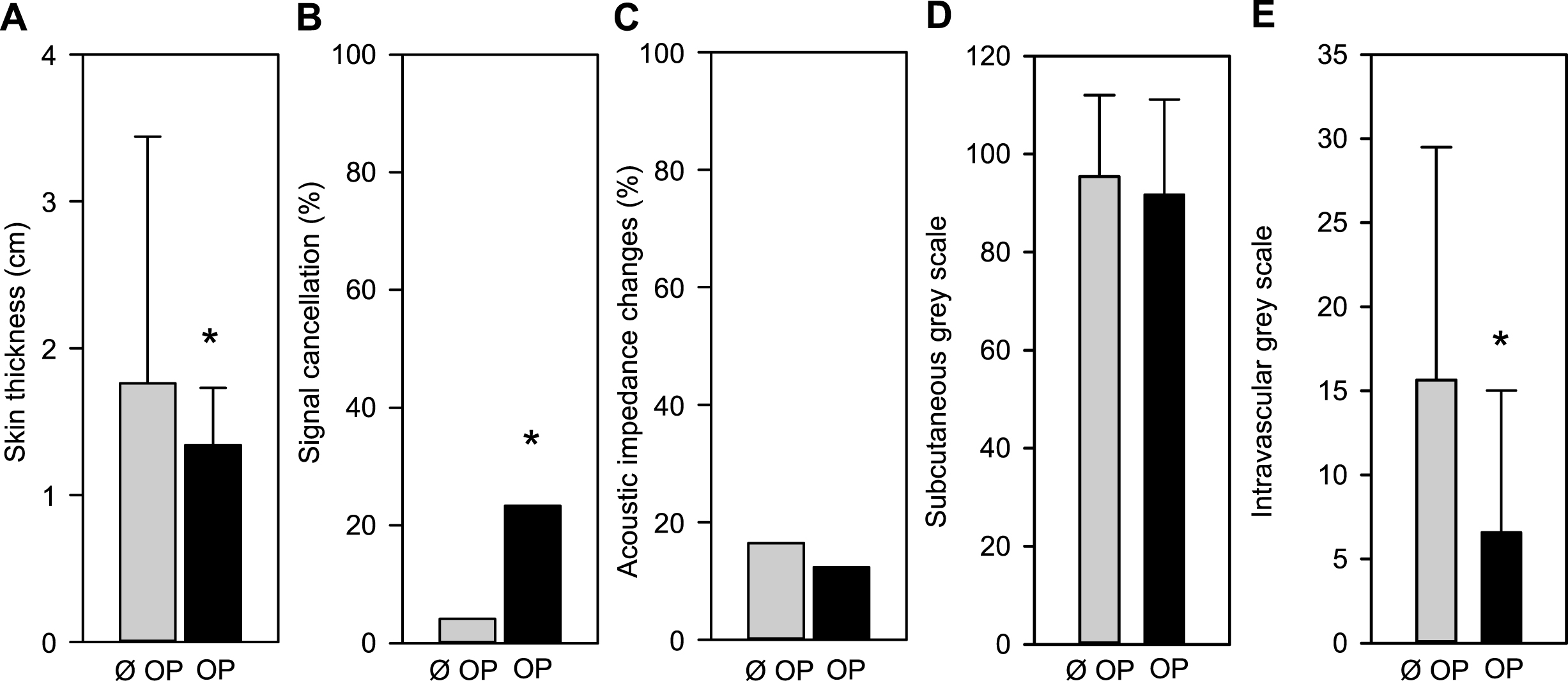
Effects of carotid endarteriectomy (CEA) on skin thickness (A), the occurrence of signal loss (B) and on acoustic impedance changes (C). Impedance was further quantified by subcutaneous (D) and intravascular (E) grey scale analysis. Ø OP - non-operated and OP - operated side of the neck. Values are given as mean±SD. *p < 0.05 vs. non-operated side of the neck.
A comparison of EEA and TEA regarding the assessed parameters above revealed no significant differences (Fig. 2). Additionally, neither the application of a fibrin-coated sealant nor the chosen patch material revealed any significant effect regarding skin thickness (p = 0.2838), appearance of signal loss (p = 0.1935) or impedance changes (p = 0.526) as well as subcutaneous and intravascular grey scale levels (p = 0.4266 and p = 0.3043 respectively). Detailed information is given in Table 3.
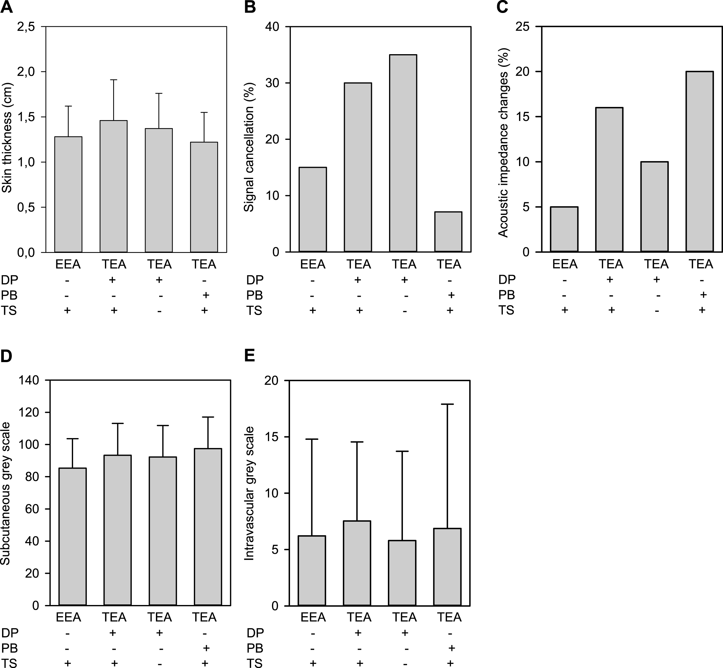
Quantitative assessment of skin thickness (A), the occurrence of signal loss (B) and on acoustic impedance changes (C) as well as subcutaneous (D) and intravascular (E) grey scale analysis after CEA. The parameters were evaluated depending on the operation technique EEA or TEA. Thereby TEA was performed either with a Dacron (DP) or bovine pericardial patch (BP), with or without TachoSil® (TS) respectively. Values are given as mean±SD.
Comparison of different operation techniques, patch materials and the use of TachoSil® regarding duplex ultrasound quality in 75 patients (55 TEAs and 20 EEAs)
Values are means±SD. Statistical analysis of skin thickness (p = 0.2838), incidence of signal losses (p = 0.1935), acoustic impedance changes (p = 0.5260), subcutaneous grey scale (p = 0.4266) and intravascular grey scale (p = 0.3043) revealed no significant differences between the tested subgroups.
The incidence of both un-operated ICA disease progression to high-grade lesions and number of recurrent stenoses after CEA found in our study was within the reported range of 4 to 22% [6]. Due to these events DUS is recommended for both pre- and postoperative as well as postinterventional routine as a non-invasive surveillance [7, 12]. DUS is safe, allows simultaneous acquisition of morphologic and hemodynamic informations with high spatial and temporal resolution and has an appropriate accuracy detecting vessel wall characteristics and abnormalities like hemodynamic relevant stenosis of the carotid artery. However, the significance of DUS depends on the experience of the performer, why all examinations within this study were performed just by one well-experienced angiologist.
The ultrasound signal can be attenuated or extinguished by calcifications or scar formations. It is known that glutaraldehyde, used to fix bovine pericardial patches that might be applied during TEA, induces a perivascular calcification [9]. In contrast to bovine pericardium, Dacron patches are not stored in glutaraldehyde. However, the minimal time span between CEA and this study was three years and sixteen days. After this time, wound healing can be assumed to be complete and no further changes at the operation site could be expected. The range of timespans from CEA to DUS surveillance within the study is explained by modifications in surgical techniques over the years. For example, since 2007 we used TachoSil® in an increasing rate as a local haemostatic sealant positioned around the carotid arteries to avoid postoperative bleeding in the suture line. However, within our own patients a clear effect could not been shown [13]. Since 2010 the use of bovine pericardial patches rapidly increased in our department while Dacron patches were barely applied during TEA.
With respect to individual anatomical characteristics that might affect ultrasound quality, the BMI as a value for obesity, the gender distribution and the neck length and perimeter were assessed showing no significant differences between the subgroups. Thus, it can be resumed that further findings by DUS are independent of these characteristics. Comparison of the non-operated and the operated side of the neck revealed a general effect of CEA regarding the quality of DUS. Since skin thickness and both subcutaneous and intravascular grey scale levels were reduced on the operated side of the neck it can be assumed that tissue remodelling and scar formation induced by surgery affect DUS. This is further supported by the significantly increased occurrence of areas with signal losses on the operated side of the neck.
The haemostatic sealant TachoSil® includes a collagen sponge coated with human fibrinogen, thrombin and riboflavin on one side. The carrier of the substances is equine collagen, which is free of immunogenic epitopes [14]. It is routinely used in microneurosurgery [15] as well as in various other procedures such as lung, kidney, spleen, liver and cardiovascular surgery [14, 16–20]. TachoSil® is known to induce fibrovascular matured granulation tissue after twelve days in its surrounding [10]. Therefore, it was reasonable that the choice of the patch material or the application of local haemostatic sealants might affect the postoperative quality and therefore the significance of DUS. The analysis of grey scale values and acoustic impedance was performed to prove tissue remodelling and formation of scar tissue. Differences were expectable due to calcification or fibrotic remodelling at the surgical site.
Surprisingly comparing the effect of the different surgical techniques assessed in this study, it was seen that local application of TachoSil® did not alter the ultrasound signal comparing patients after TEA with and without the local haemostatic sealant. Since no significant difference was found, it was further analysed whether the surgical techniques of EEA and TEA, each performed applying TachoSil®, affect the quality of DUS. This comparison showed no differences between the surgical techniques were as well. However, neither the use of glutaraldehyde fixed bovine pericardial patches altered DUS quality compared to non-glutaraldehyde fixed Dacron patches. Thus it can be assumed that the glutaraldehyde-induced perivascular calcification, although not directly assessed in this study, does not affect ultrasound quality following CEA.
However, the only statistically significant differences were found between the operated and the non-operated side of the neck regarding the distance between the surface of the skin and the carotid bifurcation due to physiologic scar formation, areas showing a loss of signal as well grey scale levels in the subcutaneous tissue and intravascular.
Thus, it can be summarized that the CEA per se affects the quality of DUS and therefore might make postoperative ultrasound surveillance more difficult. However, neither the use of different patches in TEA, different surgical techniques nor the application of a fibrin-coated sealant like TachoSil® compromises the quality and significance of DUS after CEA. Therefore, the presented data reveal that the compared techniques can be assumed equal and do not interfere with postoperative ultrasound surveillance.
Declaration of interest
The authors report no declarations of interest.
Footnotes
Acknowledgments
The authors like to thank all patients for their participation in this study.