






Abstract
Drag-reducing polymers (DRPs) was previously demonstrated to increase blood flow, tissue perfusion, and reduce vascular resistance. The purpose of this study was to investigate the effect of DRPs on pulmonary vascular remodeling and right ventricular dysfunction in a rat model of chronic hypoxia-induced pulmonary hypertension (HPH). A total of forty male Wistar rats were randomly and equally assigned into four experimental groups (Group I: normoxia + saline, Group II: normoxia + PEO, Group III: hypoxia + saline, Group IV: hypoxia + PEO) and maintained in normoxia (21% O2) or hypobaric hypoxia (10% O2). After four weeks, comparisons were made of the following aspects: the mean pulmonary arterial pressure (mPAP), right ventricular systolic pressure (RVSP), right ventricular hypertrophy, wall thickness of pulmonary trunk and arteries, internal diameter of pulmonary arteries, cardiomyocyte cross-sectional area (CM CSA), and ultrastructure of right ventricular. Treatment with PEO in Group IV attenuated the increases in RVSP and mPAP (40.5±7.2 and 34.7±7.0 mmHg, respectively, both P < 0.05), compared with Group III. Distal vascular remodeling was visible as a significant increase in medial wall thickness (64.2±12.3% vs. 43.95±7.0%, P < 0.01) and a remarkable decrease in internal diameter of small pulmonary arteries (35.2±9.7μ m vs. 50.4±14.7μ m, P < 0.01) in Group III, to a greater extent than that detected in Group IV. Nevertheless, no significant histopathological differences in medial wall thickness was observed in pulmonary trunk between Group III and Group IV (P > 0.05), denoting that PEO chiefly attenuated the remodeling of small pulmonary arteries rather than main arteries in hypoxic environment. Infusion of DRPs (intravenous injection twice weekly) also attenuated the index of right ventricular hypertrophy, protected against the increase of cardiomyocyte cross-sectional area, and provided protection for cardiac ultrastructure. DRP treatment with intravenous injection elicited a protective effect against pulmonary vascular remodeling and right ventricular dysfunction in the rat model of HPH. DRPs may offer a new potential approach for the treatment of HPH, which may have theoretical significance and application value to society.
Keywords
Introduction
Hypoxic pulmonary hypertension (HPH) represents a complex pathophysiological disorder with high mortality rates that may involve a variety of clinical conditions and can complicate the majority of respiratory and cardiovascular diseases [1, 2]. According to ESC/ERS Guidelines, the specific drug therapies of pulmonary hypertension contain calcium channel blockers, endothelin receptor antagonists, phosphodiesterase type 5 inhibitors, prostacyclin analogues and prostacyclin receptor agonists [3]. Nevertheless, clinical trials of current therapies have failed to demonstrate a significant reduction in mortality and were associated with a series of side effects, underscoring the need for development of novel therapies and therapeutic strategies for HPH [4, 5].
While acute hypoxia gives rise to pulmonary vasoconstriction, chronic hypoxia leads to pulmonary vascular remodeling including medial hypertrophy and intimal aberrant proliferation [6, 7]. HPH is characterized by an abnormal increase in the mean pulmonary artery pressure (mPAP), elevated right ventricular (RV) dysfunction and remodeling, that ultimately results in life-threatening RV failure [8]. Therefore, reducing pulmonary artery resistance and attenuating RV remodeling are the key points to prevent and treat HPH. Drag-reducing polymers (DRPs) are long-chain, soluble macromolecules that can increase blood flow and reduce hydrodynamic resistance in turbulent flow, a phenomenon known as the Toms effect [9]. DRPs are well-known as industrial drag reducer in petroleum, firefighting, irrigation, navigation, and other industrial pipelines [10, 11]. There are many similarities between blood circulation system and industrial pipeline. Therefore, the application of drag reducer in circulation system can refer to the theory of “industrial anti-drag”, which is a potential approach to the treatment of coronary heart disease, atherosclerosis, hypertension, hemorrhagic shock, breast cancer and other medical diseases [12–18].
Based on our previous researches, we chose Polyethylene oxide (PEO) as the DRPs reagent for the current study [12, 13]. Our previous study in monocrotaline-induced pulmonary hypertension has demonstrated that PEO can significantly increase the internal diameter of small pulmonary arteries, reduce medial wall thickness, consequently leading to the decline of pulmonary artery pressure [13]. Unfortunately, the effect and the mechanism of DRPs on large pulmonary arteries and right ventricular remains poorly understood. The study was thus designed to mainly investigate the effect of DRPs on pulmonary vascular remodeling and RV dysfunction in a rat model of chronic hypoxia-induced pulmonary hypertension.
Materials and methods
Preparation of DRPs
Polyethylene oxide with an average molecular weight (MV) of 5×10⁶ Da (Sigma-Aldrich Co., St Louis, MO, USA) was chosen to be the DRP in current study. PEO was carefully dissolved in normal saline with the magnetic stirrer at a constant speed (60r/min) until the concentration reaches 1×10⁻³ g/ml, and then dialyzed against saline for 24 h using a membrane (Regenerated Cellulose dialysis membrane, Spectra; Spectrum Laboratories Inc., NJ, USA) with 50kDa MW cutoff. Bacteria and impurities were removed by the 0.2μm sterilizing filter. After dialysis, the PEO solution was diluted to 5×10⁻⁵ g/ml and stored at 4°C.
Animal model and drug administration
All experimental procedures were performed in strict accordance with the NIH “Guide for the Care and Use of Laboratory Animals” and the animal housing and experimental protocol (SYXK2017-0044) is approved by Yangzhou University Institutional Animal Care and Use Committee. Forty male Wistar rats (230–260 g) were obtained from the Experimental Animal Center of Yangzhou University. Rats were randomly assigned and equally into four experimental groups (Group I: normoxia + saline, Group II: normoxia + PEO, Group III: hypoxia + saline, Group IV: hypoxia + PEO) and maintained in normoxia (21% O2) or hypobaric hypoxia (10% O2) for four weeks. Rats were fed in a commercial chamber (BioSpherix, USA), in which the oxygen concentration can be adjusted dynamically by a feedback-control system of O2 sensor. All the rats were housed under an artificial 12 h∼12 h light cycle at a constant temperature of 22±2C and a relative humidity of 65%. PEO and Saline were administered by dorsal penile vein injection twice weekly according to our previous study [13].
Hemodynamic measurements
All animals survived until study completion.The rats were anesthetized with intraperitoneal injection of pentobarbital sodium (45 mg/kg). A polyethylene-50 catheter was inserted via the right jugular vein into the right ventricle to measure the right ventricular systolic pressure (RVSP), which was connected to the physiological signal acquisition system (Power Lab, Australia). The catheter was then placed to the pulmonary artery, and the mean pulmonary arterial pressure (mPAP) was calculated.
Histological analysis of pulmonary arteries
The right pulmonary trunk and arteries were taken and prepared into 5μm thick sections and stained with Hematoxylin-eosin (HE) and Masson trichrome to observe the pulmonary vascular morphology. Percentage of medial wall thickness (% WT) was used to evaluate remodeling of small pulmonary arteries with diameter less than 100μm according to the formula:% WT = [(external diameter-internal diameter)/external diameter]×100%.We calculated internal diameter of small pulmonary arteries according to the formula:(D1 + D2)/2, where D1 and D2 were the shortest diameter and the longest diameter of the artery. Medial wall thickness (WT) of pulmonary trunk was quantified using ImageJ software by measuring the mean thickness (minimum of 5 rats/group, minimum of 10 slides/rat).
Measurement of right ventricular hypertrophy
After all hemodynamic measurements had been performed, the hearts of rats were isolated and weighed. The right ventricle (RV) was dissected from the left ventricle plus septum (LV + S), and the ratio of RV to (LV + S) was calculated as the index of right ventricular hypertrophy.
Histopathological staining of right ventricle
The RV tissues were fixed with buffered 4% paraformaldehyde, embedded in paraffin, sectioned, and HE staining was performed as described previously. Cardiomyocyte cross-sectional area (CM CSA) of the RV (minimum of 5 rats/group, minimum of 60 cardiomyocytes/rat) was measured with Image Pro-Plus software (version 6.0; Media Cybernetics, Rockville, MD, USA).
Ultrastructure of right ventricle under electron microscope
Right ventricle was fixed in 2.5% glutaraldehyde and 1% osmic acid, dehydrated in ethanol and acetone, embedded in epoxy resin, then processed into 50 nm thick sections and stained with uranyl acetate and lead citrate. Finally, the morphological changes of right ventricle were observed and photographed under electron microscopy.
Statistical analysis
All values were presented as mean±SD. All statistical computations were processed using SPSS 24.0 software package (Chicago, IL, USA). The statistical differences among treatment groups were evaluated by one-way ANOVA, followed by Tukey test for multiple comparisons. A value of P < 0.05 was accepted as an indication of statistical significance.
Results
PEO improves hemodynamic parameters
Firstly, we investigated the therapeutic effect of PEO on the hemodynamics of HPH. Compared to group I, hypoxia significantly increased the RVSP (56.6±9.8 mmHg vs 25.4±5.0 mmHg) and the mPAP (42.3±9.3 mmHg vs 19.4±5.4 mmHg) respectively (all P < 0.01) in group III, indicating successful HPH rat model. The RVSP and mPAP, major features of HPH, were substantially reduced in rats of group IV administrated with PEO compared to rats of group III treated with saline (Fig. 1, P < 0.05, respectively).
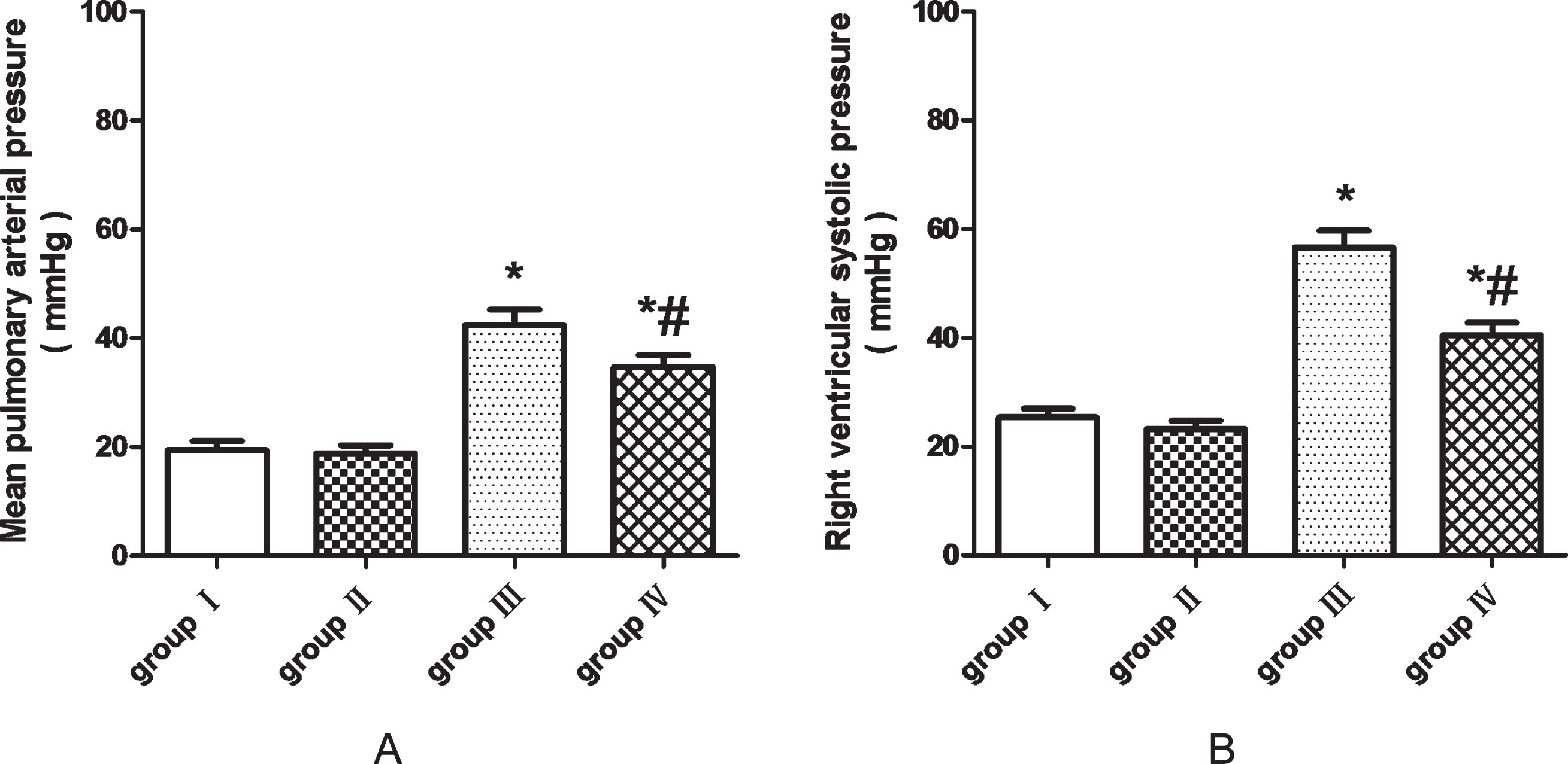
Effects of PEO on hemodynamic parameters in the rat model of chronic hypoxia-induced Pulmonary hypertension (n = 10 in each group). A polyethylene-50 catheter was inserted via the right jugular vein into the right ventricle to measure the RVSP, which was connected to the physiological signal acquisition system. The catheter was then placed to the pulmonary artery, and the mPAP was calculated. Therapeutic application of PEO in Group IV significantly attenuated the RVSP and mPAP (40.5±7.2 and 34.7±7.0 mmHg, respectively, both P < 0.05), in comparison with Group III. Data are presented as the mean±SD. *P < 0.01 compared with group I, #P < 0.05 compared with group III.
As illustrated in Fig. 2, distal vascular remodeling was visible as a significant increase in percentage of medial wall thickness (64.2±12.3% vs. 43.95±7.0%, P < 0.01) and a remarkable decrease in internal diameter of small pulmonary arteries (35.2±9.7μ m vs. 50.4±14.7μ m, P < 0.01) in Group III, to a greater extent than that detected in Group IV. After exposure to hypoxia, the medial and adventitial layers of the pulmonary trunk thickened in Group III and IV, compared with Group I and II. However, no significant histopathological differences were observed in medial wall thickness of pulmonary trunk between Group III and Group IV (Fig. 3, both P > 0.05). These results suggest that PEO chiefly attenuated the remodeling of small pulmonary arteries rather than main arteries in hypoxic environment.
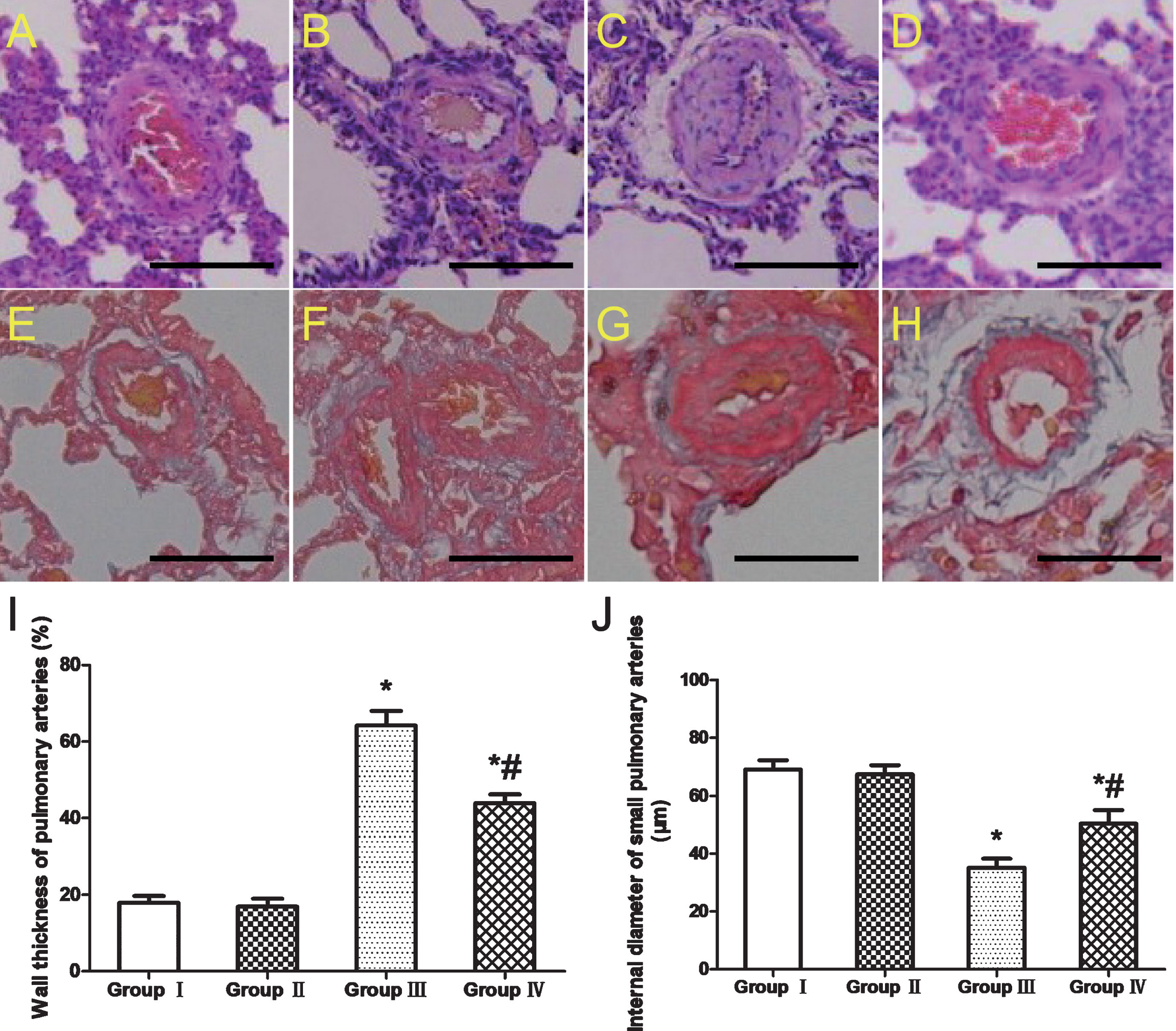
Morphometry of small pulmonary arteries with diameter less than 100μm. (A) Group I, HE staining; (B) Group II, HE staining; (C) Group III, HE staining;(D) Group IV, HE staining;(E) Group I, Masson trichrome staining; (F) Group II, Masson trichrome staining; (G) Group III, Masson trichrome staining; (H) Group IV, Masson trichrome staining; (I) Percentage of medial wall thickness (% WT). (J) Comparison of internal diameter of small pulmonary arteries. Distal vascular remodeling was visible as a significant increase in medial wall thickness and a remarkable decrease in internal diameter of small pulmonary arteries in Group III, to a greater extent than that detected in Group IV (P < 0.01). Data are mean±SD. Magnification×400; scale bar = 50μm.
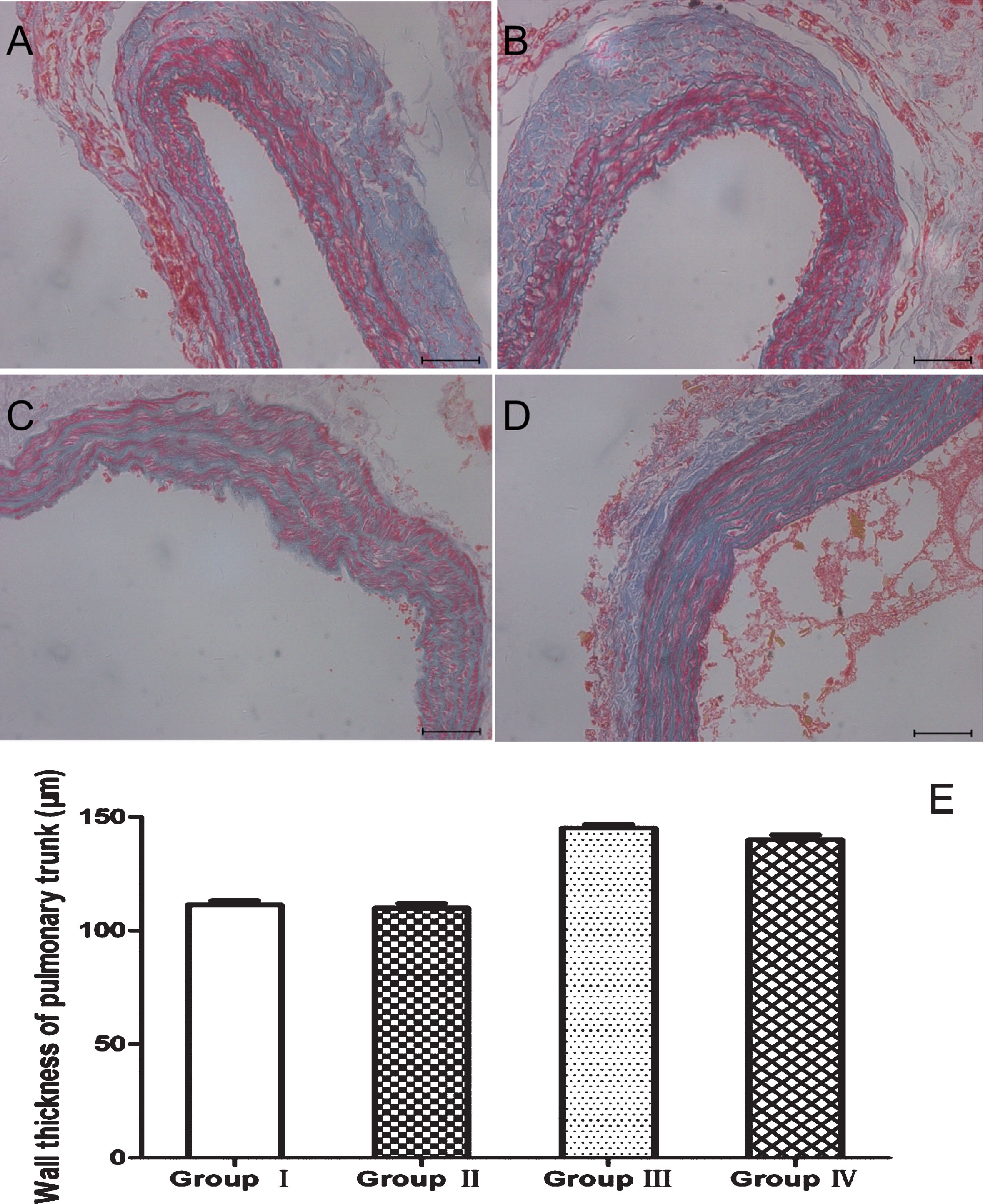
Morphometry of pulmonary trunk. (A) Group I, Masson trichrome staining; (B) Group II, Masson trichrome staining; (C) Group III, Masson trichrome staining; (D) Group IV, Masson trichrome staining; (E) Medial wall thickness of pulmonary trunk (WT). After exposure to hypoxia, the medial and adventitial layers of the pulmonary trunk thickened in Group III and IV, compared with Group I and II. However, no significant histopathological differences were observed in medial wall thickness of pulmonary trunk between Group III and Group IV (P > 0.05). Data are mean±SD. Magnification×200; scale bar = 100 μm.
To identify the role of PEO in pathological myocardial hypertrophy, Wistar rats underwent hypoxic exposure to induce pathological cardiac hypertrophy or normoxic exposure as a negative control. As illustrated in Fig. 4, the rats in Group III demonstrated a remarkable increase in right ventricle and index of right ventricular hypertrophy (P < 0.01), compared with Group I. Moreover, we observed that the index of right ventricular hypertrophy was significantly inhibited by administration of PEO in Group IV, compared to Group III (P < 0.05). The weight of (LV + S) and total heart weight make no statistically significant difference among the experimental groups. Our results demonstrated that PEO protects against cardiac hypertrophy induced by hypoxia.
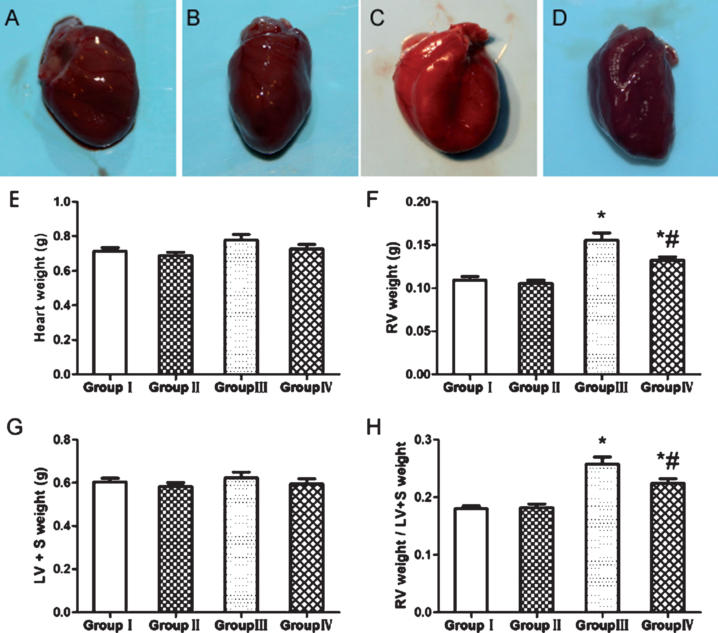
PEO mitigates right ventricular hypertrophy in a hypoxia-induced PH rat model. (A)Group I.(B)Group II. (C)Group III. (D)Group IV. (E) Heart weight. (F) Right ventricular weight. (G)Left ventricle plus septum (LV + S) weight. (H) The index of right ventricular hypertrophy. Group III presented significant increase in right ventricular weight (P < 0.01) and the index of right ventricular hypertrophy (P < 0.01). Conversely, Group IV revealed suppression of the increases in right ventricular weight and the index of right ventricular hypertrophy (P < 0.05) by administration of PEO, compared with Group III. The ratio of RV to (LV + S) was calculated as the index of right ventricular hypertrophy. *P < 0.01 compared with group I, #P < 0.05 compared with group III.
Furthermore, the morphological features of remodeling in right ventricular were identified via H&E staining (Fig. 5). What captured our attention is that cardiomyocyte cross-sectional area is significantly increased in accordance with the presence of right ventricular hypertrophy induced by hypoxia in Group III. However, the effect was obviously attenuated in Group IV treated with PEO. No such apparent changes were detected in the RV cardiomyocytes in normoxia whether injected with PEO or saline.
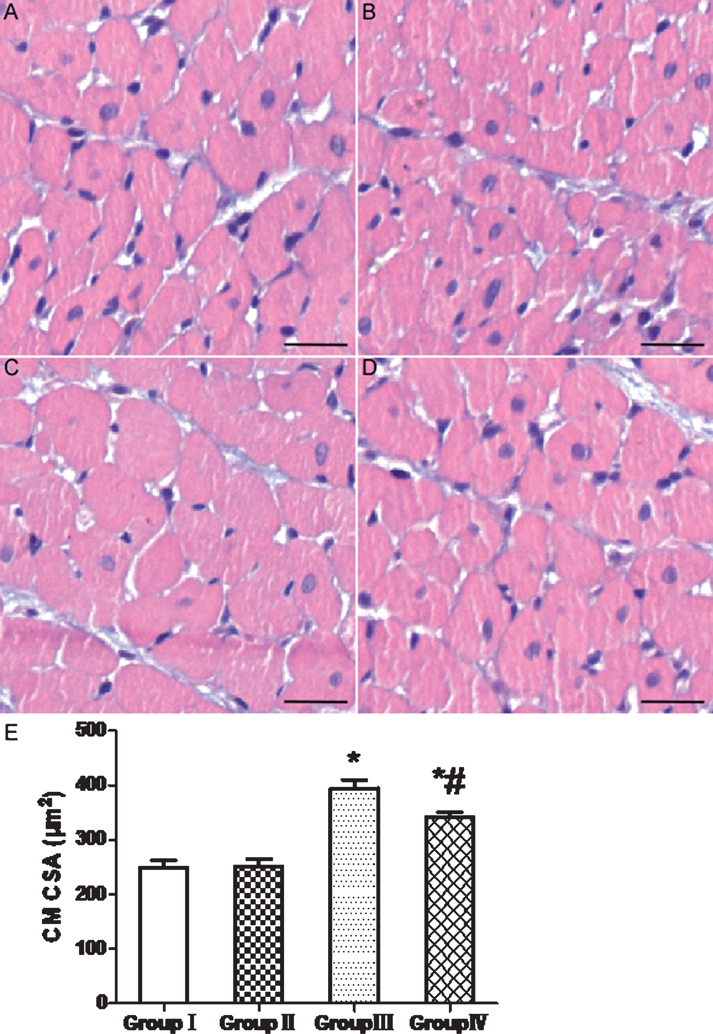
Pathology changes of right ventricular triggered by hypoxia. (A)Group I.(B)Group II. (C)Group III. (D)Group IV. (E) RV cardiomyocyte cross-sectional area (CM CSA) analysis. Cardiomyocyte cross-sectional area is significantly increased in accordance with the presence of right ventricular hypertrophy in Group III. However, the effect was obviously attenuated in Group IV treated with PEO. Scale bar = 50μm; *P < 0.01 compared with group I, #P < 0.05 compared with group III.
An examination was carried out by electron microscope to assess the favorable effects of PEO on ultrastructural architecture of right ventricular (Fig. 6). Integral sarcolemma, well-arranged myofilaments, clear dark and light bands, and normal mitochondrial structure were observed in the control group. As demonstrated in Group III, hypoxia results in a host of changes as follows: incomplete sarcolemma, irregular myofilaments, sparse cytoplasm with numerous vacuoles, damaged mitochondria, and scattered autophagosomes. Ruptured mitochondria appear degenerated with cristae disorganization, apparent swelling in their size, and multiple vacuolization. Compared with Group III, PEO elicits its higher protective effect on cardiac ultrastructure in Group IV, with less disruption of mitochondria, fewer cytoplasmic vacuoles, largely intact sarcomere.
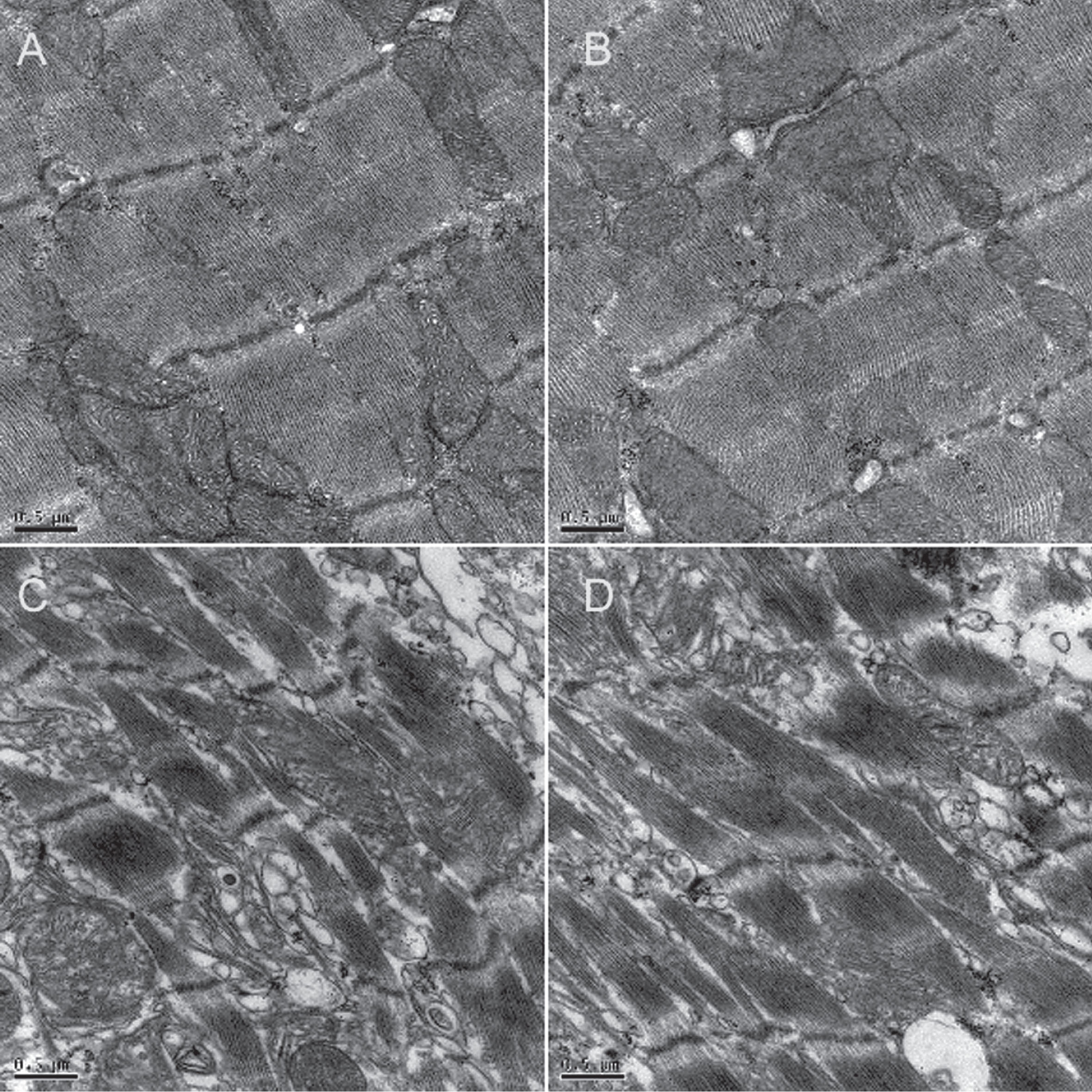
Effect of PEO on hypoxic pulmonary hypertension at ultrastructure level (Magnification×20000). (A) Group I. (B) Group II. (C) Group III. (D) Group IV. The ultrathin sections of right ventricle tissues were observed under a Japanese JEM1230 transmission electron microscopy. Irregular myofilaments, damaged mitochondria, and scattered autophagosomes were detected in Group III. Compared with Group III, PEO elicits its higher protective effect on cardiac ultrastructure in Group IV, with less disruption of mitochondria, fewer cytoplasmic vacuoles, largely intact sarcomere.
As demonstrated in the current study, infusion of PEO regularly in Group IV significantly mitigated the increase in the mean pulmonary arterial pressure (mPAP) and the right ventricle systolic pressure (RVSP), compared to Group III. The remarkable improvements of DRPs on the hemodynamic effects could be responsible for suppression of HPH and RV remodeling. The underlying mechanisms by which DRPs improves circulation and modulates hemodynamics are still puzzling, and may be related to the following aspects:(1) The effect of DRPs on blood flow is associated with altered shear stress [19]. Blood shear stress can act on signaling pathways of vascular endothelial cells, regulating the expression of a series of cytoactive factors and various adhesion molecules [20–23]. (2) Recently, a redistribution in RBCs (e.g., an increase in the number of RBCs in a branch or reduction in plasma skimming) was observed, thus facilitating the gas exchange between blood and tissue [14, 16].
Interestingly, distal vascular remodeling was visible as a significant difference in medial wall thickness and internal diameter of small pulmonary arteries, whereas no significant histopathological dissimilarity was observed in pulmonary trunk between Group III and Group IV. To the best of our knowledge, Bragin DE reported that DRP-RF restored collapsed capillary flow, improved impaired microvascular cerebral blood flow, reduced hypoxia and protected neurons [14, 16]. These findings may contribute to provide novel insight into our further research of DRPs on microvascular mechanisms, instead of main channel. To shed light on the microvascular mechanism, Judith Brands et al. have demonstrated that DRPs decreased plasma skimming, enhanced arteriolar side branch hematocrit, therefore leading more RBCs flux into the microvasculature [24]. This redistribution of RBCs may explain our current observation of a DRPs-mediated enhancement of microvascular blood flow, heralding an improvement in oxygen transportation ability of microvascular perfusion by DRPs.
It is noteworthy that hypoxia exacerbates right ventricular hypertrophy that is associated with a series of cardiovascular events that included heart failure, ventricular tachyarrhythmia, and sudden cardiac death [25, 26]. Infusion of PEO was detected to protect against cardiac hypertrophy induced by hypoxia, but the precise mechanisms are still unclear. Improving tissue oxygen supply may be one of the possible mechanisms of PEO to inhibit right ventricular hypertrophy. As demonstrated in one original discovery, DRPs reduce the resistance of red blood cells through microcirculation, thereby increasing the supply of oxygen to local tissues [14, 24]. Simultaneously, DRPs can enhance the deformability of RBCs, change RBCs into oval shape to increase their plasticity, and divert more RBCs into the capillary bed, therefore facilitating the release of oxygen to the tissues [27].
As a result of hemodynamic destabilization, hypoxia poses a considerable challenge to right ventricular cytoarchitecture [28]. Compared with Group III, PEO displayed its higher protective effect on cardiac ultrastructure in Group IV. In a word, the current study revealed that PEO could mitigate the injury induced by hypoxia by protecting the integrity of myocardial ultrastructure. The changes of mitochondria at ultrastructural level treated with saline or PEO under hypoxic conditions indicated that the mitochondria dependent signal pathway might be involved in the regulatory process. It was reported that shear stress mitigates vascular endothelial dysfunction by promoting mitochondrial biogenesis, while the increase in blood viscoelasticity caused by DRP may strengthen non-Newtonian patterns of the axial velocity profile [19, 29].
However, our current study has several limitations that should be taken into consideration. In the first place, limited by the time and cost, the study failed to further monitor the steady-state blood drug concentration during intravenous infusion in the treatment group, as it is associated with the therapeutic effects of DRPs. Secondly, in the future, the experiment groups should be further refined according to the drug concentration in order to find the optimal drug concentration and the speed of drug administration, because it was reported that the DRPs efficacy was concentration-dependent [24]. Additionally, the underlying in-depth mechanisms and signaling pathways by which DRPs improves circulation and modulates hemodynamics are still puzzling [30]. Moreover, because of the limitations of current technology, we are unable to observe the changes of shear stress caused by the application of DRPs in the hypoxia-induced HP rats, as that is what we are interested in for future research. The next step is to further explore its mechanisms through in vitro researches by Bioflux system, which offers a unique solution to bridge the gap between in vitro and in vivo assays by combining the ease of use and throughput of a well plate with the data quality of a shear flow assay [31–33].
Conclusion
The study revealed that treatment with DRPs elicited a protective effect against right ventricular dysfunction and remodeling in the rat model of hypoxia-induced pulmonary hypertension. DRPs may offer a new potential approach for the treatment of HPH, but further study is called for to explore its in-depth mechanisms through in vitro researches.
Disclosure
The authors confirm that there are no conflicts of interest.
Footnotes
Acknowledgments
The study was partly supported by the National Natural Science Foundation of China (81800050), and Natural science fund of Yangzhou City (YZ2017119), and Science and Technology Innovation Cultivation Program of Yangzhou University (2017CXJ122).