






Abstract
BACKGROUND:
Elevated blood viscosity has been reported as a risk factor for cerebrovascular disease.
OBJECTIVE:
The relationship between blood viscosity and outcomes of mechanical thrombectomy (MT) for large artery occlusion (LAO) were investigated in the present study.
METHODS:
A total of 238 patients were enrolled and systolic blood viscosity (SBV) and diastolic blood viscosity (DBV) were measured using the scanning capillary tube viscometer. Receiver operating characteristic (ROC) analysis was performed to specify the association of viscosity with the first-pass reperfusion (FPR). Multivariable and regression analyses were performed to evaluate the relationship of viscosity with FPR and various variables.
RESULTS:
Based on ROC analysis, the best DBV cutoff value was 10.55 (cP). In multivariable analysis, high DBV was associated with FPR failure (odds ratio 2.82, 95% confidence interval 1.64–4.22; p = 0.001). Increased DVB could be associated with elevated SBV, hematocrit level, and blood urea nitrogen/creatinine ratio (p = <0.001, 0.004, and 0.002, respectively).
CONCLUSIONS:
Elevated DBV was associated with FPR failure. Patients with high DBV had longer thrombus length and required more stent passages than patients with low DBV.
Introduction
Blood viscosity plays a major role in atherosclerosis and has been demonstrated as a risk factor or predictor of cardiovascular and neurovascular disease [1–10]. Blood viscosity is determined by various hemorheological components, such as fibrinogen, white blood cells, red blood cells, platelets, and may be affected by plasma concentration, erythrocyte permeability and aggregation, and dehydration [6, 11–14]. Elevated blood viscosity affects the deterioration of tissue perfusion with an increase of shear stress and inflammatory reaction to blood vessels [3]. Shear rate is an important factor regulating blood viscosity as a non-Newtonian fluid, and blood viscosity measured at a high shear rate ≥300 s–1 is defined as systolic blood viscosity (SBV) [3, 15–17]. In addition, the role of diastolic blood viscosity (DBV), which is the blood viscosity measured at a low shear rate ≤5 s–1, is emphasized more than SBV as a risk factor for cardiovascular disease [18–21].
In previous studies, the association of blood viscosity with stroke was reported [4–7, 22–26]. Similar to coronary artery disease, elevated DBV may play an important role in the development of neurovascular disease as well as acute and chronic lacunar manifestations of cerebral small vessel disease [24]. Blood viscosity level in patients with small artery occlusion was higher than in other stroke subtypes with large artery atherosclerosis or cardioembolism [6, 7]. Flow resistance may be proportional to blood viscosity and vessel length and inversely proportional to vessel diameter, thus, flow resistance is markedly increased in narrow vessels such as stenotic lesions or small perforating arteries [6, 27]. This is only a hypothesis and whether blood viscosity affects large arteries other than small vessels has not been proven. In addition, there are few papers analyzed the association of blood viscosity and results of mechanical thrombectomy (MT) which is a standard treatment for large vessel occlusion (LAO) with acute ischemic stroke (AIS) [28].
Based on previous studies, the authors hypothesized that blood viscosity may affect the results of MT for LAO. Thus, in the present study, the association of blood viscosity with outcomes of MT for LAO was analyzed and factors that affect the blood viscosity were identified.
Materials and methods
Patients and mechanical thrombectomy
This study was approved by the local Institutional Review Board (IRB No. VC17RESI0048) of each participating center. Data were retrospectively reviewed from each institution’s database of consecutive patients who underwent MT for LAO between January 2016 and December 2019, and a total of 238 patients were enrolled. In all patients, AIS and LAO were confirmed using diffusion-weighted magnetic resonance imaging and computed tomography angiography, respectively. Before the MT, intravenous thrombolysis (IVT) with tissue plasminogen activator (alteplase) was administered within 4.5 hours after stroke onset at a maximum dose of 0.9 mg/kg in accordance with the European Cooperative Acute Stroke Study (ECASS) III trial [29, 30]. MT with stent retriever or combined technique were performed in patients with LAO, including occlusion of the intracranial carotid artery, middle cerebral artery, anterior cerebral artery, or posterior circulation (vertebral artery or basilar artery) [31].
Blood viscosity measurements
The scanning capillary tube viscometer (Hemovister, Pharmode Inc., Seoul, Korea) was used to measure whole blood viscosity including SBV and DBV. Whole blood viscosity was analyzed at high (≥300 s–1) and low (≤5 s–1) shear rates for SBV and DBV, respectively [15, 24]. Whole blood (3 mL) was collected in an ethylenediaminetetraacetic acid (EDTA) tube separately by peripheral venous puncture within 3 hours after admission and stored in a refrigerator at 4°C until measurement. All venous samples were obtained during the initial blood sampling procedure in the emergency room before any medical or interventional treatment was given to reduce the diluting effect of blood viscosity due to hydration [6, 24], and all measurements were performed within 12 hours of blood sampling.
Baseline assessments and laboratory findings
Multivariable factors were reviewed, including age, sex, risk factors, stroke etiology based on TOAST criteria [32], arterial occlusion site, application of IVT, Alberta Stroke Program Early CT Score (ASPECTS), and time intervals affecting viscosity, such as symptom onset to groin puncture time and procedure time (time from groin puncture to reperfusion) [33]. Risk factors included history of hypertension, diabetes mellitus, atrial fibrillation, coronary artery disease, prior stroke or transient ischemic attack (TIA), current smoking status, dyslipidemia, and body mass index (BMI: weight divided by height) ≥25 kg/m2. Laboratory findings which could affect blood viscosity and prognosis of patients were included, such as white blood cells, red blood cells, hemoglobin, hematocrit, platelets, total protein, blood urea nitrogen (BUN), creatinine (Cr), BUN/Cr ratio, fibrinogen, D-dimer, fibrin degradation products, high-sensitivity C-reactive protein, and lipoproteins [23, 34–39].
Clinical and radiological outcomes
All patients underwent a clinical investigation including National Institutes of Health Stroke Scale (NIHSS; range, 0–42, with higher score indicating more severe neurological deficit), the modified Rankin Scale (mRS) score (favorable clinical outcome was defined as mRS score of 0–2 at 3 months), symptomatic intracerebral hemorrhage (any hemorrhage with an increase of ≥4 points on the total NIHSS score after MT) [29], and mortality at 3 months. Regarding radiological outcomes, a successful recanalization was defined as a Thrombolysis in Cerebral Infarction (TICI) grade of 2b or 3, the first-pass reperfusion (FPR) indicates achievement of modified TICI (mTICI 2b or 3) with the first pass of thrombectomy devices [40, 41], and total number of stent passages for successful recanalization were analyzed. The length of the thrombus was measured in the patient’s angiography images and was defined as the length of the lesion artery not contrasted on angiography.
Statistical analyses
All statistical analyses were conducted using statistical software package SPSS 25.0 for Windows (IBM, Armonk, NY, USA). Receiver operating characteristic (ROC) analysis was performed to specify the associations of viscosities (systolic and diastolic) with the FPR and determine the best cutoff values for FPR prediction. All patients were dichotomized based on the first-pass effect (FPR vs. non-FPR) and best cutoff value of diastolic viscosity. Differences between FPR and non-FPR were analyzed using Student’s t-test or Mann-Whitney U test for continuous variables and the χ2 test or Fisher’s exact test for categorical variables. In addition, multivariable analysis with binary logistic regression was performed to determine independent variables for non-FPR and p-values < 0.20 in univariable analysis were entered into a backward multivariable analysis. Multiple linear regression analyses were performed to evaluate the independent association of DBV with other variables. Two-tailed p-values≤0.05 were considered to indicate statistically significant difference.
Results
Association of systolic and diastolic blood viscosity with first-pass reperfusion
In ROC analysis, the best cutoff threshold of SBV was 3.65 (cP) to discriminate between FPR and non-FPR (area under the curve, AUC 0.772, 95% confidence interval, CI 0.710–0.833; p < 0.001, FPR: SBV < 3.65, 49/107 (45.8%) vs. SBV≥3.65, 44/131 (33.6%); p = 0.002). In the same category, 10.55 (cP) was the best cutoff value for FPR (AUC 0.792, 95% CI 0.730–0.853; p < 0.001, FPR: DBV < 10.55, 52/104 (50.0%) vs. DBV≥10.55, 41/134 (30.6%); P < 0.001) (Fig. 1).
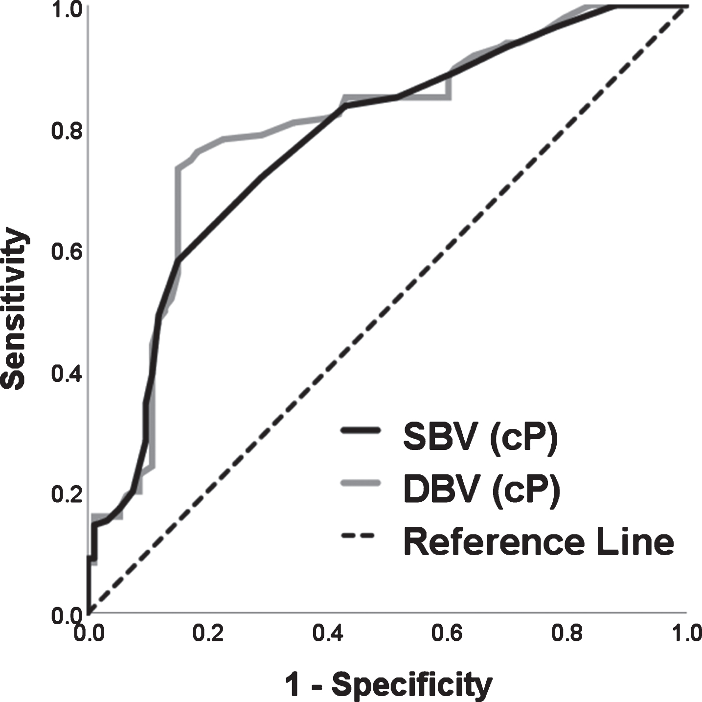
Receiver operating characteristic (ROC) analyses of the association between systolic blood viscosity (SBV) and diastolic blood viscosity (DBV) with first-pass effect were performed to discriminate between first-pass reperfusion (FPR) and non-FPR. The best cutoff values were identified at SBV of 3.65 (cP) and DBV of 10.55 (cP), respectively; area under the curve, (AUC) 0.772, 95% confidence interval, CI 0.710–0.833; p < 0.001, and AUC 0.792, 95% CI 0.730–0.853, p < 0.001, respectively.
Among 238 patients, 93 had FPR (39.1%, mean age 69.6 years, 54.8% male) and the other 145 patients had non-FPR (60.9%, mean age 68.0 years, 56.6% male). Mean SBV and DBV values were significantly higher in non-FPR patients than in FPR patients (non-FPR:FPR, SBV = 4.00 (standard deviation, SD 0.71):3.43 (SD 0.47), DBV = 12.59 (SD 4.43):9.50 (SD 2.21), p = < 0.001 and <0.001, respectively; Fig. 2A and B). However, significant differences in other laboratory findings, risk factors, stroke etiology, and pre-interventional details were not observed between FPR and non-FRP patients. Median procedure time was shorter in FPR patients (25 min, interquartile range, IQR 13–37) than in non-FPR patients (57 min, IQR 29–85) because more stent retrievals were attempted in non-FPR patients (P < 0.001). Non-FPR patients (24.8 mm, SD 9.8) showed significantly longer mean thrombus length than FPR patients (19.3 mm, SD 7.7; p < 0.001; Fig. 2C). The prevalence of successful recanalization and favorable clinical outcome were higher in FPR patients (100% and 68.8%) than in non-FPR patients (93.1% and 53.1%; p = 0.002 and 0.004, respectively; Table 1). Subjects were classified into high (≥10.55 cP) or low (<10.55 cP) DBV patients based on the best cutoff DBV value in ROC analysis; 134 (56.3%) patients had high DBV and 104 patients (43.7%) had low DBV. The mean hemoglobin and hematocrit levels were significantly elevated in patients with high DBV (13.81 g/dL, SD 2.19, and 40.57%, SD 6.00) compared with patients with low DBV (13.19 g/dL, SD 1.96, and 38.91%, SD 5.89; p = 0.023 and 0.033, respectively). Patients with high DBV showed significantly longer median procedure time (51 min, IQR 17–80) and mean thrombus length (25.7 mm, SD 9.4) than patients with low DBV (36 min, IQR 16–61; 18.7 mm, SD 7.9; p = < 0.001 and < 0.001, respectively). Patients with high DBV had higher median number of stent passages (3, IQR 1–5) and lower FPR rate (30.6%) compared with patients with low DBV (1, IQR 1–3 and 50.0%; p = < 0.001 and <0.001, respectively) (Table 2).
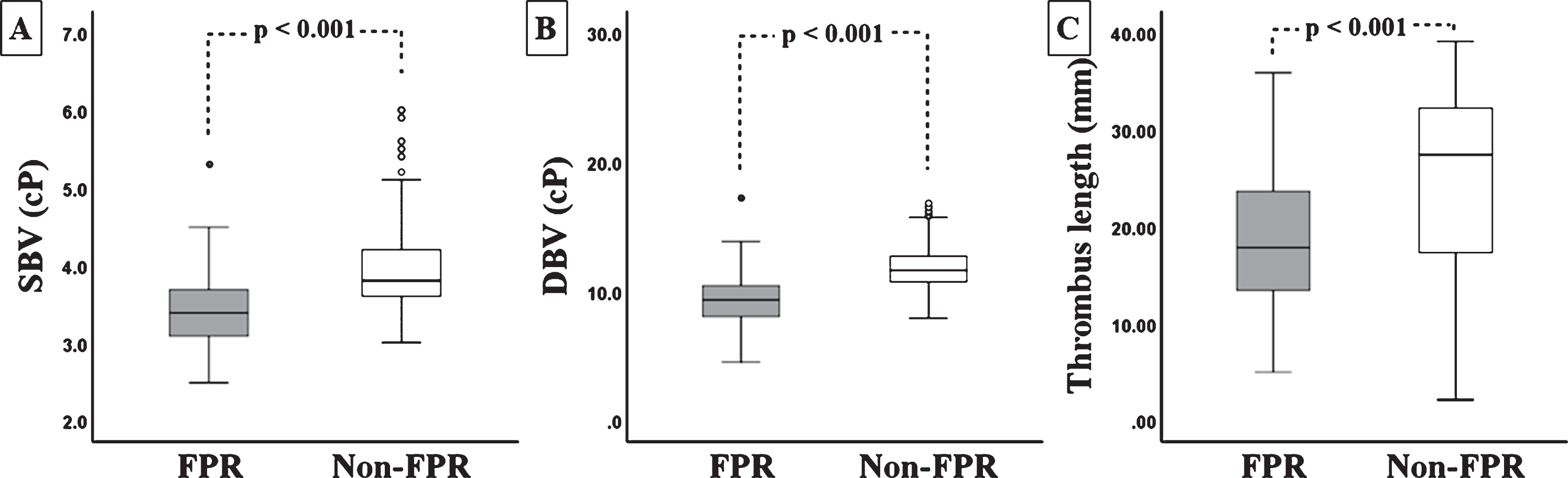
Box plots of systolic blood viscosity (SBV), diastolic blood viscosity (DBA), and thrombus length between first-pass reperfusion (FPR, gray square) and non-FPR (white square). (A) The non-FPR patients had a higher mean SBV level than FPR patients (p < 0.001). (B) The box plots of the non-FPR patients show a significantly elevated DBV compared with FPR patients (p < 0.001). (C) The mean thrombus length was significantly longer in non-FPR patients than in FPR patients (p < 0.001).
Comparisons of baseline characteristics and outcomes in patients with mechanical thrombectomy, according to first-pass reperfusion
*p≤0.05.
Comparisons of demographics in patients with mechanical thrombectomy, according to level of diastolic blood viscosity (DBV)
*p≤0.05.
To identify parameters independently associated with FPR failure, univariable and multivariable binary logistic regression analyses were performed. In univariable analysis, longer thrombus length (≥20 mm), high SBV (≥3.65 cP), and high DBV (≥10.55 cP) were associated with non-FPR. In multivariate analysis, only high DBV (≥10.55 cP) was independently associated with FPR failure (odds ratio, OR 2.82, 95% CI 1.64–4.22; p = 0.001) (Table 3). In simple correlation analysis, DBV was significantly associated with SBV, hematocrit level, and BUN/Cr ratio. Similarly, SBV, hematocrit level, and BUN/Cr ratio had a significant predictive power for increased DBV in multiple regression analysis (p = < 0.001, 0.004, and 0.002, respectively) (Table 4).
Multivariable analysis of parameters associated with failure of first-pass reperfusion (FPR)
Multivariable analysis of parameters associated with failure of first-pass reperfusion (FPR)
*p≤0.05.
Multiple regression analysis for correlation between diastolic blood viscosity and other variables
*p≤0.05.
In the present study, a strong association between elevated DBV and FPR failure was observed in the cohort of patients who underwent MT for LAO. High DBV (≥10.55 cP) values contributed to increased non-FPR. Furthermore, non-FPR patients had higher SBV and DBV levels than FPR patients. Patients with high DBV had a higher median number of stent passages and lower FPR rate than patients with low DBV. The co-related factors for the elevated DBV were increased SBV, hematocrit level, and BUN/Cr ratio.
The association of blood viscosity with the occurrence of stroke was reported in previous studies that included healthy subjects or patients at high risk of ischemic stroke. Song et al. reported that DBV is correlated with lacunar infarct (adjusted OR 1.01, CI 1.00–1.02, p = 0.033) and baseline DBV was significantly higher in patients with small artery occlusion (274.7 mP = 27.47 cP) than in patients with LAO (214.5 mP) or cardio-embolic stroke (252.0 mP; p = 0.037). [24] Grotemeyer et al. showed subjects with small vessel disease had significantly higher mean plasma viscosity values (1.29 mPas) than patients with large vessel disease or cardio-embolic stroke (1.22 mPas; p < 0.001). [7] Li et al. suggested that silent infarct patients had higher DBV (10.34 mPas) and SBV (4.46 mPas) values than patients without silent infarct (8.98 and 4.29 mPas), and increased DBV was associated with risk of silent cerebral infarct (OR 2.025, CI 1.750–2.343, p < 0.001). [42] In the present study, only patients with MT for LAO were evaluated and difference in viscosity based on the difference of stroke subtypes was not observed. Despite the differences in study population and classification criteria, higher DBV (≥10.55 cP) in the present study was an independent factor for inhibition of FPR. Failed FPR patients had higher mean DBV (12.59 cP) and SBV (9.50 cP) values than FPR patients. Furthermore, patients with high DBV (≥10.55 cP) had more prevalence of FPR than patients with low DBV (<10.55 cP).
Increased blood viscosity at low or high shear stress (equivalent DBV or SBV, respectively) may affect the various pathogenesis in cerebral vessels. First, erythrocyte aggregation with deformability could increase blood viscosity, and elevated blood viscosity might decreases the cerebral blood flow [43, 44]. In patients with small vessel disease with lacunar infarction, elevated blood viscosity can aggravate the flow disturbance and trigger endothelial remodeling and luminal occlusion, and DBV may be more responsible than SBV in this process [24, 42]. Similar pathogenesis could be occurred in cases with occlusion of large artery; elevated DBV in LAO may induce more flow stasis and blood cell accumulation thereby increasing the thrombus integration. Increased thrombus length by thrombus integration had no relevant effect on recanalization or neurological outcome. However, it is associated with increased number of stent passages and failure of FPR, and result in increase of the procedure time [45–47]. In the present study, the mean thrombus length in patients with high DBV (25.7 mm) was longer than in patients with low DBV (18.7 mm). In addition, patients with high DBV required more stent passages and longer reperfusion procedure time, resulting in reduced FPR prevalence. The FRR is known as a factor related to good clinical outcome in patients with MT [48]. In contrast, more number of stent passage result in increase of parenchymal hematoma after MT [49]. Therefore, blood viscosity with predictability for FPR can be a useful prognostic factor in patients with MT. Second, inflammatory reactions which are positively correlated with blood viscosity, could induce vascular damage, formation of pathologic erythrocytes, and further contribute to reduced deformability and thrombus formation [50–52]. However, there were no significant differences in inflammatory markers of our study, such as white blood cell count and high-sensitivity C-reactive protein, based on blood viscosity.
The most important determinants of blood viscosity are fibrinogen and hematocrit levels, which play pivotal roles in aggregation of red blood cells and erythrocyte deformability [17, 24]. In the present study, patients with high DBV had higher hemoglobin and hematocrit levels than patients with low DBV, however, fibrinogen levels were not different. Furthermore, in multiple regression analysis, increased SBV, hematocrit level, and BUN/Cr ratio were associated with elevated DBV. Hematocrit level and BUN/Cr ratio are indicative of dehydration and represented by increased blood viscosity [53, 54]. Therefore, dehydration in stroke patients should be avoided because not only poor outcome but also elevated DBV may occur [54]. In addition, lipoproteins were reported to influence plasma or blood viscosity in previous studies, and viscosity correlated with increased low-density lipoprotein/high-density lipoprotein ratio and triglycerides [35, 56]. However, there was no significant the association between lipoproteins and blood viscosity in our study, and further analyses are needed.
The present study had several limitations. First, the retrospective non-blinded analysis of a relatively small sample size without randomization is prone to introduction of bias and can allow errors in data interpretation. Second, plasma viscosity, which influences the whole blood viscosity with tissue perfusion, and is a well-documented as a cardiovascular risk factor [43, 58]. However, our study is lacking in information about plasma viscosity. Third, patients did not fast before venous blood samples were obtained, thus, blood viscosity could be easily affected by hydration therapy or drinking water. Fourth, dynamic changes with repeated measurements of blood viscosity were not included. Other unmeasured and residual confounding data may have contributed to the results. However, every effort was made to adjust for the possibility of false results.
Conclusion
In the present study, elevated blood viscosity on hospital admission was associated with FPR failure in patients who underwent MT for LAO. In particular, DBV may play a role in FPR and elevated DBV could be a predicting factor for FPR failure. In addition, longer thrombus length and requirement of increased number of stent passages for reperfusion were identified in patients with high DBV compared with patients with low DBV. Furthermore, increased SBV, hemoglobin level, and BUN/Cr ratio resulted in elevated DBV. Further investigations are required to verify the results of the present study and identify the underlying mechanisms.
Footnotes
Acknowledgments
This research did not receive any specific grant from funding agencies in the public, commercial, or not-for-profit sectors. All study protocols received full approval from local ethical committee. This study was approved by institutional review board