






Abstract
OBJECTIVE:
Glypican-3 (GPC3) has emerged as a significant marker for the diagnosis and prognosis of hepatocellular carcinoma (HCC) and has garnered considerable attention as an immunotherapeutic target. In this study, we propose a combination of preoperative contrast-enhanced ultrasound (CEUS) imaging features and clinical factors to predict the positive expression of GPC3 in HCC patients.
METHODS:
We retrospectively included 30 cases of GPC3-negative HCC and 115 cases of GPC3-positive HCC patients who underwent conventional ultrasound and CEUS evaluation. We assessed and compared the clinical characteristics, conventional ultrasound features, and CEUS features between the two groups of HCC patients. Based on the clinical and ultrasound features between the two groups, we developed a binary logistic regression model for predicting GPC3-positive HCC.
RESULTS:
A total of 145 HCC patients were included in this study. Binary logistic regression analysis showed that AFP > 20 ng/mL (OR = 4.047; 95% CI: 1.467-11.16; p = 0.007), arterial phase hyperenhancement (APHE) (OR = 12.557; 95% CI: 3.608-43.706; p < 0.001), and asynchronous perfusion (OR = 4.209; 95% CI: 1.206-14.691; p = 0.024) were predictive factors for GPC3-positive HCC. Receiver operating characteristic (ROC) analysis was conducted to predict GPC3-positive expression. The model combining the three independent predictive factors showed good predictive performance (AUC 0.817, 95% CI: 0.731-0.902, sensitivity: 91.3%, specificity: 60.0%). This combined model demonstrated excellent discriminatory ability to predict GPC3-positive HCC.
CONCLUSION:
Preoperative integration of CEUS features and clinical factors can non-invasively and effectively identify GPC3-positive HCC patients, providing valuable assistance in making personalized treatment decisions.
Introduction
According to the latest global cancer survey, liver cancer is ranked fifth and seventh in terms of mortality rates among males and females, respectively. Despite recent stabilization, liver cancer remains a significant global health burden [1]. Hepatocellular carcinoma (HCC) is the most common type of primary liver cancer, which is characterized by a hidden onset, high recurrence and metastasis rates, and overall suboptimal survival [2]. Failure to regularly monitor high-risk HCC patients and delays in early diagnosis can increase the risk of poor prognosis and mortality. However, accurate diagnosis and appropriate treatment for early-stage HCC patients can achieve a median five-year survival rate of 50% –70% [3].
To achieve early diagnosis and improve treatment strategies for HCC, recent research has focused on HCC-specific biomarkers and targeted therapies. Extensive studies have consistently demonstrated that glypican-3 (GPC3) serves as both a highly specific diagnostic biomarker for HCC and a clinically significant target for immunotherapy [4, 5]. Notably, GPC3 garners attention in HCC due to its prevalent expression in over 70% of HCC patients while being absent in healthy individuals [6]. In line with this, Llovet et al. [7] observed positive GPC3 immunostaining in all HCC cases, while dysplastic nodules displayed negative staining. GPC-3, a 70 kDa protein, is tethered to the cell membrane via glycosylphosphatidylinositol attachment. Its involvement in HCC development encompasses various mechanisms, including the stimulation of Wnt signaling pathway, interaction with growth factors, recruitment of macrophages, and promotion of epithelial-mesenchymal transition (EMT) [8]. The upregulation of the Wnt/β-catenin signaling pathway in HCC is commonly associated with the maintenance of tumor-initiating cells, immune evasion, and resistance [9]. In animal experiments, Heparan sulfate 20 has demonstrated the inhibition of HCC xenograft growth by disrupting the interaction between Wnt3a and GPC3, thus blocking the Wnt3a/β-catenin signal [10]. Moreover, GPC3 potentially facilitates HCC progression and metastasis by influencing M2 macrophage functions and reducing E-cadherin expression [11, 12]. In summary, GPC3 plays a role in HCC-related signaling pathways, exhibiting upregulated expression. Conversely, the downregulation of GPC3 impedes the migration and invasion of HCC cells. This suggests that GPC3 may serve as a potential prognostic factor for adverse outcomes in HCC, establishing a connection between GPC3 expression and HCC invasion, recurrence, and metastasis. Recent studies have demonstrated the inhibitory effects of GPC3-targeted chimeric antigen receptor (CAR) T cells on HCC cell growth [13, 14]. Furthermore, the combined utilization of immunohistochemical markers such as GPC3, heat shock protein 70, and glutamine synthetase can enhance the precision of HCC diagnosis.
GPC3 expression can be detected in serum tests and surgical pathology specimens, suggesting its potential as an indicator for HCC. While high serum levels of GPC3 are more commonly observed in late-stage HCC patients, detecting GPC3 in early-stage cases remains challenging [15]. Although histopathological biopsy effectively assesses GPC3 expression, its invasiveness, limited tissue availability, sampling errors, and patient tolerance are important considerations. Consequently, researchers have explored non-invasive imaging techniques for evaluating GPC3. Previous studies have demonstrated the efficacy of MRI utilizing specific superparamagnetic iron oxide anti-GPC-3 molecules to assess GPC-3 expression in HCC tissue [16]. Furthermore, correlations have been established between various MRI morphological features, quantitative parameters, and GPC-3 expression[17, 18]. To the best of our knowledge, no studies have investigated the association between ultrasound features, particularly contrast-enhanced ultrasound (CEUS), and GPC3 in HCC. CEUS is widely used in focal liver lesions, as it not only provides standard CEUS protocols for the diagnosis of HCC but also achieves satisfactory results in the characterization of liver lesions through advanced high-resolution techniques, including the high frame rate (HiFR) mode of CEUS [19]. Furthermore, the combination of CEUS and radiomics has demonstrated certain predictive value in assessing adverse prognosis and invasiveness in HCC [20]. Therefore, this study aims to evaluate the predictive value of combined CEUS features and clinical factors in determining GPC3-positive expression in HCC patients.
Materials and methods
Study population
This retrospective study received approval from the Institutional Review Board of Lanzhou University Second Hospital (approval number: 2022A-330) and was conducted in accordance with the principles of the Helsinki Declaration. Prior to undergoing CEUS, written informed consent was obtained from all participating patients. Our study included patients diagnosed with HCC confirmed by liver resection or needle biopsy. These patients underwent both conventional ultrasound (CUS) and CEUS examinations within two weeks of obtaining the pathological specimens. The study period spanned from January 2021 to March 2023 at Lanzhou University Second Hospital.
The inclusion criteria were as follows: (a) age≥18; (b) HCC confirmed by liver resection or needle biopsy; (c) undergoing CUS and CEUS examinations within 2 weeks prior to surgery; (d) availability of complete immunohistochemical staining results for GPC3. The exclusion criteria were as follows: (a) missing preoperative CUS and CEUS images; (b) history of local regional treatments, including partial hepatectomy, radiofrequency ablation, and transarterial chemoembolization; (c) poor quality of CUS and CEUS images; (d) incomplete or missing clinical information; (e) uncertain GPC3 expression according to the pathological report.
As shown in Fig. 1, after evaluating the immunohistochemical staining results for GPC3, we included a total of 145 patients with HCC in our study. Among them, 115 patients were classified as GPC3-positive, while 30 patients were classified as GPC3-negative.
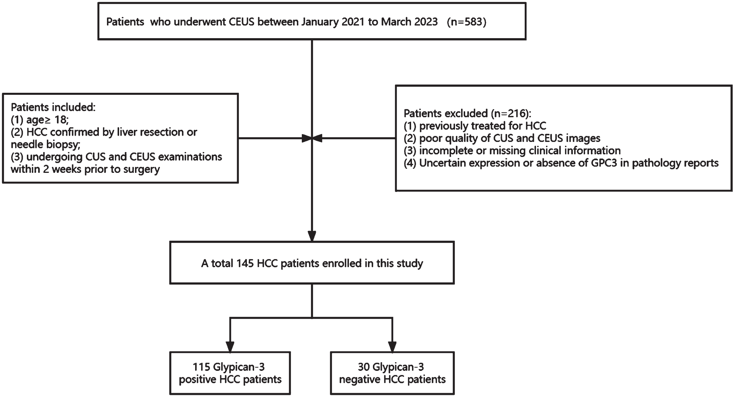
Flowchart shows the patients inclusion process used in this study.
Clinical information, including gender and age, was routinely recorded for each patient. Hepatitis, serum alpha-fetoprotein (AFP) levels, total bilirubin, albumin, CA199, CA125, Child-Pugh stage, Barcelona Clinic Liver Cancer (BCLC) stage, and China liver cancer staging (CNLC) stage were obtained from the electronic medical record system of the patients. US-guided core needle biopsy was performed by radiologists using an 18-gauge automatic biopsy needle (MN1820, Bard Medical) in an avascular path through normal liver tissue before puncturing the target nodule.
CUS and CEUS scans were performed by a radiologist with more than 5 years of experience in CEUS. CEUS was performed in low mechanical index mode using Philips IU22/ EPIQ 7 and Siemens ACUSON Sequoia color Doppler ultrasound diagnostic instruments with convex array probes C5-1 and5C1(both at 1–5 MHz), respectively. During the CEUS examinations, a mechanical index of less than 0.08 was maintained. The liver was initially comprehensively scanned using CUS, including grayscale and color Doppler, to identify the target lesions. In cases of multiple lesions, the tumor with the largest diameter was selected for further examination. For the CEUS procedure, the contrast agent (SonoVue; Bracco) was administered via the peripheral vein after dilution with 5 mL of 0.9% saline solution, followed by a rapid flush with 5 mL of saline solution. Time recording was initiated, and the lesions were continuously observed for a duration of 4-6 minutes, with the final images being saved as dynamic videos. In accordance with the guidelines provided by the European Federation of Societies for Ultrasound in Medicine and Biology (EFSUMB) for CEUS, the liver CEUS procedure consists of distinct phases: the arterial phase, the portal venous phase, and the delayed phase [21]. The arterial phase commences 20 seconds after the injection of the contrast agent through the peripheral vein and lasts until 30-45 seconds. Subsequently, the portal venous phase extends from 31 to 120 seconds, followed by the delayed phase, which continues from 121 to 360 seconds.
CUS and CEUS analysis
The final analysis of CUS and CEUS dynamic videos was conducted by two experienced radiologists with over 5 years of expertise in CEUS. They carefully reviewed the images and summarized the lesion characteristics. The radiologists possessed knowledge of the patients’ clinical history and relied on image analysis to make the definitive diagnosis. It should be noted that they were blinded to the pathological results to maintain objectivity. In instances where there was a discrepancy in opinions between the two readers, a third reviewer, who had 18 years of experience in CUS and CEUS diagnosis, was consulted to achieve a consensus.
We evaluated the ultrasound features of each HCC patient, extracting lesion characteristics from the US images, including: (a) location (left/right lobe of the liver), (b) number (solitary/multiple), (c) maximum diameter (<3 cm/≥3 cm), (d) shape (irregular/regular), (e) echogenicity (isoechoic/hyperechoic/hypoechoic/mixed echogenicity), (f) margin (clear/indistinct), (g) halo sign (present/absent), and (h) color Doppler flow imaging (rich/poor blood supply).
The extraction of lesion features in CEUS images encompasses several aspects: (a) Arterial phase hyperenhancement (APHE, presence/absence), defined as overall or partial high enhancement of the lesion in the arterial phase, with intensity higher than the surrounding liver parenchyma (neither showing a ring-like enhancement nor peripheral discontinuous nodular enhancement); (b) portal venous phase enhancement level (high enhancement/iso-enhancement/low enhancement); (c) delayed phase enhancement level (iso-enhancement/low enhancement);(d) Intralesional necrosis (presence/absence), defined as non-enhancing areas within the tumor visible in both arterial and venous phases; (e) Intralesional arterial vessels (presence/absence), defined as persistent enhancement of arterial vessels observed in the tumor arterial phase, while portal veins and late phases are washed out; (f) Washout time (no washout/early washout/late washout), where early washout is defined as the time to clearance starting from contrast agent injection being < 60 s, and if≥60 s, it is considered as late washout; (g) Degree of washout (no washout/mild washout/significant washout), where significant washout is defined as complete clearance of the lesion presenting as low echogenicity (“black hole-like change”) within 2 minutes of contrast agent injection, and mild washout is defined as the lesion being enhanced less than the liver parenchyma but not completely cleared (i.e., some enhancement still present within the lesion); (h) Perfusion pattern (synchronous/asynchronous). Synchronous perfusion indicates nearly simultaneous entry of the contrast agent into the lesion and surrounding liver tissue, while asynchronous perfusion indicates inconsistent timing of contrast agent entry into the lesion and surrounding liver tissue.
Histopathological analyses
The expression of GPC3 was evaluated using the comprehensive scoring system proposed by Takai et al. [22]. This scoring system incorporates three evaluation parameters: the percentage of positive cells, staining intensity, and staining pattern. The proportion of positive cells is graded from 0 to 3 + . Grade 0 indicates tumor cells with less than 5% positivity, 1 + corresponds to 5-10% positivity, 2 + represents 10-50% positivity, and 3 + signifies over 50% positivity. Staining intensity is categorized as weak, moderate, or strong. The staining pattern is classified based on cell membrane integrity: Type I (complete absence), Type II (partial or irregular staining), and Type III (complete and uniform staining). GPC3 positive expression is defined as tumor cells with a positivity rate of≥5% and staining intensity other than weak. Negative results include tumor cells with a positivity rate < 5%, or weak staining at the 1 + level and a positivity rate of≥5%.
Statistical analyses
All analyses were performed using SPSS 26.0 statistical software (IBM Corp., Armonk, NY, USA). The Kolmogorov-Smirnov test was used to assess the normality of continuous variables. Student’s t-test and Mann-Whitney U test were employed for normally distributed and non-normally distributed continuous variables, respectively. Results were reported as mean±standard deviation (SD) for normally distributed variables and as median with interquartile range for non-normally distributed variables. Categorical variables were evaluated using the chi-square test. Significant variables identified from the univariate analysis were included in the multivariable logistic regression analysis. Receiver operating characteristic (ROC) analysis was conducted using the optimal variables derived from the multivariable logistic regression analysis to evaluate the model’s performance. A two-tailed p-value of < 0.05 was considered statistically significant.
Results
Patient’s clinical characteristics
The study included 145 patients diagnosed with HCC, comprising 115 GPC3-positive and 30 GPC3-negative patients. Among them, 24 were female and 121 were male, with a mean age of 56.25±9.82 years. Clinical characteristics of all 145 patients are summarized in Table 1. Significant differences in AFP levels were observed between the GPC3-positive and GPC3-negative groups (P = 0.001). The incidence of HBV infection was higher in the GPC3-positive HCC group compared to the GPC3-negative HCC group (82.6% vs. 73.3%). However, no significant differences were found in ALB levels, TIBL levels, CA199, CA125, Child-pugh stage, BCLC stage, and CNLC stage between the GPC3-positive and GPC3-negative groups.
Clinical characteristics of HCC patients
Clinical characteristics of HCC patients
AFP, Alpha-fetoprotein; ALB, Albumin; TIBL, total bilirubin; BCLC stage, Barcelona Clinic Liver Cancer stage; CNLC stage, China liver cancer staging stage. * indicates significant values, P<0.05.
Tables 2 and 3 summarize the CUS and CEUS imaging characteristics of the GPC3-positive and GPC3-negative groups. There was no significant difference in CUS imaging characteristics of the tumors between the GPC3 positive and GPC3 negative groups. Among the 145 HCC, 107 patients had a maximum tumor diameter≥3 cm (107/145 [73.8%]), but there was no significant difference between the GPC3-positive and GPC3-negative groups (p = 0.072). Compared to the GPC3-negative group, the GPC3-positive group was associated with APHE (p < 0.001), delayed-phase hypo-enhancement (p = 0.019) and asynchronous perfusion (p = 0.037) were significantly correlated, while other features were not significantly correlated (p > 0.05, Table 3). Images typical of GPC3-positive and GPC3-negative HCC are shown in Figs. 2 3. Among the 145 HCC patients, the presence of vascular stray images in the arterial phase was observed in 86 patients (86/145, [59.3%]) and intratumoral necrosis in 79 patients (79/145, [54.5%]), while there was no difference between the GPC3-positive group and the GPC3-negative group (p = 0.931, p = 0.887).
CUS characteristics of lesions in HCC patients
CUS characteristics of lesions in HCC patients
CEUS characteristics of lesions in HCC patients
* indicates significant values, P<0.05.
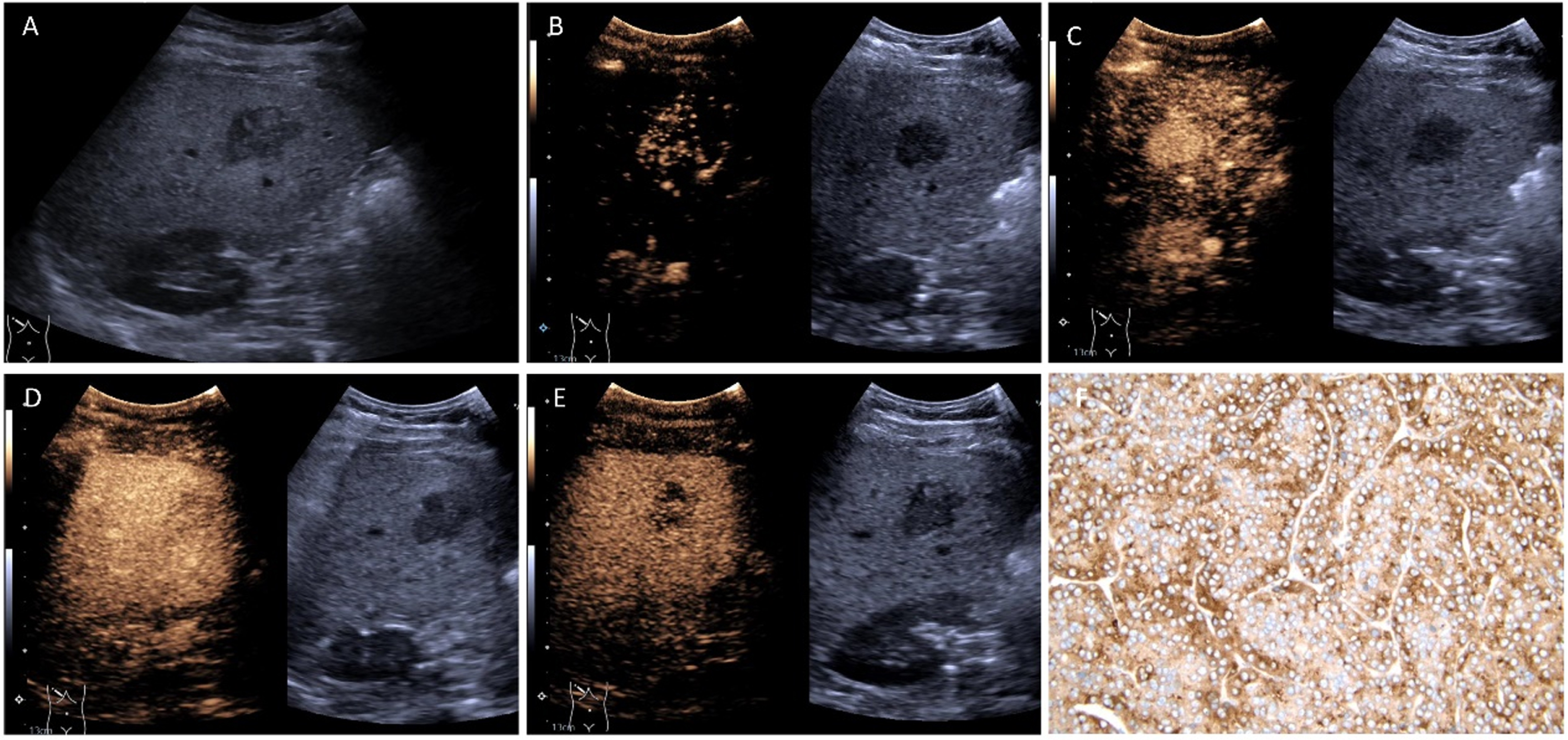
A 59-year-old male with confirmed Glypican-3 positivity after surgical resection with an AFP of 58.4 ng/mL. (A) A hypoechoic lesion measuring approximately 2.6x2.2 cm was located in liver S6 with clear borders and irregular morphology. (B) The lesion showed heterogeneous arterial phase enhancement (APHE) 9 seconds after contrast agent injection, with asynchronous perfusion compared to surrounding liver tissue. (C) The lesion reached peak enhancement 14 seconds after contrast agent injection. (D) Contrast agent within the lesion slowly washed out 30 seconds after injection, resulting in low enhancement. (E) In the delayed phase, the contrast agent continued to wash out within the lesion, showing heterogeneous and even lower enhancement. (F) Pathological examination confirmed GPC3-positive HCC after surgical resection.
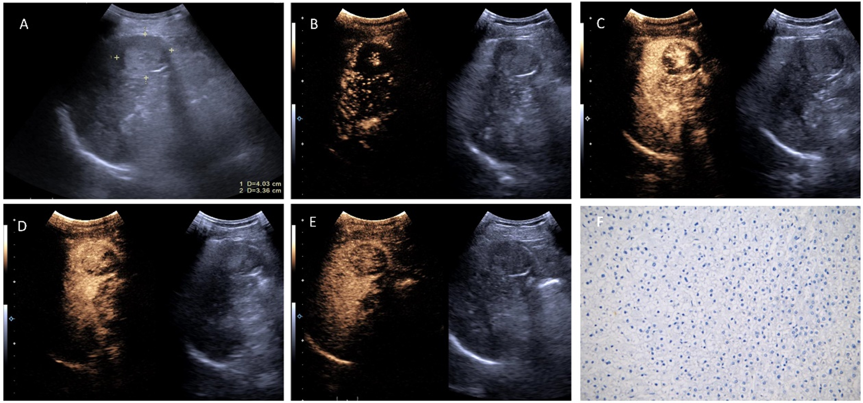
A 72-year-old male with confirmed Glypican-3 negativity after surgical resection with an AFP level of 4.57 ng/mL. (A) A hypoechoic lesion measuring approximately 4.03x3.36 cm was located in liver S5 with clear borders and regular morphology. (B) the peripheral and central areas of the lesion showed homogeneous hyper-enhancement 17 seconds after contrast agent injection, with synchronous perfusion compared to surrounding liver tissue. (C) The lesion reached peak enhancement 27 seconds after contrast agent injection. (D) Contrast agent within the lesion slowly washed out 76 seconds after injection, resulting in low enhancement. (E) In the delayed phase, the contrast agent continued to wash out within the lesion, showing heterogeneous and even lower enhancement. (F) Pathological examination confirmed GPC3-negative HCC after surgical resection.
Based on the results of the univariate analysis, variables with p values < 0.05 were included in the multifactorial logistic regression model, and the multifactorial logistic regression analysis showed that AFP > 20 ng/mL, APHE, and asynchronous perfusion were independent predictors of GPC3 positivity (Table 4). Regression modeling:
Multivariate logistic regression analysis of CEUS and clinical factors predicting GPC3 positive expression
Multivariate logistic regression analysis of CEUS and clinical factors predicting GPC3 positive expression
X1 indicates AFP (AFP>20 ng/mL=1, AFP<20 ng/mL=0); X2 indicates APHE (presence = 1,absence=0); X3 indicates perfusion pattern (asynchronous perfusion = 1, synchronous perfusion = 0).
We developed combined models to predict GPC3-positive expression using subject operating characteristic (ROC) analysis, as illustrated in Fig. 3. The model that combined the three independent predictors exhibited strong predictive performance (AUC 0.817, 95% CI: 0.731-0.902), demonstrating a sensitivity of 91.3% and specificity of 60.0%. In comparison to AFP > 20 ng/mL, APHE, and asynchronous perfusion alone, the combined model demonstrated significant improvements (p < 0.001). Refer to Table 5 for further details. Overall, the combined model effectively distinguished GPC3-positive HCC from GPC3-negative cases.
Performance of optimal features for predicting GPC3 positive expression
Performance of optimal features for predicting GPC3 positive expression
In recent years, GPC3 has emerged as a key player in the development and progression of HCC tumors. Its elevated expression is associated with an increased likelihood of postoperative recurrence and metastasis in HCC patients. Additionally, GPC3 has gained considerable recognition as a viable therapeutic target for HCC [23, 24]. Consequently, the exploration of non-invasive methods for detecting GPC3 expression in early-stage HCC patients can greatly support clinicians in optimizing treatment strategies. Currently, imaging studies predominantly concentrate on utilizing GPC3 molecular imaging probes and preoperative gadoxetic acid (EOB) -enhanced MRI image feature analysis to anticipate GPC3 expression [25, 26]. In a groundbreaking approach, we explored the integration of CEUS imaging features with clinical factors for preoperative evaluation of GPC3 expression in patients with HCC. We discovered that AFP > 20 ng/mL, APHE, and asynchronous perfusion independently predict GPC3 positive expression. The combination of these three indicators exhibited a satisfactory predictive performance with an area under the curve (AUC) of 0.817 (95% CI: 0.731-0.902), sensitivity of 91.3%, and specificity of 60.0%.
GPC3 plays a crucial role in cell proliferation and tissue morphogenesis, particularly in the mesenchymal tissue of embryos. While it is nearly absent in normal adult liver tissue, its expression is upregulated in HCC, indicating its significance in HCC prognosis. Studies have demonstrated that suppressing the GPC3 gene inhibits the migration and invasion of liver cancer cells [12], further emphasizing its relevance in HCC. One notable example is its involvement in the classical Wnt signaling pathway, where GPC3 binds to membrane Wnt and its receptor Frizzled in HCC cells. This binding stimulates the formation of signaling complexes and activates Wnt signal transduction, thereby promoting the proliferation and survival of specific genes. Remarkably, this pathway is potentially activated in up to 95% of HCC cases [27]. Given the specific expression of GPC3 in HCC, researchers have extensively explored targeted therapies focusing on GPC3. Notably, GPC3-targeted CAR-T cell therapy has shown promising results in solid tumors. In a mouse experiment, CAR-T cells targeting GPC3 effectively eliminated GPC3-positive HCC cells by inducing cell apoptosis and reducing Wnt signal transduction in tumor cells [5]. This exciting finding suggests a potential application for GPC3 targeting in HCC patients.
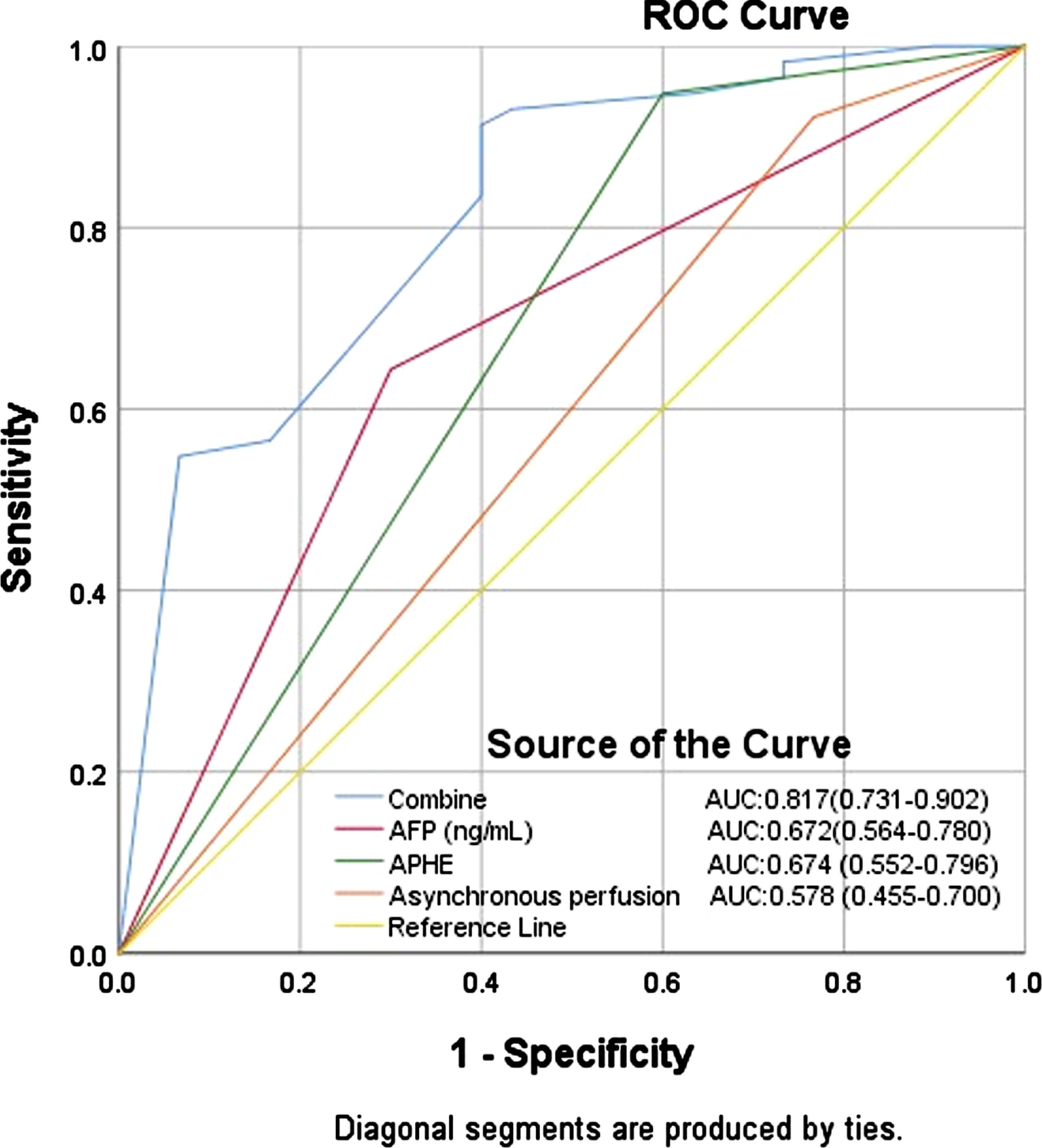
Receiver operating characteristic (ROC) curve of independent predictors and the combined model to predict Glypican-3 positive expression in HCC patients.
Zhao et al. [18] compared MRI features of 43 GPC3-negative and 100 GPC3-positive HCC patients and found no enhancing capsule, peritumoral enhancing appearance on the arterial phase, and apparent diffusion coefficient (ADC) values at the 75th percentile were significantly associated with GPC3-positive expression. The enhancement of the lesion capsule in the arterial phase is a crucial imaging characteristic for diagnosing HCC in CT/MRI LI-RADSv2017. This phenomenon occurs as a result of tumor cell infiltration and the destruction of surrounding liver tissue, leading to a blurred boundary between the surrounding liver parenchyma and the lesion. Consequently, this blurred boundary may be mistakenly interpreted as capsular enhancement in CT or MRI scans. Additionally, in cases where HCC invades adjacent arteries, arterial contrast enhancement may create a ring-shaped structure around the lesion, resembling capsular enhancement. However, it is important to note that ring enhancement similar to capsular enhancement in the arterial phase of CEUS is categorized as an important sign of LR-M in the CEUS LI-RADS classification and should not be considered as an inclusion criterion for LR-5. APHE is an important imaging characteristic for diagnosing HCC in the CEUS LI-RADS classification. It refers to the lesion showing overall or partial high enhancement in the arterial phase (APHE, excluding non-rim enhancement and peripheral discontinuous nodular enhancement), with intensities higher than the surrounding liver parenchyma [28]. Unlike the normal hepatic blood supply pattern, APHE reflects the early abnormal arterial blood supply to the tumor. HCC is a highly vascularized tumor, and during the arterial phase, the tumor cells exhibit increased metabolic activity and require a significant blood supply. This leads to evident vascular contrast enhancement during the arterial phase. Additionally, during the growth of HCC, tumor cells can release various angiogenic factors, such as vascular endothelial growth factor (VEGF), which promotes the growth and expansion of surrounding blood vessels. This process involves the activation of BMP9-ID1 and certain long non-coding RNAs, further facilitating HCC progression and angiogenesis [29, 30]. This positive feedback mechanism contributes to the appearance of abnormal vascular enhancement in the arterial phase of HCC. GPC3 may promote angiogenesis by influencing the expression of vascular growth factors and the activity of signaling pathways. In our study, there was a significant difference in APHE between the GPC3-positive and GPC3-negative groups (p < 0.001). Furthermore, APHE was identified as an independent predictive factor for GPC3 positivity (AUC = 0.674, sensitivity = 94.8%, specificity = 40.0%). Among the 145 HCC patients included in this study, 86 patients exhibited arterial phase vascular enhancement (86/145, [59.3%]), however, intralesional arterial vascularity was not an independent predictor of GPC3-positive HCC in our study, most likely due to the small sample size and the lack of significance between the GPC3-negative and GPC3-positive groups.
Additionally, our study revealed a significant difference in delayed-phase hypo-enhancement between the GPC3-positive and GPC3-negative groups. However, when incorporated into a multivariate logistic regression model, delayed-phase hypo-enhancement did not emerge as an independent predictor for GPC3 positivity. Delayed-phase hypo-enhancement indicates a late washout (>60 s) of HCC during CEUS. A study by CHEN et al. [26] also demonstrated the significant relevance of non-peripheral washout in distinguishing GPC3-positive HCC patients. This is attributed to the lower local blood volume and extracellular matrix capacity in most HCCs compared to the liver parenchyma. As a result, both pure intravascular and extracellular contrast agents manifest a “washout” phenomenon. Furthermore, in a prospective DEGUM study, Meitner-Schellhaas et al. [31] found that only 50% of HCC cases exhibited the typical LR-5 features, including late (60 s) and mild washout. The authors suggested that this might be attributed to perfusion changes caused by tumor infiltration into liver vessels. In our study, 33% (38/115) of GPC3-positive HCC cases showed a washout phenomenon exceeding 60 seconds. This may be due to the relatively small sample size included in our analysis.
Furthermore, our study revealed a significant difference in serum AFP levels between the GPC3-positive and GPC3-negative groups. Serum AFP > 20 ng/mL were identified as an independent predictive factor for GPC3 positivity, which is consistent with the findings of Zhao et al. [18]. Gu et al. [17] investigated MR-based radiomics to predict GPC3 expression and found that combining AFP levels with radiomic features improved the prediction of GPC3-positive HCC. These findings suggest that AFP serves as a specific diagnostic marker for HCC, and there is a certain correlation between GPC3 expression and AFP in HCC, as they both indicate microvascular invasion, poor prognosis, and recurrence. Lower GPC3 expression may result in better survival outcomes when compared to patients with high GPC3 expression, both in the subgroup with AFP 20μg/L and in the subgroup with AFP 20μg/L [32].
Our results indicate that tumor diameter≥3 cm cannot be regarded as an independent predictive factor for positive GPC3 expression in predicting HCC. This finding contradicts the results of Chen et al. [26], who suggested a significant correlation between tumor size > 3.0 cm and GPC-3 positive expression. The possible reasons for the discrepancy in these results may be as follows: MRI scans have fixed imaging planes, and operators tend to select clear and distinct lesions that can be measured. On the other hand, ultrasound examinations involve scanning the lesion from different angles and perspectives. During this process, operators choose the most favorable angles to display the lesion while excluding interference from respiration and rib shadows, especially in the case of the liver. These differences in imaging techniques may contribute to the disparity in results. Additionally, Sonovue may not be able to provide a clear diagnosis for tumors with a diameter less than 1 cm, and additional imaging using the liver-specific contrast agent MRET may be required. According to a recent study, high-resolution contrast imaging, parametric false-color imaging, and the microvascularization of tumors can all be improved by using CEUS parameter analysis, offering substantial diagnostic potential for thorough examination of tiny tumors [33]. Therefore, further investigation is warranted in large-scale prospective studies to explore the impact of tumor diameter on GPC3 expression.
Current imaging techniques have limitations in identifying tumor heterogeneity, tumor microenvironment, and cancer phenotypes. In contrast, radiomics, utilizing high-throughput extraction algorithms, can capture multidimensional features with significant exploitable potential. Gu et al. [17] proposed that radiomic features derived from contrast-enhanced MR images can serve as preoperative predictive factors for identifying GPC3-positive HCC. By combining these radiomics features with AFP, a radiomic model was constructed, achieving impressive predictive ability with AUCs of 0.926 and 0.914 in the training and validation cohorts, respectively. CEUS emerges as a promising imaging tool for HCC diagnosis, offering clear visualization of tumor microcirculation using pure blood pool contrast agents. The distinct perfusion patterns exhibited at different vascular phases highlight the increasing importance of CEUS in HCC diagnosis and prognostic evaluation. Hemodynamic differences revealed by new CEUS perfusion patterns may further distinguish HCC from other malignancies [19]. Future research should focus on prospective studies and radiomic features based on ultrasound imaging to identify GPC3-positive HCC, showing great potential and application.
Our study has several limitations. Firstly, it was a retrospective, single-center study with a small sample size, potentially introducing selection bias. Secondly, a portion of the HCC patients included in our study were based on the results of needle biopsy, which poses risks such as limited sample collection and sampling errors. However, the criteria for biopsy of liver-occupying lesions are based on the practice guidelines for the pathological diagnosis of primary liver cancer [34]. Thirdly, the majority of enrolled patients had a history of hepatitis (127/145, [87.6%]), which may not fully represent the broader population of HCC patients. Therefore, the results of this study require validation through larger prospective population studies. Furthermore, additional research is needed to explore the application of quantitative analysis, radiomics, and artificial intelligence in identifying GPC3-positive expression.
In conclusion, we found that the combination of AFP, APHE and asynchronous perfusion is helpful for the diagnosis of GPC3-positive HCC. Combined preoperative CEUS and clinical factors can help clinicians identify HCC patients at high risk of recurrence and metastasis and provide options for precise treatment of clinical treatment options.
Footnotes
Acknowledgments
We thank all participants for their contributions to this study.
Funding
This work is jointly supported by Science and Technology Department of Gansu Province, China (No. 21YF5FA122).
Conflict of interest
The authors declare they have no potential conflicts of interest.