






Abstract
OBJECTIVE:
This article aims to clarify pitfalls and find strategies for the detecting and diagnosing hyperechoic liver metastases (LMs) using Sonazoid-contrast enhanced ultrasonography (Sonazoid-CEUS).
METHODS:
This study was a prospective self-controlled study. Patients with hepatic lesions suspected as LMs or benign lesions were included in the study. Baseline ultrasonography (BUS) and Sonazoid-CEUS were performed on every patient. Characteristics of LMs and benign nodules were compared by chi-square test and fisher test. Factors influenced the CEUS were demonstrated by univariate analysis and multivariate logistic regression analysis.
RESULTS:
54 patients were included in this study. CEUS found additional 75 LMs from 19 patients in Kupffer phase. We found hyperechoic focal liver lesions and deep seated in liver are main confounding factors in CEUS diagnosis. Sensitivity would be improved from 16.67% to 78.57%, negative predictive value (NPV) would be improved from 28.57% to 76.92% and accuracy would be improved from 37.5% to 87.50% when using rapid “wash-in” and “wash-out” as main diagnostic criteria.
CONCLUSIONS:
Hyperechoic LMs especially deeply seated ones are usually not shown typical “black hole” sign in Kupffer phase. Quickly “wash-in and wash out” shows high accuracy in diagnosing malignant nodules. We highly recommend CEUS as a routing exam to detect and diagnose LMs.
Background
Correctly diagnosing metastases is important in choosing treatment strategy for patients with malignant tumor [1, 2]. Liver is one of the common organs prone to be metastasized [3]. Baseline ultrasonography (BUS) is a useful tool to detect hepatic lesions in routine following up of tumor patients because of its simple, non-invasive nature and high temporal resolution with real-time imaging. Especially, contrasted enhanced ultrasound (CEUS) upgraded diagnostic performance of detecting and characterizing focal liver lesions (FLLs) with comparable diagnostic performance to that of Computed Tomography (CT) and Megnetic Resonance Imaging (MRI) with fewer adverse reactions and no risk of radiation exposure [4–9].
Sonazoid is one of second generation ultrasonographic contrast agents and composed of microbubbles of perfluorobutane gas coated with hydrogenated egg phosphatidylserine sodium (H-EPSNa). Besides vascular phases like other traditional microbubbles, the agents are accumulated in Kupffer cells by phagocytosis in 10 mins when the agents are nearly removed from circulation [10–13]. Then, additional and specific phase is available with Sonazoid CEUS, and this is referred to as the postvascular phase which is also known as Kupffer phase lasting for several hours after injection [10, 14]. For liver metastases (LM), rim-like arterial phase hyper-enhancement appearing in the peripheral portions of the tumor and black hole in Kupffer phase are considered as characteristic feature [15].
Although Sonazoid-CEUS showed great diagnostic performance in differentiating LM, according to our practice and observation, hyperechoic malignant lesions appeared in BUS usually may not exhibit classic perfusion defects or obvious hypoenhancement during the Kupffer phase, especially for those hyperechoic nodules deeply seated as 10 cm to the surface in the liver [16, 17]. Besides, hypoenhancement could also be observed in benign lesions during Kupffer phase. These appearances could be pitfalls which will mislead diagnosis of hepatic lesions.
In this study, we will focus on detecting hepatic lesions, differentiating metastatic hepatic lesions from benign lesions using Sonazoid-CEUS, summarizing enhancement characters of vascular phase and Kupffer phase in LMs of different types, finding pitfalls and strategies for diagnosing difficult lesions.
Methods
Patient recruitment
This study was approved by the Independent Ethics Committee of Shanghai Sixth People’s Hospital (Approval No.: 2021-197) in accordance with the Declaration of Helsinki. We prospectively recruited all patients with suspected LMs or benign lesions reported by BUS or other imaging modalities in Shanghai Jiao Tong University Affiliated Sixth People’s Hospital between December 2021 and February 2022. Inclusion criteria including: 1. Hepatic lesions suspected as liver metastases or benign lesions by contrast enhanced magnetic resonance imaging (CEMRI); 2. Have done or scheduled CEMRI examination within 1 months; 3. Sign an informed consent form; 4. Age 18–75y; Exclusion criteria including: 1. Patients with liver metastases after local treatment such as ablation or transcatheter arterial chemoembolization; 2. Patients with a history of allergy to egg or any type of ultrasound contrast agent; 3. Pregnant or lactating women; 4. Severe heart disease, hypertension, severe liver and kidney dysfunction; 5. Pulmonary dysfunction.
One doctor served as coordinator were responsible for recruiting patients with suspected LMs or benign hepatic lesions. The resources of patients could be from clinical ultrasound exam or Department of oncology. Some of them might have done CEMRI within 1 month. Every patient would be given a unique trial number which would go along with the patients within all the investigation procedure. The names of the patients would be hidden until all images were uploaded and reports written by examiners were finished and closed.
Examining process
All of patients meeting the inclusion criteria and not conform to the exclusion criteria were recruited in this study and then accepted regular ultrasound scan and CEUS done by one ultrasonogrpaher. CEMRI within 1 month was also required. Sonographers and radiologists responsible for CEMRI were blinded to other imaging results. If biopsy or surgery would be performed, pathological results would be golden standard. If histological results are not available, follow-up imaging (number and size) combined with clinical records would be standard of reference.
Conventional ultrasound
All patients in the group will undergo conventional ultrasound first, and once any lesion is detected, the number, size, location according to Couinaud’s classification and sonographic features will be recorded. The ultrasound examination will be performed on PHILIPS EPIQ Elite (Philips Healthcare, Bothell, WA) with a 6-1 MHz convex transducer.
Sonazoid-CEUS
All patients underwent CEUS with Sonazoid right after BUS. All examinations were performed using PHILIPS EPIQ Elite (Philips Healthcare, Bothell, WA) with a 6-1 MHz convex transducer. CEUS acoustic power was at the default setting with a mechanical index of 0.18 and a dynamic range fixed at 45–65 db. Sonazoid was reconstituted in 2 mL sterile water for injection. The injection dose was 0.01 mL of encapsulated gas per kg of body weight. Vascular phase images were obtained 0–5 minutes after Sonazoid injection. For the lesions found from conventional ultrasound, the vascular-phase characters of them would be recorded, Kupffer phase (10 to 15 minutes) will be observed, and the relevant characteristics will also be recorded on the CRF. If more than one nodule is found in pre-contrast conventional ultrasound, the largest one will be selected as target lesion. Kupffer phase scan were obtained at least 10 minutes after Sonazoid injection. If finding any additional defect in KPWLS, Sonazoid-CEUS would be repeated to observe the vascular character (for each patient, at most twice in one exam). CEUS images (pre-contrast, arterial, and Kupffer phase) were prospectively interpreted by consensus between the two doctors with 6 years’ experiences respectively who were blinded to the other imaging. According to 2020 consensus [15], the diagnostic reference of LMs is rim-like arterial phase hyperenhancement in the peripheral portions of the tumor, wash-out quickly and “black hole” sign or obvious hypoenhanced in the postvascular phase. Hemangioma exhibits peripheral rim enhancement and peripheral nodular enhancement patterns and filling in a centripetal direction in the vascular phase, isoenhancement or slight hypoenhancement in postvascular phase. Focal nodular hyperplasia (FNH) displayed pathognomonic imaging patterns such as central artery and stellate vascularity and, isoechoic or slightly hyperechoic appearance compared with the surrounding liver parenchyma in postvascular phase.
MRI
All MRI examinations were performed using a 3.0 T MR system (Magnetom Skyra; Siemens Medical Solutions, Erlangen, Germany) and 18-channel coils. The patients were examined in the supine position, and the receiver coil was positioned to cover the upper abdomen. The patients underwent a routine liver MRI and contrast imaging protocol. For contrast enhancement, a bolus 0.1 mmol/kg body weight) of Gadodiamide (GE Healthcare, Shanghai, China) was administered into the antecubital vein at a rate of 3 ml/s. Two radiologists specialized on abdomen MRI would review the CEMRI profile slide by slide retrospectively and conclude a report after agreement.
Data analysis
A commercially available software package (SAS version 9.2, SAS Analytics, Marlow, 260 UK) will be used for all the statistical analysis. For continuous variables, t-test was performed. For For categorical variables, Pearson’s Chi-squared test with Yates’ continuity correction and Fisher test would be used to find significant indicators. Efficacy analyses of sensitivity, specificity, positive predictive value, negative predictive value and accuracy will be performed. Factors influenced the CEUS would be demonstrated by univariate analysis and multivariable logistic regression analysis. Significance level was 0.05.
Results
Patient characteristics
We enrolled 60 patients with FLLs that had been suspected as LMs or benign lesions. Among whom, 4 were diagnosed as hepatocellular carcinoma (HCC) pathologically, 2 died before obtaining conclusive diagnosis of FLLs in following up. Finally, 54 patients were included in this study and 33 of them had malignant tumor history out of liver. The primary lesion of the tumor was colorectal in 11 patients, cervix in 7, bile duct in 1, lung in 2, pancreas in 1, ovary in 4, breast in 2 and mesenchymal tissue in 7. Among the 33 patients with outer-liver malignant tumor, 27 had LMs. Other 27 patients had only benign lesions including hemangioma, focal nodular hyperplasia (FNH) and liver abscess (Fig. 1 and Table 1).
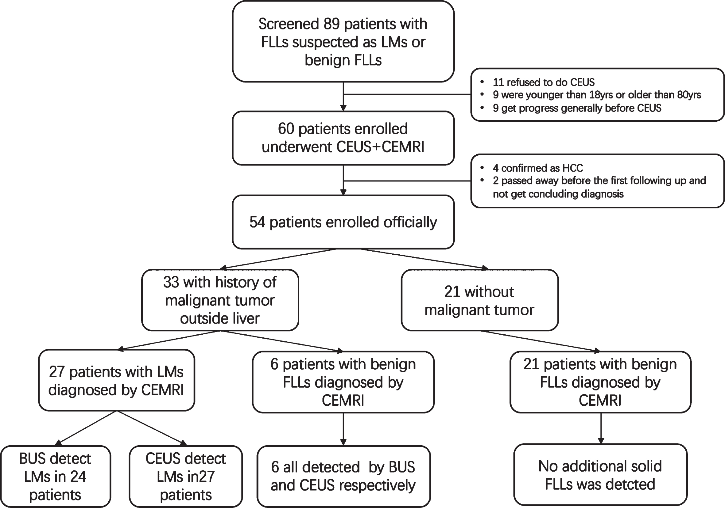
Study design and patient inclusion.
Patient and procedural characteristics (n = 54 patients)
Data are shown as n (%) unless noted otherwise with an asterisk (*). x: missing data. *Numbers show years and not percentages. **Numbers show kg/m2 and not percentages.
3.2.1 CEUS detected LMs in 27 patients with LMs while BUS missed 3 patients. So, the detection rate of LMs is 100% in CEUS and BUS missed 11.11% of them compared to CEMRI. In the group of benign nodules BUS and CEUS detect all the lesions.
3.2.2 In the group of LMs, 70.37% (19/27) of them were detected additional lesions after CEUS (mainly depended on seeking for black hole in Kupffer phase). 0–9 additional lesions were found among the 19 patients. BUS detected 89 LMs and CEUS detected 164 LMs. In benign group, no additional solid lesion was found (Fig. 2).
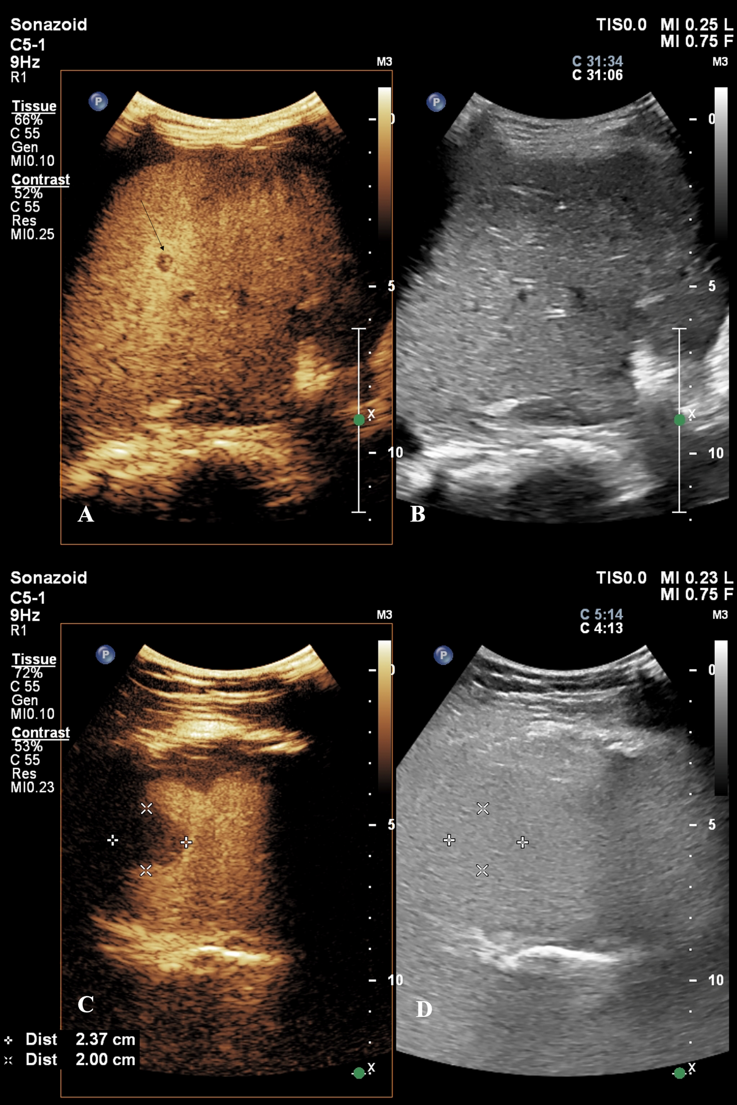
Images of 2 LMs detected by CEUS. A and B were captured from a 24 years old patient with a history of ovary cancer. A: a LM showed defect (black arrow) in the kupffer phase by CEUS but not seen any nodule in the BUS (B). C and D were captured from a 52 years old patient with a history of breast cancer. C: A: a LM showed defect (++marked) in the kupffer phase by CEUS but not seen any nodule in the BUS (B).
3.2.3 75 LMs additionally found with CEUS were 4–30 mm in diameter. 62.67% (47/75) of them were sub-centimeter and we found the most additional LMs in segment 3 (40%, 30/75), following by segment 4 (22.67%, 17/75), segment 5 (21.33%, 16/75), segment 6 (10.67%, 8/75) and segment 2 (5.33%, 4/75) (Table 1).
3.3.1 There are one or more FLLs in one patient, so there are 91 FLLs were included in the diagnostic analysis. The 91 FLLs were observed through arterial phase to late phase completely. The characteristic of the FLLs were listed in Table 1. Enhancement type in general and in different phases, and peak intensity were all significantly different in LMs group than benign group (Table 2).
CEUS characteristics of 91 FLLs
CEUS characteristics of 91 FLLs
*p < 0.05 is considered as significant different statistically.
3.3.2 Misdiagnosis cases analysis
12 nodules were misdiagnosed by CEUS. 11 of them were false negative ones. According to univariate analysis and multivariable logistic regression analysis, hyperechoic nodules and deeply seated ones (far away from the probe) would cause more misdiagnosis (p = 0.02 and p = 0.00) (Table 3).
Multivariable logistic regression analysis of the factors resulting in misdiagnosis
*p < 0.05 is considered as significant different statistically.
There are 11 LMs did not show defect or obvious hypoenhancement in Kupffer phase. Among the LMs, 72.73% (8/11) were hyperechoic in BUS, and average distance between probe and the 8 LMs was 8.31 cm while this distance in other 3 LMs were 4.47 cm in average. When the distance between LMs and probe was fewer than 6 cm, typical defect or obvious hypoenhancement in Kupffer phase would be seen in 95% (40/42) of the LMs, 42.86% (3/7) in 6 cm to 8 cm, and 28.57% (2/7) in LMs deep seated farther than 8 cm. In 2 patients, we were luckily able to switch the lesions from far field to near filed through change the position of the probe to see the lesion from different approach, and we can see that the “black hole” appeared (Fig. 3).
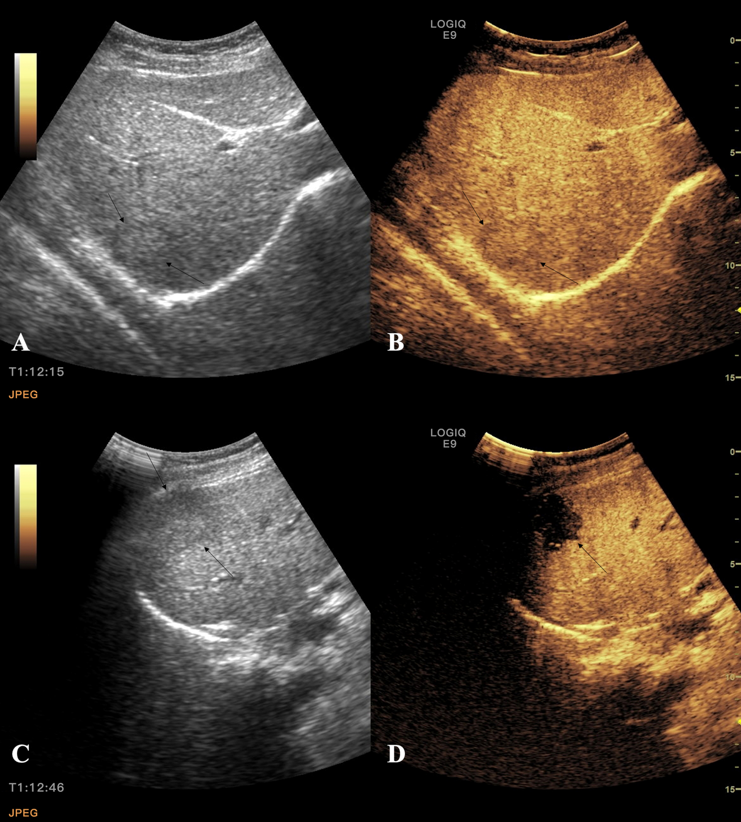
A and B were captured from a 47 years old patient with a history of rectum cancer. A, an isoechoic LM (black arrows) deep seated in 10 cm of segment 7 showed on the BUS and it showed isoenhancement in the kupffer phase in CEUS which may cause misdiagnosis a benign nodule. But when the position of the probe was changed and adjusted the position of the nodule in the near field, we can see a clear defect of the very same lesion in C and D (black arrows).
3.3.3 Among the 56 LMs, 94.64% (53/56) showed “wash in and wash out” in 1 minute and 42.86% (24/56) showed it within 30 seconds. 3 LMs show wash out a bit in portal vein phase and showed hypoenhancement or isoenhancement during portal vein phase to post vascular phase. The 3 LMs was all deep seated faraway from probe (>8 cm). Within the distance of 8 cm, all LMs showed typical rapid “wash in and wash out”.
Hyperechoic FLLs and deep seated in liver are main confounding factors in CEUS diagnosis. We divided all FLLs into different group according to these 2 factors and used 2 different criteria to diagnose. Criteria A: meeting both rapid “wash in and wash out” and defect in Kupffer phase would be diagnosed as LMs. Criteria B: meeting only rapid “wash in and wash out” would be diagnosed as LMs, defect in Kupffer phase is not essential. Sensitivity would be improved from 16.67% to 78.57%, NPV would be improved from 28.57% to 76.92% and accuracy would be improved from 37.5% to 87.50% (Table 4).
Diagnose performance of criteria A and criteria B
Diagnose performance of criteria A and criteria B
Criteria A: meeting both rapid “wash in and wash out” and defect in kupffer phase would be diagnosed as LMs. Criteria B: meeting only rapid “wash in and wash out” would be diagnosed as LMs, defect in kupffer phase is not essential. PPV: Positive predictive value. NPV: Negative predictive value. ACC: accuracy. *Near field: distance between probe and nodule is less than 8 cm. **Distant field: distance between probe and nodule is more than 8 cm.
CEUS with Sonazoid is a very effective way to differentiate LM from benign FFLs which has been investigated and proved by many studies [18, 19]. Some relative studies are still going on [20].We found that LMs do not always share the same “Black hole” sign in Kupffer phase, it could be hypoenhancement, isoenhancement, or even hyperenhancement when comes to hyperechoic FLLs. The finding would not change the existing cognition of Sonazoid-CEUS in Kupffer phase but what we found could be clinical useful for sonographer and doctors better understanding the imaging characters of LMs and avoiding misdiagonsis.
LMs could also show hyper-or iso enhancement during Kupffer phase which has not been mentioned in Guideline yet. Some researchers had reported similar situation before, but the dilemma has not been solved properly [21–23]. According to our study, we found “wash in” and “wash out” within one 1 minute in the arterial phase is more crucial to distinguish LMs. Apparent hypoenhancement or defect in Kupffer phase is common in hypoechoic FLLs but for hyperechoic nodules it is not an essential indicator.
As most of sonographers are still used to look for defect in Kupffer phase to find potential LMs and consider it is an important way to distinguish LMs from benign nodules, we had to remind that it could lead to some mistake especially with deep-seated nodules. According to our experience, when the nodule is 8 cm away from the transducer, only 28.57% (2/7) of those nodules show classic defect in Kupffer phase. Some doctors believe it was due to influence of the background B mode. But in our study, we have degraded gain as to make sure the hyperechoic lesions would not appear hyperechoic in contrast mode before UCB injection. But most of the hyperechoic lesions still showed hyperenhancement or isoenhancement during Kupffer phase. For those lesions deep seated, one strategy to decrease the misdiagnose rate is to change the position of the probe, choose different path to see the lesion, trying best to observe the nodule in near field (Fig. 3). What we should do is to scan the liver as comprehensive as we can from different angles. In our study, 50 FLLs show defect in Kupffer phase, but 10% (5/50) of them are benign. Including three hemangiomas, one FNH and one cyst (4 mm in diameter). The dignity of cyst and were confirmed promptly by repeated CEUS with completely no enhancement in arterial phase. FNH was also been diagnosed correctly because unique centrifugal type enhancement in arterial phase [24]. But one hemangioma caused some controversy when undergoing CEUS, because bubbles started washed out from 78 s after injection of the UCA that seems a little bit earlier than classic hemangioma [15]. We suggested the patient receive liver biopsy and pathological results affirmed the dignity of the hemangioma.
We also found that the defect of LMs and benign nodules in Kupffer phase were a little different: In LMs, the defect would demonstrate a hyperenhanced “rim” around the defect, but in benign nodules the “rim” is rare. One study has shared the pathological explanation of the “rim” and they found it due to the gathering of macrophages around LMs and it also related to necrosis inside the tumor [25, 26].
Considering high cost of CEMRI and radiation exposure of CECT, we highly recommend CEUS as a routing exam for patients to detect and diagnose LMs, According to our study, CEUS with Sonazoid has great accuracy (86.81% for method A and 95.6% for method B) when differentiating LMs from benign ones which is be superior to CECT and comparable to CEMRI [4]. Sub-centimeter FLLs could be more sensitive to detect in Kupffer phase. Moreover, we could observe interested nodule from the very beginning (0 s) since injection of microbubbles which means we can acquire more abundant and comprehensive information of the early arterial phase. To our knowledge, although CECT and CEMRI could also show arterial phase enhancement, it would begin at 30 seconds after injection of contrasted agent. So, it could miss some important enhancing characteristics of FLLs in early 30 s.
We also want to recommended using Sonazoid-CEUS to detect LMs in following occasions: Preoperative exam in colorectal cancer to find submillimeter LMs that could be missed by CECT or BUS. Accurate localization of LMs is helpful to complete resection of the lesions in early phase; Routing following up of liver in breast cancer patients to find small LMs which might earn more time to change therapy plan (Small LMs could be easily missed by BUS). More occasions where Sonazoid-CEUS could be involved are exploring [27–29].
However, there are some pitfalls in using CEUS which should be kept in mind of sonographers. First, in S1, S7 and S8, there are some blind areas which are relatively hard to be displayed by ultrasound which means nodules in those areas could be missed. Especially in patients underwent right lung lobectomy, dome of diaphragm is elevated, it is often difficult to visualize small lesions (even big) near the dome of diaphragm with B-mode ultrasound or CEUS. Secondly, Contrast images are still subject to various limitations, such as frame rate, penetration depth, and spatial resolution, which are due to the physical characteristics of US. So, better understanding the limitation of regular US and CEUS would help clinicians and surgeons to choose proper imaging modality for different patients.
There are several limitations in this study. Firstly, the standard of reference was a combination of pathology and follow-up imaging findings. The presence of multiple metastases and the small size of many of the nodules made biopsy of individual lesions unnecessary or impractical. Secondly, due to the small volume of the cohort, we did not explore the value of Sonazoid-CEUS in LMs among different original tumor which call for large scale and multicenter prospective study.
Conclusions
CEUS with Sonazoid is an effective way to detect and diagnose LMs. Hyperechoic nodules especially deeply seated ones are usually not shown typical “black hole” sign in Kupffer phase. Quickly “wash-in and wash out” shows high accuracy in diagnosing malignant nodules. We highly recommend sonographer who performed liver Sonazoid-CEUS be aware of the pitfalls and skillfully using strategies we suggested.
Footnotes
Acknowledgments
The authors would like to thank the Independent Ethics Committee of Shanghai Jiao Tong University Affiliated Sixth People’s Hospital for the guidance of ethics.
Funding
This study was supported by College Project of Shanghai Jiao Tong University Affiliated Sixth People’s Hospital (Grant number Ynts202104).
Conflict of interest
The authors have no relevant financial or non-financial interests to disclose.
Ethics approval
This study was performed in line with the principles of the Declaration of Helsinki. Approval was granted by the Ethics Committee of Shanghai Jiao Tong University Affiliated Sixth People’s Hospital (Approval No: 2021-197).
Consent to participate
Informed consent was obtained from all individual participants included in the study.
Consent to publish
The authors affirm that human research participants provided informed consent for publication of the images in Fig. 2 and  .
.
Data availability statement
The datasets used and/or analysed during the current study are available from the corresponding author on reasonable request.
Author contributions
Li Yi, Zhang Wei: Conceptualization, Writing-Original Draft Wei Xiaoer, Liu Beibei, Jiao Qiong: Investigation Li Yi, Wu Qiong: Methodology, Formal analysis Liu Yilun, Zhang Rui: Data Curation, Writing-Review & Editing Hu Bing, Ying Tao: Supervision, Project administration, Li Yi: Funding acquisition. All authors have read and approved the final manuscript. The corresponding authors have full access to all the data in the study and have final responsibility for the decision to submit for publication.