






Abstract
BACKGROUND:
The precise association between lncRNA H19 and ferroptosis in the context of atherosclerosis remains uncertain.
OBJECTIVE:
This study is to clarify the underlying process and propose novel approaches for the advancement of therapeutic interventions targeting atherosclerosis.
METHODS:
Assessment of ferroptosis, which entails the evaluation of cell viability using CCK-8 and the quantification of intracellular MDA, GSH, and ferrous ions. Simultaneously, the protein expression levels of assessed by western blot analysis, while the expression level of lncRNA H19 was also determined. Furthermore, HAECs that were cultured with ox-LDL were subjected to Fer-1 interference. HAECs were exposed to ox-LDL and then transfected with H19 shRNA and H19 overexpression vector pcDNA3.1. The level of ferroptosis in the cells was then measured. Then, HAECs were subjected to incubation with ox-LDL, followed by transfection with H19 shRNA and treated with Erastin to assess the levels of ferroptosis, cell viability, and inflammatory factor production. and the ability for blood vessel development.
RESULTS:
The survival rate of HAECs in the ox-LDL group was much lower. Ox-LDL resulted in an upregulation of ACSL4 expression in HAECs, while the expression of SLC7A11 and GPX4 decreased.
CONCLUSIONS:
lncRNA H19 enhances ferroptosis and exacerbates arterial endothelial cell damage induced by LDL.
Introduction
Cardiovascular disease (CVD) is a prominent contributor to global mortality and morbidity, encompassing significant conditions such as coronary artery disease and cerebrovascular illness [1, 2]. Atherosclerosis (AS) is a prevalent contributor to the elevated death rates associated with cardiovascular disease [3]. AS is a persistent inflammatory condition characterised by the presence of lipid deposition, arterial wall fibrosis, and the development of susceptible plaques. The gradual induction of arterial lesions and stenosis [4]. Prior research has demonstrated that the buildup of low-density lipoprotein (LDL) can lead to impaired functionality and alterations in the lining cells of internal arteries. Consequently, these cells release inflammatory chemicals that attract additional inflammatory cells, ultimately contributing to the development or worsening of atherosclerotic disease [5]. The role of endothelial cell dysfunction in the initiation of atherosclerosis is evident. Hence, an investigation into the molecular mechanism underlying the detrimental effects of oxidized (ox)-LDL on artery endothelial cells would yield novel.
Ferroptosis is widely recognized as a mechanism via which endothelial cells are vulnerable to harm caused by the external environment. Ferroptosis is a type of controlled cell death that occurs without apoptosis, and is mostly triggered by aberrant iron metabolism and lipid peroxidation. Research has indicated that advanced plaques of atherosclerosis exhibit notable lipid peroxidation, intra-plaque haemorrhage, and iron deposition [6]. This shows evidence that ferroptosis occurs in the atherosclerotic process. Vinchi et al. conducted research that demonstrated how excessive iron levels drive the creation of reactive oxygen species (ROS) and promote the recruitment of monocytes, resulting in oxidative stress in blood vessels and the formation of plaque [7]. Furthermore, empirical research has shown evidence that the suppression of ferroptosis in aortic endothelial cells can effectively mitigate lipid peroxidation and endothelial dysfunction, hence mitigating the progression of atherosclerosis [8]. Kordi et al. reviewed the relationship between ferroptosis and aerobic training in aging, suggesting that regular physical activity might modulate ferroptosis pathways, thereby offering protective benefits against age-related cardiovascular decline [9]. These studies emphasize the significance of ferroptosis in the pathogenesis of AS, indicating a potential avenue of modulating ferroptosis to protect against atherosclerosis and other cardiovascular diseases.
Non-coding RNA is a vital component in signal control. One example of a biomolecule is long non-coding RNA (lncRNA), which is an RNA transcript exceeding 200 nucleotides in length. It has been documented to have a role in diverse biological and pathological mechanisms [10]. Long non-coding RNA (lncRNA) plays a role in both transcriptional and post-transcriptional regulation. Additionally, it acts as an internal signal to control the activities of other non-coding RNAs, such as miRNA or circRNA. Studies indicate that a diverse range of lncRNAs have the ability to control lipid metabolism, inflammatory response, and angiogenesis. These lncRNAs are strongly associated with vascular disorders and atherosclerosis [3, 12]. Recent research suggests that lncRNA H19 plays a crucial role as a signaling molecule in the development of atherosclerosis. The complete length of lncRNA H19 is 2.3 kb and has a high degree of conservation throughout evolution, indicating its potential significance in biological processes [13]. The initial observation made by Han et al. in 1996 revealed that lncRNA H19 exhibited minimal expression in healthy coronary artery tissue, while demonstrating significant expression in human atherosclerotic plaques and damaged carotid arteries [14]. Recent research has indicated that higher concentrations of LncRNA-H19 in the bloodstream are linked to a heightened susceptibility to coronary artery disease [15]. Following this, a study conducted by Huang et al. proposed that the excessive expression of long non-coding RNA H19 would be associated with the development of atherosclerosis [16]. These findings indicate that lncRNA H19 may have a role in the onset and progression of atherosclerosis. Interestingly, lncRNA H19 has been observed to potentially serve as a signaling molecule involved in the regulation of ferroptosis. Certain studies argue that the upregulation of lncRNA H19 has the potential to trigger ferroptosis in hypertrophic cell lung cancer [17]. Nevertheless, certain studies propose that the down-regulation of lncRNA H19 expression can trigger ferroptosis in breast cancer cells [18]. The involvement of lncRNA H19 in the occurrence of ferroptosis is evident, however its regulatory role remains a subject of controversy.
The objective of this study was to provide a comprehensive understanding of the regulatory function of lncRNA H19 and ferroptosis in the context of atherosclerosis, as well as to discover potential molecular targets for treating this condition.
Materials and methods
Cell culture and processing
The HAECS obtained from ATCC were cultured in an endothelial cell growth medium supplemented with 5% foetal bovine serum (FBS), 1% endothelial cell growth supplement (ECGS), and 1% penicillin and streptomycin (1001, ScienCell, USA). The culture environment was maintained at 37°C, with 5% carbon dioxide (CO2) and humidified conditions. HAECS were routinely passaged and utilized for experiments after three consecutive passages to ensure a consistent growth rate and phenotype. The artery endothelial cell injury model was established by inoculating HAECs into a 6-well plate at a density of 5×10⁴ cells per well using fresh medium. The medium was subsequently replaced with serum-free medium and incubated for a duration of 12 hours. Upon reaching approximately 80% confluency, which typically took about 24 hours, the cells were used for subsequent experiments.
In order to investigate the involvement of ferroptosis in the damage of artery endothelial cells, HAECS cells were exposed to a concentration of 100μg/mL of ox-LDL (20605ES10, Yeasen, China) to induce AS model and subsequently treated with 10μM Fer-1 for a duration of 24 hours [19–21]. To investigate the impact of lncRNA H19 expression on arterial endothelial cell damage, transfection was performed using Lipofectamine® 2000 (Invitrogen) with negative shRNA, H19 shRNA, negative overexpression vector pcDNA3.1, and H19 overexpression vector pcDNA3.1 (Sangon, China). After 24 hours, incubate HAECS in the logarithmic phase of development with 100μg/mL ox-LDL for a further 24 hours. To investigate the correlation between long non-coding RNA H19 and ferroptosis, we altered the H19 expression level of cells. Subsequently, we introduced 100μg/mL of ox-LDL and 5μM Erastin to incubate HAECS for a duration of 24 hours.
CCK-8 assay
HAECS, either treated or untreated, were placed in 96-well plates at a density of 5×10⁴ cells per well and allowed to culture overnight. Afterwards, the CCK-8 technique was employed to ascertain cell viability. In each well, a volume of 10μL of CCK-8 solution (C0038, Beyotime, China) was introduced. Following a 4-hour incubation period at a temperature of 37°C, the supernatant was extracted. Subsequently, a volume of 150μL of dimethyl sulfoxide (DMSO) was introduced into each well and subjected to a shaking table oscillator for a duration of 10 minutes at a low velocity, ensuring the full dissolution of the crystals. To determine the absorbance of HAECS at a wavelength of 450 nm, employ a microreader (Thermo Fisher Scientific, USA). By considering the viability of HAECS cells in the absence of any treatment as 100%, the subsequent cell viability of the remaining groups can be calculated.
Blood vessel formation assay
A pre-coated basement membrane extract (354230, BD Biosciences, USA) was applied to a 24-well cell plate for a duration of 24 hours. A total of 1v105 HAECS, both treated and untreated, were injected into the culture plate. Following a 6-hour incubation period, every cell plate was examined using a microscope. In order to assess the cellular morphology and blood vessel formation within each group, three randomly selected fields of view were obtained for each treatment group. The degree of tubule formation was then evaluated using ImageJ software.
Biochemical index assay
To analyze the levels of MDA, GSH, and Fe2+ ions, HAECS were seeded at a density of 5×10⁵ cells per well in 6-well plates and allowed to culture overnight. Following the completion of the HAECS treatment, the cell culture medium was aspirated, and the cells were rinsed twice with pre-chilled PBS. Subsequently, the cells were detached by scraping, collected into a centrifuge tube, and kept on ice for further processing. Cell lysis was carried out as per the manufacturer’s protocols using specific detection kits for MDA (S0131M, Beyotime, China), GSH (S0053, Beyotime, China), and Fe2+ ions (YuanYe, R22185, China). Following the chromogenic reactions, the levels of MDA, GSH, and Fe2+ were quantified using a microplate reader (Thermo Fisher Scientific, USA) at wavelengths of 532 nm, 412 nm, and 562 nm, respectively.
Extracellular TNF-α, IL-6, IL-1β Assay
In a manner akin to the aforementioned techniques employed for identifying biochemical markers, the levels of extracellular TNF-α, IL-6, and IL-1β were assessed. HAECS were seeded at a density of 5×10⁵ cells per well in 6-well plates and allowed to culture overnight. Subsequent to the completion of HAECS treatment, the cell culture was harvested through centrifugation at 4°C, 12000 rpm for 5 minutes, and the resulting supernatant was preserved on ice for subsequent analysis. The ELISA kits for TNF-α (0122H1, MEIMIAN, China), IL-6 (0049H2, MEIMIAN, China), and IL-1β (0181H2, MEIMIAN, China) were utilized as per the manufacturer’s instructions, with a microreader (Thermo Fisher Scientific, USA) employed to measure absorbance at 450 nm. The concentrations of TNF-α, IL-6, and IL-1β were determined post-establishment of a standard curve.
qRT-PCR
The processed HAECS cells were retrieved following removal of the medium, and the cell plate was placed on ice. The cells were then rinsed with pre-chilled PBS buffer, followed by the addition of 1 mL of TRIzol reagent. After a 10-minute incubation period, the cells were scraped off the plate, transferred to a centrifuge tube, and lysed by repeated pipetting. Subsequently, the lysate was centrifuged at 4 °C at 12,000 rpm for 10 minutes, and the supernatant was collected. One-third of the volume was mixed with chloroform to facilitate RNA extraction, and the aqueous phase containing RNA was isolated. An equal volume of isopropanol was added to precipitate the RNA, which was then dried and dissolved in RNase-free water. The RNA concentration was determined using a microspectrophotometer NanoDrop (840-317400, ThermoFisher, USA) for each group. Subsequently, 1μg of total RNA was reverse transcribed into cDNA using a reverse transcription kit (BL696A, Biosharp, China). The expression level of lncRNA H19 in cells subjected to different treatments was quantified using a fluorescence quantification kit (BL698A, Biosharp, China) and fluorescence quantitative PCR was conducted on a CFX384 Touch instrument (Bio-Rad, USA). The mRNA expression level of GAPDH served as the standard for normalizing the expression of lncRNA H19. Primers for gene quantification were procured from Sangon Bioengineering (Shanghai) Co., Ltd. lncRNA H19: F: 5′-ATCGGTGCCTCAGCGTTCGG-3′, R: 5′-CTGTCCTCGCCGTCACACCG-3′; GAPDH: F: 5′-GTCTCCTCTGACTTCAACAGCG-3′,R: 5′-ACCACCCTGTTGCTGTAGCCAA-3′.
Western blotting
HAECS cells, whether untreated or treated, were harvested and subjected to a series of procedures. Following removal of the cell culture medium, the cells were washed thrice with pre-chilled PBS. Subsequently, 1′ml of RIPA buffer supplemented with protease inhibitors (P0013C, Beyotime, China) was added to the cells, which were then lysed thoroughly through repeated pipetting. The resulting lysate was centrifuged at 4°C at 12,000 rpm for 10 minutes to collect the supernatant. The concentration of total protein in each sample was determined using the BCA protein assay kit (P0009, Beyotime, China), and 20μg of total protein was combined with 5× SDS loading buffer, followed by boiling for 10 minutes. The protein samples were separated via 10% SDS-PAGE electrophoresis, and the proteins of interest were transferred onto a polyvinylidene fluoride membrane (PVDF) through electroporation. The membrane was then incubated in 5% skim milk for 2 hours, followed by overnight incubation at 4°C with primary antibodies targeting SLC7A11 (1 : 1000, ab307601, abcam, UK), acyl-CoA synthetase long-chain family member 4 (ACSL4, 1 : 1000, ab155282, ab32373, abcam, UK), GPX4 (1 : 1000, ab252833, abcam, UK), and GAPDH (1 : 1000, ab9485, abcam, UK). Visualization of the proteins on the PVDF membranes was achieved using Enhanced chemiluminescence (SuperSignal ECL, ThermoFisher) for 1 hour with secondary antibodies (1 : 3000, ab6721, ab205719, abcam). The expression levels of the proteins were documented using the Gel Doc XR+Gel Documentation System (Bio-Rad, USA), and the relative expression level of the target protein was determined based on the expression level of GAPDH.
Statistics
The data underwent analysis through SPSS16.0 software and were visualized using GraphPad Prism 9.2.0 software. ANOVA was employed to assess distinctions among groups, t-tests were utilized for comparisons involving two or more independent samples, and one-way ANOVA was applied for analyses involving multiple independent samples. A significance level of p < 0.05 was adopted as the threshold for determining statistical differences.
Results
ox-LDL induces ferroptosis in HAECS
In this investigation, HAECS cells stimulated with ox-LDL were utilized as an in vitro representation of atherosclerosis, with a focus on assessing the extent of ferroptosis in HAECS. The study initially examined the impact of ox-LDL on the viability of HAECs through a CCK-8 assay, revealing a significant decrease in cell survival rates in the ox-LDL-exposed group compared to the control. However, the introduction of a ferroptosis inhibitor (Fer-1) resulted in a notable reduction in ox-LDL-induced ferroptosis levels (Fig. 1A). Subsequent evaluations were conducted to investigate the influence of ox-LDL on oxidative stress levels in HAECs, demonstrating that ox-LDL triggered the accumulation of MDA and a decline in GSH content in HAECs relative to the control group. Notably, the oxidative stress induced by ox-LDL was mitigated by Fer-1 (Fig. 1B–C). Furthermore, ox-LDL was found to promote the accumulation of ferrous ions, while Fer-1 facilitated the elimination of ferrous ions (Fig. 1D). Western blot analysis revealed that ox-LDL down-regulated the protein levels of SLC7A11 and GPX4, while inducing the expression of ACSL4 compared to the control group. Conversely, Fer-1 up-regulated the protein levels of SLC7A11 and GPX4, while inhibiting ACSL4 protein expression compared to the ox-LDL group (Fig. 1E–F). These findings indicate that ox-LDL induces damage to HAECS through the induction of ferroptosis.

ox-LDL induces ferroptosis in HAECS. A, CCK-8 assay was used to detect the survival rate of HAECs in the Control group, ox-LDL group and ox-LDL + Fer-1 group. B-D, The biochemical kit was used to detect the levels of MDA (B), GSH (C) and Fe2+ (D) in HAECs in Control group, ox-LDL group and ox-LDL + Fer-1 group. E–F, Western blot was used to detect the protein expression levels of SLC7A11, ACSL4 and GPX4 in HAECs in Control group, ox-LDL group and ox-LDL + Fer-1 group. **indicates comparison with Control, and p < 0.01; ##indicates comparison with ox-LDL, and p < 0.01.
lncRNA H19 is a potential diagnostic marker for coronary artery disease [22, 23]. Therefore, we further investigated how changes in H19 expression affect ferroptosis induced by ox-LDL in HAECs. Based on qRT-PCR analysis, the induction of ox-LDL was found to significantly enhance the expression of H19 in HAECs. Conversely, the down-regulation of H19 by sh-H19 led to a decrease in its expression in HAECs, while the introduction of an expression vector resulted in a notable increase in H19 expression levels (Fig. 2A). Subsequent assessment of cellular oxidative stress levels using a commercial kit revealed that the malondialdehyde (MDA) content and glutathione (GSH) levels were notably diminished in the ox-LDL + sh-H19 group compared to the ox-LDL + shNC group. Additionally, in comparison to the ox-LDL + vector group, the MDA content was significantly elevated in the ox-LDL + H19 group, whereas the GSH level was notably reduced in the ox-LDL + H19 group (Fig. 2B–C). Furthermore, the ferrous ion levels in HAECs were significantly increased in the ox-LDL + sh-H19 group when compared to the ox-LDL + shNC group, while the ferrous ion content in HAECs was notably decreased in the ox-LDL + H19 group relative to the ox-LDL + vector group (Fig. 2D). Western blot analysis indicated that the protein levels of solute carrier family 7 member 11 (SLC7A11) and glutathione peroxidase 4 (GPX4) were significantly elevated in the ox-LDL + sh-H19 group compared to the ox-LDL + shNC group. Conversely, the protein expression of ACSL4 was notably reduced in the ox-LDL + sh-H19 group. Moreover, in comparison to the ox-LDL + vector group, the protein levels of SLC7A11 and GPX4 were notably decreased in the ox-LDL+H19 group, while the protein expression of ACSL4 was significantly increased (Fig. 2E–F). These findings collectively suggest that the lncRNA H19 plays a role in promoting ferroptosis in HAECs.
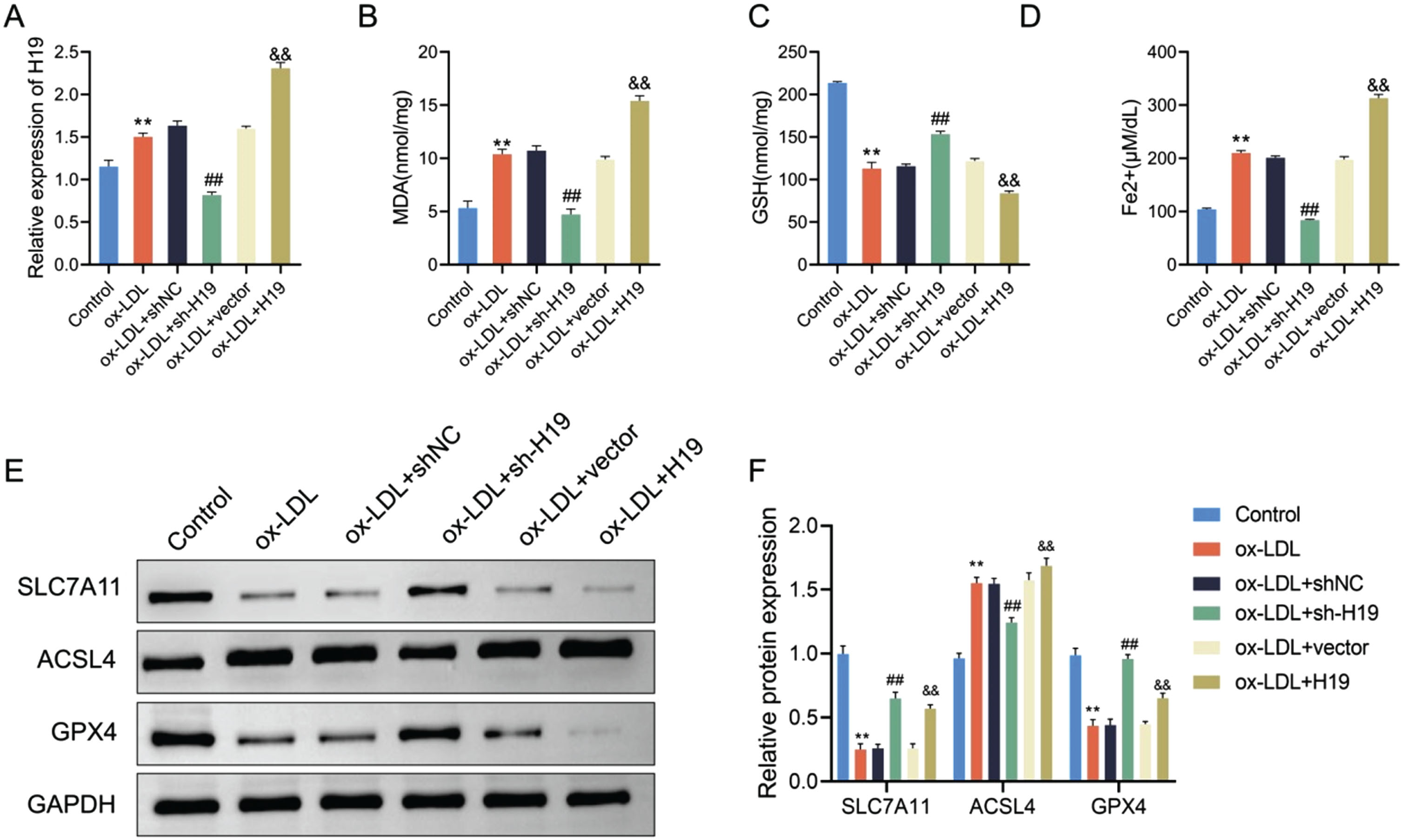
Role of lncRNA H19 in ox-LDL-treated HAECs ferroptosis. A, The expression level of H19 in control group, ox-LDL group, ox-LDL + shNC group, ox-LDL + sh-H19 group, ox-LDL + vector group and ox-LDL + H19 group was detected by qRT-PCR. B-C, The biochemical kit was used to detect the levels of MDA (B) and GSH (C) in HAECs in Control group, ox-LDL group, ox-LDL + shNC group, ox-LDL + sh-H19 group, ox-LDL + vector group and ox-LDL + H19 group. D, The level of Fe2+ in HAECs in Control group, ox-LDL group, ox-LDL + shNC group, ox-LDL + sh-H19 group, ox-LDL + vector group and ox-LDL + H19 group was detected. E-F, Western blot was used to detect the protein expression levels of SLC7A11, ACSL4 and GPX4 in HAECs in Control group, ox-LDL group, ox-LDL + shNC group, ox-LDL + sh-H19 group, ox-LDL + vector group and ox-LDL + H19 group. **indicates comparison with Control, and p < 0.01; ##indicates comparison with ox-LDL + shNC comparison, and p < 0.01; & & comparison with ox-LDL + vector, and p < 0.01.
The aforementioned results indicated that the upregulation of H19 in response to ox-LDL may serve as a signaling factor triggering ferroptosis. To corroborate this hypothesis, Erastin, a ferroptosis inducer, was utilized in this investigation to incubate ox-LDL-treated HAECs, aiming to elucidate the interplay between H19 expression and ferroptosis. After silencing H19 in ox-LDL-treated HAECs, alterations in cellular oxidative stress levels were observed. The findings indicated that compared to the ox-LDL + sh-H19 group, the ox-LDL + sh-H19 + Erastin group exhibited a notable increase in MDA levels in HAECs, alongside a significant decrease in GSH levels (Fig. 3A–B). Additionally, the concentration of ferrous ions in HAECs within the ox-LDL + sh-H19 + Erastin group was markedly higher compared to the ox-LDL + sh-H19 group Fig. 3C. Western blot analysis revealed that the protein levels of SLC7A11 and GPX4 were notably reduced in the ox-LDL + sh-H19 + Erastin group in contrast to the ox-LDL + sh-H19 group, while the protein expression of ACSL4 was significantly elevated Fig. 3D–E. Consequently, Erastin was found to counteract the decline in ferroptosis levels induced by lncRNA H19 in ox-LDL-treated HAECs.
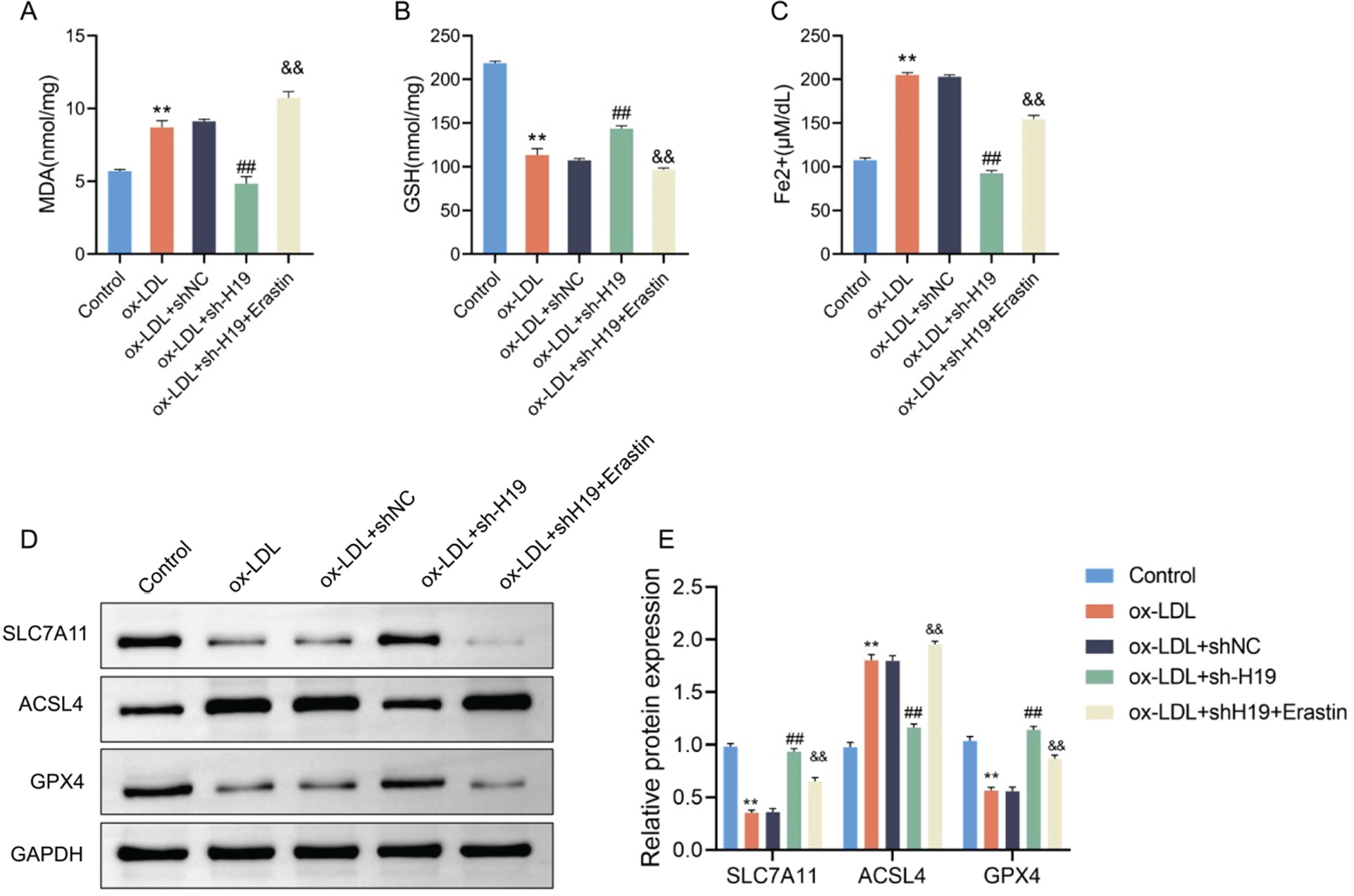
The effect of lncRNA H19 knockdown on ferroptosis in ox-LDL-treated HAECs can be reversed by Erastin. A–B, The biochemical kit was used to detect the levels of MDA (A) and GSH (B) in the control group, ox-LDL group, ox-LDL + shNC group, ox-LDL + sh-H19 group, and ox-LDL + sh-H19 + Erastin group. C, The level of Fe2+ in HAECs in Control group, ox-LDL group, ox-LDL + shNC group, ox-LDL + sh-H19 group, ox-LDL + sh-H19 + Erastin group was detected. D-F, Western blot was used to detect the protein expression levels of SLC7A11, ACSL4 and GPX4 in HAECs in Control group, ox-LDL group, ox-LDL + shNC group, ox-LDL + sh-H19 group, and ox-LDL + sh-H19 + Erastin group. **indicates comparison with Control, and p < 0.01; ##indicates comparison with ox-LDL + shNC comparison, and p < 0.01; & & comparison with ox-LDL + sh-H19, and p < 0.01.
During the process of AS progression, imbalanced levels of inflammatory cytokines serve as triggering elements for dysfunction in arterial vascular endothelial cells. The findings presented in this study suggest that H19 plays a crucial role as a signaling molecule in facilitating ox-LDL-induced ferroptosis in HAECs. Consequently, the study examined the impact on HAECs’ functionality, ability to form tubes, and inflammatory reaction following alterations in H19 expression and ferroptosis levels. Initially, as per the CCK-8 assay results, the viability of HAECs in the ox-LDL + sh-H19 group exhibited a notable increase compared to the ox-LDL + sh-H19 group, while it demonstrated a significant decrease in comparison to the ox-LDL + sh-H19 group (Fig. 4A). Subsequently, the tube formation assay indicated a substantial enhancement in the tube formation ability of HAECs within the ox-LDL + sh-H19 group when contrasted with the ox-LDL + shNC group, and a marked reduction in the ox-LDL + sh-H19 + Erastin group in relation to the ox-LDL+sh-H19 group (Fig. 4B). Furthermore, the ELISA analysis revealed a significant decrease in the levels of TNF-α, IL-6, and IL-1β in the supernatant of HAECs culture within the ox-LDL + sh-H19 group as opposed to the ox-LDL + sh-H19 group, whereas a substantial increase was observed in the levels of TNF-α, IL-6, and IL-1β in the supernatant of HAECs culture within the ox-LDL+sh-H19 + Erastin group compared to the ox-LDL + sh-H19 group (Fig. 4C–E). These findings indicate that the downregulation of H19 mitigates dysfunction and inflammation levels in ox-LDL-treated HAECs by suppressing ferroptosis.

LncRNA H19 knockdown attenuates the dysfunction and inflammation levels of ox-LDL-treated HAECs by inhibiting ferroptosis. A, CCK-8 assay was used to detect the survival rate of HAECs in the Control group, ox-LDL group, ox-LDL + shNC group, ox-LDL + sh-H19 group, and ox-LDL + sh-H19 + Erastin group. B, Tube formation assay was performed to detect the tube formation ability of HAECs in Control group, ox-LDL group, ox-LDL + shNC group, ox-LDL + sh-H19 group, and ox-LDL + sh-H19 + Erastin group. C-E, The levels of TNF-α (C), IL-6 (D) and IL-1β (E) in the supernatant of HAECs culture in Control group, ox-LDL group, ox-LDL + shNC group, ox-LDL + sh-H19 group, and ox-LDL + sh-H19 + Erastin group were detected by ELISA. **indicates comparison with Control, and p < 0.01; ##indicates comparison with ox-LDL + shNC comparison, and p < 0.01; & & comparison with ox-LDL + sh-H19, and p < 0.01.
Endothelial cells found in the arterial vasculature are extensively spread across the vascular system and play a crucial role in preserving cardiovascular balance. It is believed that programmed cell death is linked to various pathological conditions [24]. Endothelial dysfunction resulting from the programmed cell death of arterial vascular endothelial cells, along with localized endothelial detachment and unregulated thrombotic processes, facilitates the accumulation of lipid and fibrous materials within the walls of major and intermediate arteries, thereby fostering the development of atherosclerosis [25, 26]. It is evident that the modulation of arterial vascular endothelial cell death regulation will be pivotal in the prevention and management of atherosclerosis. This study focuses on the role of lncRNA H19 in ferroptosis and its impact on endothelial cell injury induced by ox-LDL, a key factor in atherosclerosis progression.
Ferroptosis, a type of regulated cell death driven by iron accumulation and lipid peroxidation [27, 28]. Current research indicates that ferroptosis plays a significant role in the progression of atherosclerosis [29], however, the precise molecular pathways involved remain to be fully elucidated. To investigate this further, ox-LDL-induced HAECs were employed as a model for atherosclerosis. Our findings demonstrate that ox-LDL exposure reduces HAEC viability and induces ferroptosis, evidenced by increased MDA levels and decreased GSH levels. The ferroptosis inhibitor Fer-1 mitigated these effects, confirming the involvement of ferroptosis. Ferroptosis is a process that enhances oxidative reactions leading to an oxidative stress imbalance, resulting in the collapse of cell membranes and mitochondrial membranes [8]. The buildup of ferrous ions from ox-LDL exposure facilitates the generation of lipid hydroperoxides from polyunsaturated fatty acids via ACSL4, generating harmful lipid radicals that damage cells [30]. The ferroptosis process also impedes the activation of GPX4, the primary protective mechanism against lipid peroxidation [31]. Likewise, SLC7A11 functions as a cystine/glutamate counter transporter, providing intracellular reducing power to counteract excessive oxidation induced by ferroptosis [32]. Our study assessed the expression levels of ferroptosis-related proteins (SLC7A11, ACSL4, GPX4), revealing that ox-LDL reduced SLC7A11 and GPX4 protein levels while increasing ACSL4 expression. Introducing a ferroptosis inhibitor mitigated the damage caused by ox-LDL, indicating ferroptosis’s role in atherosclerosis progression.
LncRNAs are significant regulators in diverse cellular processes, influencing ferroptosis [33]. Previous research found that LINC00336 functions as a ceRNA to suppress ferroptosis, while lncRNA P53RRA promotes ferroptosis by activating the p53 pathway [34, 35]. In our investigation, suppressing lncRNA H19 decreased ferroptosis levels in ox-LDL-incubated HAECs, while augmenting lncRNA H19 significantly elevated ferroptosis levels. Reintroducing Erastin, a ferroptosis inducer, further confirmed the role of lncRNA H19 in ox-LDL-induced damage. These findings underscore the pivotal role of the upregulation of lncRNA H19 in the induction of ferroptosis in HAECs following exposure to ox-LDL. Previous research indicated that suppressing lncRNA H19 in endothelial cells hinders angiogenesis and triggers LDL cholesterol synthesis within foam cells, suggesting that lncRNA H19 causes endothelial cell angiogenesis irregularities through ferroptosis [36, 37]. Our findings support this hypothesis, highlighting lncRNA H19 as a pivotal signaling molecule fostering ferroptosis in atherosclerosis. Furthermore, the regulatory influence of lncRNA H19 on ferroptosis exhibits a dual nature. This duality may stem from variations in gene expression levels across distinct cell types and the array of targets for lncRNA H19 [38]. In summary, lncRNA H19 emerges as a pivotal signaling molecule that fosters ferroptosis in the context of atherosclerosis.
However, this research has several limitations. First, the precise molecular mechanisms through which lncRNA H19 facilitates ferroptosis remain unclear. Although we identified changes in protein levels associated with ferroptosis, the specific pathways and interactions require further elucidation. Additionally, our study primarily focused on endothelial cells; the role of lncRNA H19 in other cell types involved in atherosclerosis, such as macrophages and smooth muscle cells, was not explored. Another limitation is the in vitro nature of our experiments. While ox-LDL-induced HAECs provide a useful model for studying atherosclerosis, in vivo studies are necessary to validate our findings and understand the systemic effects of lncRNA H19 modulation in a more complex biological environment. The influence of other factors present in the vascular milieu, such as shear stress and interaction with other cell types, should also be considered in future studies.
Conclusion
In the context of atherosclerosis, our research has revealed that lncRNA H19 plays a significant role in promoting ferroptosis within endothelial cells, thereby contributing to cellular dysfunction through the escalation of oxidative stress and inflammatory reactions. Consequently, targeting lncRNA H19 may hold promise as a viable therapeutic strategy for addressing atherosclerosis.
Ethics approval and consent to participate
Not applicable.
Consent for publication
Not applicable.
Footnotes
Acknowledgments
Not applicable.
Funding
This study is supported by Project approval number of the National Natural Science Foundation of China (82260094); Guizhou Provincial Science and Technology Foundation Project (Approval Number: Qiankehe Fundamentals ZK [2021] General 355). Guizhou Provincial Health Science and Technology Fund (gzwjkj 2020-1-065, Feng Tang).
Conflict of interest
The authors have no relevant financial or non-financial interests to disclose.
Data availability
The datasets generated during and/or analyzed during the current study are available from the corresponding author on reasonable request.
Author contributions
Feng Tang and Yongyao Yang conceived and designed the study. Feng Tang and Yongyao Yang conducted the study. Longhai Tian, and Shijia Xu contributed to data acquisition. Xiao han Zhu., Sen Yang and Huan Zeng analyzed the data. Longhai Tian, and Shijia Xu interpreted the data. Feng Tang and Yongyao Yang edited the manuscript draft. Feng Tang and Yongyao Yang reviewed and edited the manuscript. All authors have read and approved the manuscript.