






Abstract
We recently demonstrated that soluble forms of the amyloid-β protein precursor (sAβPP) assemble into multimeric complexes in cerebrospinal fluid (CSF), which contributes to the underestimation of specific sAβPP species when assessed by ELISA. To circumvent this issue, we analyzed by SDS-PAGE large fragments of sAβPP and their variants in the CSF from Alzheimer’s disease (AD; n = 20) and control (n = 20) subjects, probing with specific antibodies against particular domains. Similar levels of sAβPPα and sAβPPβ protein were found in CSF samples from AD and controls, yet there appeared to be a shift in the balance of the soluble full-length AβPP (sAβPPf) species in AD samples, with a decrease in the proportion of the lower (∼100 kDa) band relative to the upper (∼120 kDa) band. Similar differences were observed in the contribution of the major KPI-immunoreactive AβPP species. CSF samples also displayed differences in the correlations of AβPP species with classical AD biomarkers, particularly with respect to the Aβ42 peptide. The differences reveal alterations that probably reflect pathophysiological changes in the brain.
INTRODUCTION
Early and accurate diagnosis of Alzheimer’s disease (AD) is essential to establish timely and effective therapies, and to follow the progression of the disease. Due to its stipulated role in the disease [1], the amyloid-β peptide (Aβ) that accumulates in the brain parenchyma as senile plaques, which is reflected by reduced lumbar cerebrospinal fluid (CSF) concentrations of the protein, is likely to be one of the more specific biomarkers for AD. Increase in the production and deposition of Aβ42 in the AD-affected brain probably occurs in parallel decades prior to the appearance of symptoms [2]. Accordingly, longitudinal changes in CSF Aβ species are difficult to interpret and additional biomarkers are needed, especially at early disease stages. Similarly, it is difficult to gain significant information about the potential changes in CSF Aβ levels that could help assess experimental therapies aimed at reducing Aβ production [3].
Other possible core biomarkers for AD are the ecto-domain fragments of amyloid-β protein precursor (AβPP) that result from amyloidogenic and non-amyloidogenic AβPP processing by secretases. AβPP is a large type I transmembrane glycoprotein that is alternatively cleaved by α-secretase (in the non-amyloidogenic pathway) or β-secretase (the amyloidogenic pathway), which provokes the secretion of large sAβPPα and sAβPPβ fragments, respectively. The C-terminal fragments (CTF) that persist in the membrane after such cleavage are then processed by γ-secretase. In the amyloidogenic pathway, further γ-secretase cleavage also releases the Aβ peptide, whereas when AβPP molecules are cleaved by α-secretase within the Aβ domain, the generation of this Aβ peptide is precluded [4]. Apart from these fragments, soluble full-length AβPP (sAβPPf) is also known to exist in the CSF and plasma [5–7].
In this context, sAβPPα and sAβPPβ have been studied as potential biomarkers for AD, yet no consistent changes in the CSF levels of these fragments have been identified [8]. In fact, the pattern of AβPP species in the CSF is even more complex than that indicated above, as there are three AβPP isoforms produced in the brain as a result of alternative exon splicing [9]. These splice variants encode: AβPP695, the neuronal predominant variant; AβPP751, mostly expressed in astrocytes and glial cells, and harboring an amino acid insert homologous to a Kunitz-type trypsin inhibitor (KPI); and AβPP770, which is widely expressed in peripheral tissues but minimally in the brain, and that also contains the KPI domain and an additional domain with homology to the MRC OX-2 antigen [10]. Early western blot studies demonstrated that the soluble KPI-containing and KPI-free AβPP isoforms are readily detected in CSF [11, 12].
The evaluation of biomarkers in CSF presently relies on ELISA immunoassays. However, we recently demonstrated that all the sAβPP species in the CSF assemble into heteromers, including sAβPPα and sAβPPβ, and this affects their assessment by the available ELISAs [7]. In order to circumvent this issue and with the goal of evaluating the potential of CSF-sAβPP species to serve as AD biomarkers, we evaluated the levels of large sAβPP fragments and their variants using denaturing SDS-PAGE. However, the expected pattern of CSF-sAβPP fragments resolved in this way, and detected in western blots, is intricate, with several different forms and large proteolytic fragments displaying similar molecular masses of which make them mostly indistinguishable by electrophoretic analysis. Thus, we performed the characterization of CSF-sAβPP species by probing with specific antibodies for AβPP raised against different pan-specific domains.
MATERIALS AND METHODS
Patients
Lumbar CSF samples were obtained from 20 patients with AD (5 men and 15 women, mean age 69±3 years) and 20 age-matched controls (12 men and 8 women, mean age 69±2 years). The AD patients had cognitive deterioration and a CSF biomarker profile indicative of AD, including increased T-tau (>400 pg/mL) and P-tau (>70 pg/mL) together with low Aβ42 (<550 pg/mL) concentrations in CSF; while controls were patients who sought medical advice because of memory complaints but had normal levels of all three classical CSF biomarkers (T-tau <350 pg/mL, P-tau <60 pg/mL, and Aβ42 >550 pg/mL), thereby excluding early AD pathology [13]. The CSF samples used for the present study were de-identified leftover aliquots from clinical routine analyses, following a procedure approved by the Ethics Committee at University of Gothenburg. This study was also approved by the ethics committee of the Miguel Hernandez University.
Western blotting
CSF samples (30μL) were boiled at 95°C for 5 min and resolved by 7.5% SDS-PAGE. The sAβPP species in the samples were detected with the following antibodies: a polyclonal anti-AβPP C-terminal antiserum (1:2,000; Sigma Aldrich, St. Louis, MO, USA; named here Sigma-Ct); a monoclonal anti-AβPP 6E10 that reacts with both sAβPPf and sAβPPα, as well as with free Aβ (1:2,000; Covance; named here 6E10); a monoclonal anti-AβPP N-terminal (1:500; Sigma Aldrich; named here Sigma-Nt), a polyclonal anti-sAβPPβ antiserum specific to the C-terminus of sAβPPβ (1:50; IBL, Hamburg, Germany; named here IBL-β); a monoclonal anti-sAβPPα specific to the C-terminus of sAβPPα (1:50; IBL; named here IBL-α); and a polyclonal anti-KPI antiserum specific to the KPI domain of AβPP (1:500; Millipore; named here KPI). When appropriate, a control CSF sample was used to normalize the immunoreactive signal between immunoblots. Band intensities were analyzed using LI-COR software (Image Studio Lite).
To assess the specificity of the pan-specific AβPP antibodies, CSF samples were resolved to simultaneously detect the immunoreactivity of sAβPPα and sAβPPβ. The blots were then probed with the appropriately conjugated secondary antibodies (IRDye 680RD goat anti-mouse and IRDye 800CW goat anti-rabbit: LI-COR Biosciences GmbH, Bad Homburg, Germany) and analyzed on an Odyssey Clx Infrared Imaging System (LI-COR).
To test the identity of the 120 and 100 kDa KPI bands, CSF samples were resolved to simultaneously detect the immunoreactivity of two antibodies in different combinations: Sigma-Nt/KPI and 6E10/KPI. The blots were probed with the appropriate conjugated secondary antibodies (IRDye 800CW goat anti-mouse and IRDye 680RD goat anti-rabbit: LI-COR Biosciences GmbH, Bad Homburg, Germany) and analyzed on an Odyssey Clx Infrared Imaging System (LI-COR).
Measurement of T-tau, P-tau, and Aβ42 by ELISA
Total tau (T-tau), phosphorylated tau (P-tau) and Aβ1 - 42 (Aβ42) concentrations in CSF were measured using INNOTEST ELISA methods (Fujirebio Europe). All samples were analyzed as part of clinical routine, by board certified laboratory technicians following strict procedures for batch-bridging, analyses and quality control of individual ELISA plates, as described previously in detail [14].
Statistical analysis
All the data were analyzed using SigmaStat (Version 2.0; SPSS, Inc.) using a Student’s t test (two-tailed) or a Mann-Whitney U test for single pairwise comparisons, and determining the exact p values. The results are presented as means±SEM and the correlation between variables was assessed by linear regression analyses.
RESULTS
Unaltered sAβPPα and sAβPPβ levels in AD CSF
To date, studies of sAβPPα and sAβPPβ levels in CSF as potential biomarkers for AD have generated inconclusive results. These studies have generally employed ELISA to measure these proteins. Since sAβPP exists as heteromers in CSF [7], the detection of AβPPα and sAβPPβ by ELISA is likely to be compromised. To avoid potential interference of the heteromeric complexes formed in CSF, we determined the levels of sAβPPα and sAβPPβ after their separation by SDS/PAGE and detected these proteins in western blots probed whit different pan-specific antibodies (Supplementary Figure 1A).
We first measured the sAβPPα and sAβPPβ in CSF from AD and control subjects characterized for classical biomarkers (see Table 1). These soluble AβPP fragments were detected using antibodies that recognize their exclusive C-terminal domains; the specificity of which was corroborated by a simultaneous assay of fluorescence (Supplementary Figure 1B). Probing immunoblots with the antibodies against sAβPPα and sAβPPβ revealed a similar banding pattern, with a broad lower band of approximately 100–110 and a faint upper band of 120–130 kDa. These bands are referred to here always as “100 kDa” and “120 kDa” bands; nonetheless, minor differences in electrophoretic migration were detected when they were probed with different antibodies. There was moderate co-localization of only the 120 kDa but not the more abundant 100 kDa band when the membranes were probed with the sAβPPα and sAβPPβ antibodies together (Supplementary Figure 1B). There were no clear differences between AD and control subjects in the lower or upper bands for sAβPPα or sAβPPβ (Fig. 1A, B), and thus, we also attempted to assess whether there was a relative difference in the balance between the 100 and 120 kDa bands. As such, we calculated the ratio between the immunoreactivity of the lower and the upper AβPP bands (100 kDa/120 kDa ratio, or AβPPr), and again, there was apparently no difference in the AβPPr between control and AD subjects (Fig. 1A, B).
Clinical and demographic data, and classic CSF biomarkers
F, female; M, male. *p < 0.001.
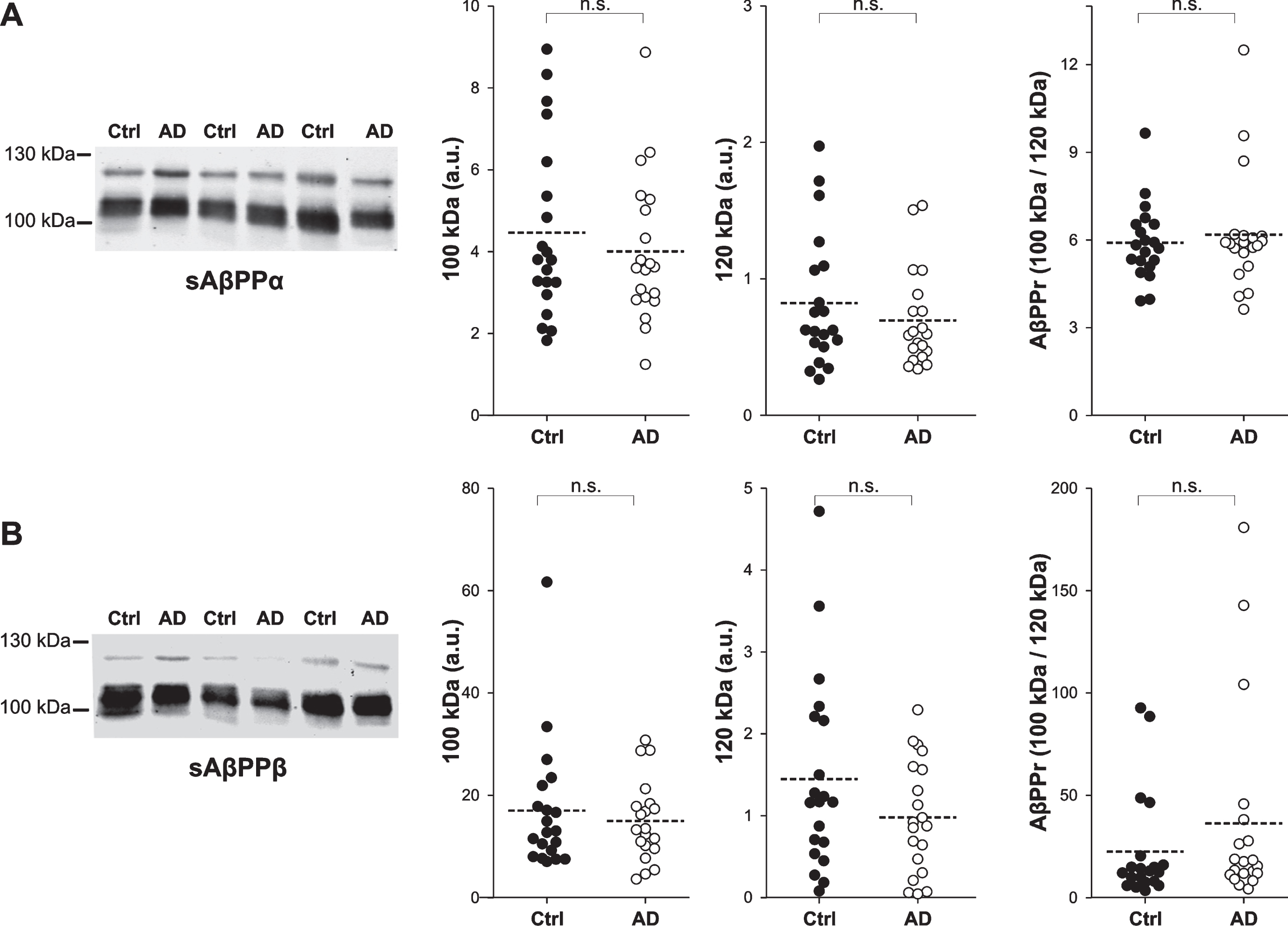
Levels of sAβPPα and sAβPPβ in AD CSF. A) Representative western blots of human CSF samples from age-matched cognitively normal controls (Ctrl, closed symbol; n = 20) and AD patients (open symbol; n = 20) probed with an antibody against (A) sAβPPα (IBL-α) or (B) sAβPPβ (IBL-β). Densitometric quantification of the lower (∼100 kDa) and the upper (∼120 kDa) AβPP bands is shown, and the ratio derived from the immunoreactivity for the lower band relative to that for the upper band in each sample (AβPPr: 100 kDa/120 kDa). The data represent the means±SEM (calculations performed in duplicate). n.s.: non-significant p value.
Interestingly, the amounts of sAβPPα or sAβPPβ appear to be tightly correlated in CSF [15]; but we had previously demonstrated that heteromers containing sAβPPβ and sAβPPα exist in CSF, which may underlie this correlation [7]. Anyhow, the SDS/PAGE data confirms the correlation between sAβPPα and sAβPPβ for the lower and upper bands, both in the control and AD groups (Supplementary Figure 2). In addition, the amount of the lower and upper sAβPPα-immunoreactive bands correlated with the levels of Aβ42 in the control group, while this correlation was not evident in the AD subjects. By contrast, the levels of sAβPPα were correlated with those of T-tau in pathological but not in control samples (Fig. 2A, B). Similarly, correlations between the lower and upper sAβPPβ bands with T-tau were only detected in AD samples (Fig. 2C, D), while there were no clear correlations between sAβPPβ and Aβ42 in either the control or AD subjects.
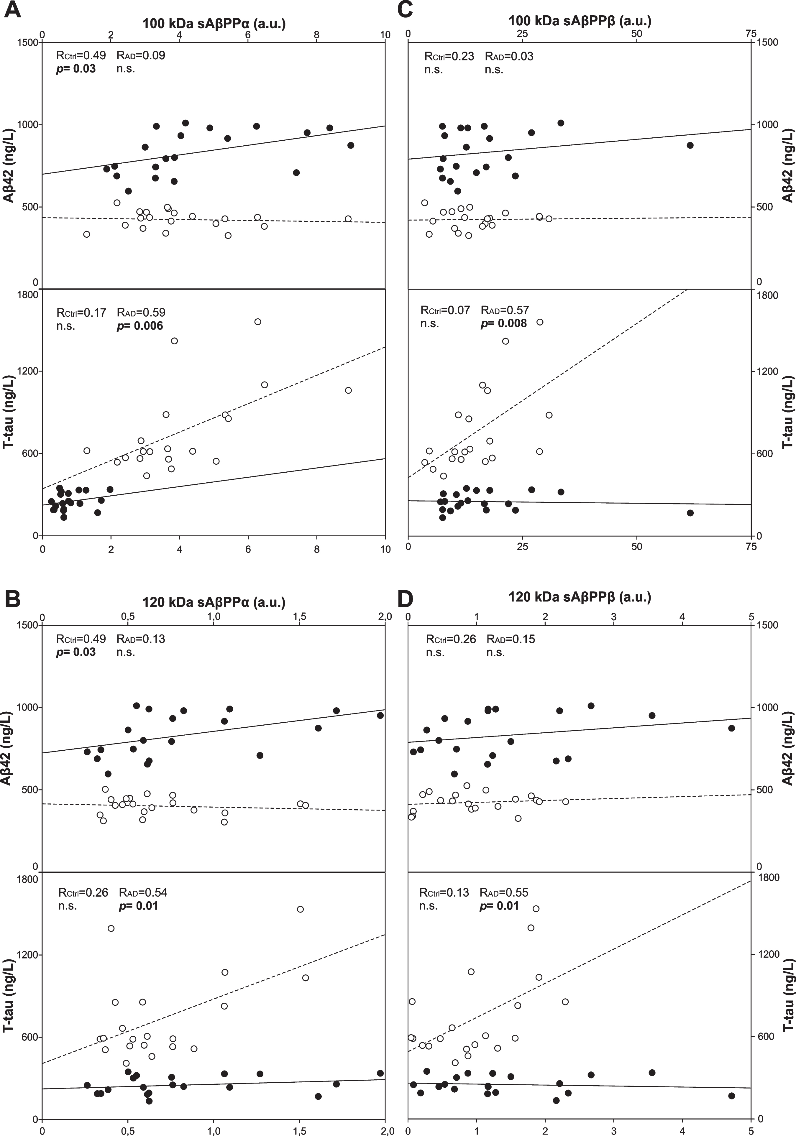
Correlation of sAβPPα and sAβPPβ with Aβ42 and T-tau levels in CSF samples. Levels for the lower (∼100 kDa; A and C) and upper band (∼120 kDa; B and D) of sAβPPα (A, B) and sAβPPβ (C, D) in the samples from age-matched cognitively normal controls (Ctrl: closed symbol, solid lines) and AD patients (open symbol, dotted lines). A linear regression was used to assess the correlation between the Aβ42 and T-tau levels obtained by ELISA (see Table 1). Correlations for P-tau were similar to those displayed for T-tau (not shown). The linear regression coefficient (R) and p values for each correlation are shown (n.s., non-significant p value).
Altered balance of 100 and 120 kDa sAβPP bands in AD CSF
In accordance with our previous study, an antibody raised against the unprocessed C-terminal of the full-length AβPP also detected proteins in CSF resolved by SDS-PAGE that were similar in molecular mass to the large sAβPP fragments (Fig. 3A). These bands of ∼100 and 120 kDa were characterized as the lower and upper sAβPPf bands [7]. When the levels of sAβPPf were determined in fresh aliquots of the control and AD CSF samples, there were no significant differences for the lower or the upper band (Fig. 3A). By contrast, an imbalance was evident in the pathological group regarding the proportion of the lower band with respect to the upper band, producing a change in the AβPPr (Fig. 3A). In both the control and AD groups, there was a correlation between the lower sAβPPf bands and the lower sAβPPα and sAβPPβ bands, and while the uppers bands were also generally related, this was not the case for the sAβPPf and sAβPPα in control CSF (Fig. 3B). None of the sAβPPf bands were correlated with the levels of Aβ42 in the control or AD groups, although the sAβPPf bands were related to the T-tau levels in the AD but not the control CSF samples (Fig. 3C).
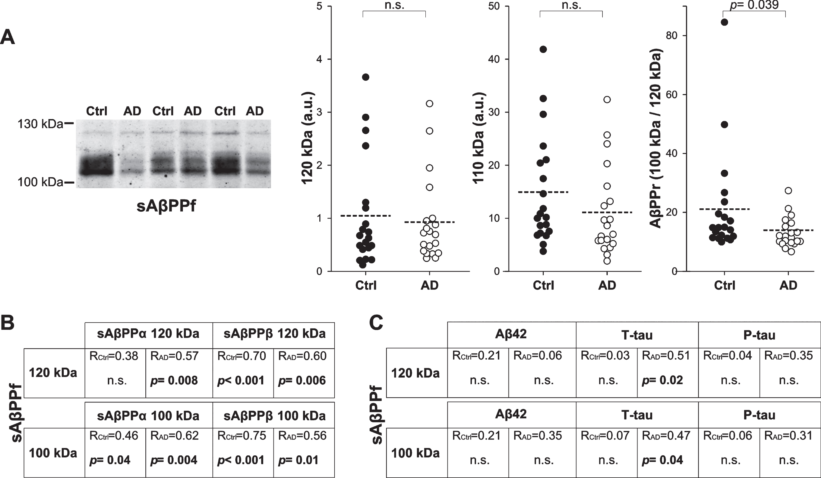
The levels of sAβPPf immunoreactivity in CSF from controls and AD subjects. The sAβPPf immunoreactivity in CSF samples from 20 controls (Ctrl; closed symbol) and 20 AD patients (open symbol). A) Representative blot of sAβPPf probed with an anti-AβPP C-terminal antibody (Sigma-Ct) and densitometric quantification for the ∼100 and 120 kDa bands. The AβPPr was calculated as described in Fig. 1. The data represent the means±SEM and the p values (performed in duplicate). B) Linear regression coefficient (R) and p values for the correlation of sAβPPf with sAβPPα or sAβPPβ (lower and upper bands). C) Linear regression coefficient (R) and p values (n.s., non-significant p value) for correlation of sAβPPf lower and upper bands with the classical AD biomarkers Aβ42, T-tau, and P-tau.
Altered balance of KPI amyloid-β proteins in AD CSF
Finally, an antibody raised against the KPI domain present in the AβPP751 and AβPP770 variants was used to probe the CSF samples. KPI immunoreactive bands of ∼100 and 120 kDa were detected in CSF, similar molecular masses to those observed with anti-AβPP antibodies (Fig. 4A). Since the KPI domain is present in several other brain glycoproteins, we first checked the identity of these bands in a simultaneous fluorescence assays. For this assay, we first used the anti KPI antibody combined with the anti AβPP N-terminal antibody, and then with the 6E10 antibody that recognized an epitope in the N-terminal region of Aβ peptide, detecting sAβPPf and sAβPPα but not sAβPPβ (Supplemental Figure 3). Co-localization between the KPI and the AβPP N-terminal antibodies confirmed the identity of the 100 and 120 kDa KPI-immunoreactive bands as sAβPP species (sAβPPKPI). As co-localization of the KPI antibody with 6E10 was only evident for the 120 kDa and not the 100 kDa band, this sAβPPKPI band probably corresponds to sAβPPβ (or another large N-terminal fragment) but not sAβPPα. Simultaneous assays of fluorescence using a pan-specific sAβPPβ antibody with the KPI antibody cannot be performed because both are rabbit polyclonal antisera and they cannot therefore be distinguished with different secondary antibodies. Nevertheless, 110-100-kDa forms of sAβPPKPI have been reported previously in human brain and cultured cells [16–18].
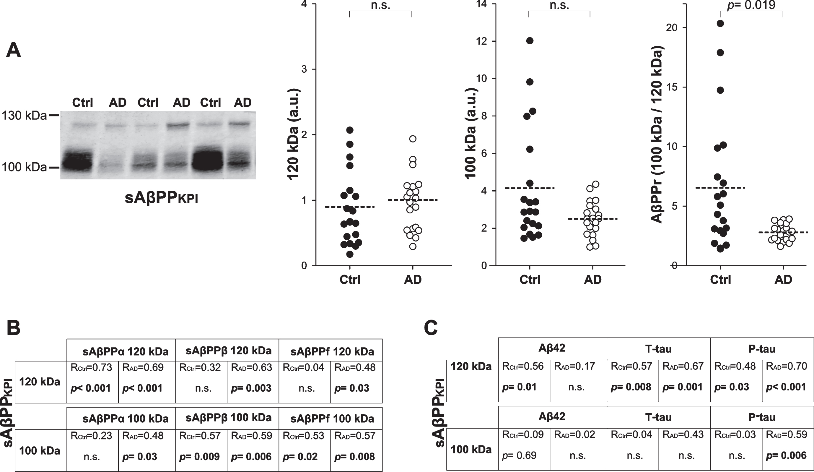
The sAβPPKPI immunoreactivity in CSF from controls and AD subjects. A) Representative blot and densitometric quantification for the ∼100 and 120 kDa sAβPPKPI bands probed with an anti-KPI antibody in 20 controls (Ctrl; closed symbol) and 20 AD patients (open symbol). The AβPPr was calculated as described in Fig. 1, and the data represent the means±SEM and p values (performed in duplicate). B) Linear regression coefficient (R) and p values for the correlation of sAβPPKPI with sAβPPα, sAβPPβ, or sAβPPf (lower and upper bands). C) Linear regression coefficient (R) and p values (n.s., non-significant p value) for the correlation of sAβPPKPI lower and upper bands with the classical AD biomarkers Aβ42, T-tau, and P-tau.
When aliquots of CSF samples were probed with the anti-KPI antibody, no significant changes were evident between AD and control subjects (Fig. 4A), although again the AβPPr for sAβPPKPI revealed a possible imbalance between the 100 kDa and the 120 kDa band in AD CSF (Fig. 4A). Moreover, when the correlation of the sAβPPKPI bands with the sAβPPα or sAβPPβ fragment or with sAβPPf was assessed, the correlations in the CSF from the control subjects were poorer than those observed in the AD group (Fig. 4B). Again, the correlations of sAβPPKPI bands with classical AD biomarker indicate a stronger relationship between these variants and T-tau or P-tau than for Aβ42 (Fig. 4C).
DISCUSSION
In this study, we attempted to define differences in the sAβPP species in the CSF of AD patients that may serve as biomarkers for this disease. Similar studies have been performed previously, as CSF levels of sAβPPα and sAβPPβ are thought to be promising candidate biomarkers. However, most of these studies were based on ELISA assays, which have produced some conflicting results. The earliest studies based on western blotting analysis indicated that the AD pathology is associated with a slight decrease in sAβPPα in the CSF [11, 19]. Similar decreases in sAβPPα in the CSF were subsequently observed using ELISA assays [12, 20–22] or no significant change [23–26], although its levels were shown to increase in other studies [15, 27–29]. Unexpectedly, most ELISA studies showed no change or even a decrease in sAβPPβ in the CSF in association with AD [21, 31], in contrast to the increase reported elsewhere [29]. A recent study of sAβPPα and sAβPPβ levels in a large number of postmortem ventricular CSF samples indicated there was a large overlap, with both large sAβPP fragments upregulated in AD CSF compared to controls [32].
Several issues may put in doubt the reliability of ELISA measurements of CSF AβPP species. First, we have described the presence of sAβPPf in human CSF [7], and some of the ELISA kits employed for assessing sAβPPα rely on the 6E10 antibody or similar antibodies that recognize the N-terminal part of the Aβ peptide [25]. Accordingly, these antibodies will bind to sAβPPα but also to sAβPPf, which may be responsible for some of the discrepancies reported with different ELISA kits. Moreover, AβPP can undergo alternative proteolytic processing, potentially generating alternative proteolytic metabolites [33]. All the canonical and non-canonical extracellular N-terminal AβPP fragments are present in human CSF [11, 34–38] and thus, other N-terminal AβPP fragments generated by alternative proteolytic processing of AβPP may also be responsible for the divergent results obtained by ELISA due to the different antibody specificities for distinct domains. Furthermore, sAβPP species associate in heteromeric complexes [7] and the existence of such heteromers may reduce the accuracy of ELISA, even if they are based on pan-specific antibodies against the exclusive C-terminus of sAβPPα and sAβPPβ. For all these reason, and despite the disadvantages of western blotting for quantitative analysis, we used a SDS-PAGE based approach to study these proteins here instead of ELISA.
Such an SDS/PAGE based analysis of sAβPP species allows the different species carrying a common epitope of the protein to be identified separately, especially if they have a distinct molecular mass. AβPP is usually detected in brain extract by SDS/PAGE as a group of 100–130 kDa proteins [16] the differences in molecular mass possibly representing splice variants of the predominant AβPP695, and of the KPI-forms AβPP751 and AβPP770. However, small differences in electrophoretic migration can also be attributed to differences in glycosylation [39] or they may reflect immature forms of the protein [40]. Moreover, sAβPPα or sAβPPβ are predicted to be only ∼5–10 kDa smaller than full-length AβPP in the CSF [34]. Accordingly, large sAβPP fragments cannot be discriminated in CSF by SDS/PAGE and specific antibodies must be used to probe the blotted proteins. Recently, pan-specific antibodies against the C-terminus exclusive to sAβPPα and sAβPPβ were validated using peptide competitors [41]. In our hands, simultaneous fluorescence assays also demonstrated the specificity of these pan-specific sAβPPα and sAβPPβ antibodies. As different sAβPP species co-immunoprecipitate due to the formation of heteromers, this restricts the verification of the immunoreactive bands by immunoprecipitation [7]. Here, we circumvent this issue by simultaneously assaying the fluorescence associated with antibody binding. However, while the failure of two antibodies to co-localize indicates they represent different proteins or species, co-location cannot be accepted as unquestionable proof of identity. Likewise, some combination of antibodies is limited by the different species in which they are generated.
In this extremely complex scenario, we affronted the biochemical dissection of sAβPPα, sAβPPβ, sAβPPf, and sAβPPKPI variants in AD CSF compared to age-matched controls. After ruling out and excluding individuals with competing causes of dementia, the selection of the controls was based on a control biomarker profile for T-tau, P-tau, and Aβ42. Follow-up studies demonstrate that stablished cut-points result >90% specific for AD [13]. No changes were observed in sAβPPα and sAβPPβ in the CSF from AD and controls. Surprisingly, we corroborated the positive correlation between the sAβPPα and sAβPPβ reported using ELISA [15, 30], and that we had initially attributed to the existence of heteromers [7]. This positive correlation was not identified in an earlier study assessing sAβPPα and sAβPPβ levels in western blots, although this may be due to the use of the 6E10 antibody to identify sAβPPα [21]. More interestingly, in control samples we found different associations of sAβPPα and sAβPPβ with classical AD biomarkers, Aβ42, T-tau, or P-tau, and interestingly these correlations were apparently disturbed in AD samples. Characterization of these differences in the association of classical AD biomarkers with sAβPPf and sAβPPKPI variants confirmed the subtle alterations in the expression and proteolytic processing of AβPP in the pathological condition, meriting more detailed analysis with refined tools and protocols.
In addition, by defining a quotient that reflects the immunoreactivity of the 100 kDa and 120 kDa bands, the AβPPr, we found differences in the sAβPPf and sAβPPKPI variants between AD and control samples. The AβPPr highlights differences in the major 100 and 120 kDa bands that could not be envisaged through ELISA. A previous report in platelets also indicated differences in the evolution of different species and the interest in defining this kind of ratio between AβPP species [42]. The alteration in the AβPPr for sAβPPf reflects only unprocessed species and since the mechanism by which sAβPPf appears in CSF is yet to be determined, this imbalance may be difficult to interpret. A more complete characterization of the different species is necessary, as the 100 and 120 kDa bands could represent different glycoforms, or mature and immature forms of the same AβPP variant, as well as different splicing variants. This altered expression may also reflect post-translational processing. Intriguingly, the AβPPr for KPI species also revealed alterations in proteolytic processing since the 100 kDa sAβPPKPI band probably represents a sAβPPβ fragment.
Here, we justify the study of CSF-sAβPP by SDS/PAGE instead of ELISA, although the disadvantages of using SDS/PAGE interfere in the translation of a biomarker for clinical purposes. Western blotting is limited in the number of samples that can be assayed, and in the means to normalize the data and compare between assays. It is also a time-consuming technique, subject to inter-assay variation and the difficulties of using calibration curves to estimate the concentrations of the analytes. Therefore, it would be useful to refine ELISA protocols in order to evaluate a single species or AβPP fragment, and to improve the performance of such analyses. The possibility of introducing chemical pre-treatment of samples aimed at disaggregating oligomers should be investigated, as reported recently for Aβ [43]. Such modifications could make ELISA a more accurate procedure to measure sAβPP, overcoming the potential interference of heteromers. Anyhow, our data about differences in the contribution of major 100 and 120 kDa bands for different AβPP species in AD CSF, as well differences in the association of classical AD biomarkers respect to controls, must be subject to confirm by another techniques with greater reliability for the quantification. Further studies should also assess the specificity of the changes in CSF from patients with variety of senile dementias.
In conclusion, our data indicate that current ELISA protocols are not suitable to evaluate CSF AβPPs as biomarkers for AD. Although AβPP has been studied extensively, our current understanding of the complex patterns of AβPP expression in the brain, taking into account the splice variants, translational modifications and its intricate proteolytic processing, are unlikely to reflect the whole picture. Discerning the changes suffered by AβPP in the pathological AD brain should favor its use as a CSF biomarker. In this regard, developing new ELISA protocols will be challenging but together with a more complete biochemical study of sAβPPs in the CSF, may serve to provide alternative core biomarkers for AD.
Footnotes
ACKNOWLEDGMENTS
This study was funded in part by the EU BIOMARKAPD-Joint Programme on Neurodegenerative Diseases (JPND), by the Instituto de Salud Carlos III (ISCIII grant PI11/03026 and PI15/00665, co-financed by the Fondo Europeo de Desarrollo Regional under the aegis of JPND), and through CIBERNED (ISCIII).
