






Abstract
Background:
Idiopathic normal pressure hydrocephalus (iNPH) is a late onset, surgically treated progressive brain disease caused by impaired cerebrospinal fluid dynamics and subsequent ventriculomegaly. Comorbid Alzheimer’s disease (AD) seems to be frequent in iNPH.
Objective:
We aim to evaluate the role of AD-related polymorphisms in iNPH.
Methods:
Overall 188 shunt-operated iNPH patients and 688 controls without diagnosed neurodegenerative disease were included into analysis. Twenty-three single-nucleotide polymorphisms (SNPs FRMD4A [rs7081208_A, rs2446581_A, rs17314229_T], CR1, BIN, CD2AP, CLU, MS4A6A, MS4A4E, PICALM, ABCA7, CD33, INPP5D, HLA_DRB5, EPHA1, PTK2B, CELF1, SORL1, FERMT2, SLC24A, DSG2, CASS4, and NME8) adjusted to APOE were analyzed between groups by using binary logistic regression analysis. Neuroradiological characteristics and AD-related changes in the right frontal cortical brain biopsies were available for further analysis.
Results:
Logistic regression analysis adjusted to age, gender, and other SNPs indicated allelic variation of NME8 between iNPH patients and non-demented controls (p = 0.014). The allelic variation of NME8 was not related to the neuropathological changes in the brain biopsies of iNPH patients. However, periventricular white matter changes (p = 0.017) were more frequent in the iNPH patients with the AA-genotype, an identified risk factor of AD.
Conclusions:
Our findings increase the evidence that iNPH is characterized by genetic and pathophysiological mechanisms independent from AD. Considering that NME8 plays a role in the ciliary function and displays SNP-related diversity in white matter changes, the mechanisms of NME8 in iNPH and other neurodegenerative processes are worth further study.
INTRODUCTION
Idiopathic normal pressure hydrocephalus (iNPH) is a progressive brain disease caused by disturbance in the cerebrospinal fluid (CSF) dynamics resulting in ventriculomegaly and compression-induced stress of periventricular parenchyma [1, 2]. Classical clinical characteristics are impaired gait, cognitive decline, and urinary incontinence [1]. The only treatment currently available is a neurosurgically implanted CSF shunt, which usually alleviates symptoms in properly-selected patients [3 –5]. However, the long-term benefit of operative treatment is only modest for a number of patients due to comorbidities like Alzheimer’s disease (AD) [3 –7].
Despite having history dating back to the 1960s, the molecular mechanisms of iNPH remain to be discovered [1 , 6–8]. Concomitant AD seems to be frequent in patients with iNPH [7, 8]. However, the prevalence of APOE ɛ 4, the most common genetic risk factor of AD, in patients with iNPH and age-adjusted controls seems to be similar [9]. Furthermore, there is no benefit in shunting patients with solitary AD [10].
Familial occurrence of iNPH was first introduced by Portenoy et al., having discovered siblings with typical neurological symptomatology, neuroradiologically confirmed ventriculomegaly, and favorable response to shunt surgery [11]. Since then a number of pedigrees with multiple affected relatives have been documented [12 –18]. According to our recent nation-wide study, the potential familial occurrence of iNPH can be up to 16% [18]. Summing up these findings, there may be inherited traits predisposing patients to suboptimal CSF dynamics and ultimately manifesting as iNPH [18]. The first genetic finding associated with iNPH-related ventriculomegaly was the copy number loss of SFMBT1, a gene that is expressed e.g. in choroid plexus, which is the primary site of CSF formation [19, 20].
Synthesizing the idea of inheritability into molecular level, several hypotheses can be assessed. The increased prevalence of AD-related pathology among iNPH patients may theoretically be caused by inadequate β-amyloid clearance due to impaired CSF circulation [8]. Disproportional subarachnoid spaces may indicate a selective block of CSF absorption and activation of potential compensatory pathways of CSF flow, which may play a key role in the pathogenesis of iNPH [21, 22]. Considering ventriculomegaly in iNPH and other forms of hydrocephalus, the ependyma with potentially altered gene expression could also play a role in iNPH [21 –24]. Whether this hypothetical dysfunction would be the primary cause or reactive to CSF flow disturbance is also intriguing.
In this study, we examined 24 established AD-related single-nucleotide polymorphisms [25 –27] in patients with iNPH and healthy controls to further provide insights into the common molecular mechanisms of AD and iNPH.
MATERIALS AND METHODS
Ethics statement
Kuopio University Hospital (KUH) Research Ethical Committee approved the study. All patients gave an informed consent to the study. Patients with lower cognitive capacity were included with the support of the next of kin but also always asking and respecting patient’s own will.
Study sample
This study included 188 iNPH patients from Kuopio University Hospital (KUH) catchment area (Table 1). Patients were shunted during the years 1993–2009. Patients underwent a careful preoperative assessment carried out by a neurologist. At least one triad symptom with characteristic findings (Evans’ index >0.3) in CT or MRI was required in order for the patient to be included. Patients were further assessed in the Neurosurgical unit with additional diagnostic procedures, including a 24-h intracerebral pressure (ICP) monitoring with a perioperative right frontal cortical biopsy [8, 9]. All iNPH patients were shunted and the shunt-response was determined clinically after three months had passed from the shunt surgery by a neurosurgeon as changes in gait, urinary incontinence, and memory (no change, improvement, regression). The control group of 688 subjects was gender-adjusted without any signs of cognitive decline in neuropsychologicalexamination [9].
Clinical characteristics and brain biopsy findings in 188 shunt-operated iNPH patients
Neuropathological samples
Frontal cortical biopsies were fixed in formalin overnight, embedded in paraffin, and then sectioned (7 μm) and stained with hematoxylin-eosin and monoclonal antibodies against Aβ (6F/3D, M0872; Dako; dilution 1:100; pre-treatment 80% formic acid 1 h) and hyperphosphorylated tau protein (Hpτ, AT8, Br-3; Innogenetics; dilution 1:30) [6, 28]. Aβ and Hpτ were evaluated either present or absent by a neuropathologist as described previously [28]. The Aβ percentage was quantified from high-resolution images and reported as the area of immunostained Aβ in square millimeters [28].
Radiological analysis
The radiological data of the iNPH patients, collected since 1990 with both CT and MRI scanners was evaluated in a subgroup analysis. The evaluation consisted of both visually-assessed and measurement-based markers by using a Sectra-PACS platform (IDS7, version 15.1.20.2, Sectra AB, Linköping, Sweden) [29]. Medial temporal lobe atrophy was evaluated by Scheltens protocol [30] and white matter changes with Fazekas grading [31]. Disproportionality of sylvian and suprasylvian subarachnoid spaces was graded from 0 to 2 (0 = none, 1 = mild, 2 = severe). With respect to the control group, no comparable radiological data wasavailable.
Genetic analyses
In addition to APOE, 23 AD-related single-nucleotide polymorphisms (FRMD4A, CR1, BIN, CD2AP, CLU, MS4A6A, MS4A4E, PICALM, ABCA7, CD33, INPP5D, HLA_DRB5, NME8, EPHA1, PTK2B, CELF1, SORL1, FERMT2, SLC24A, DSG2, CASS4) were examined between groups. Polymorphisms and their association with AD have been previously discovered in GWAS-studies with large samples [24 –26]. DNA was extracted from venous blood samples. SNPs were genotyped by using a Sequenom iPlex platform (Sequenom, Hamburg, Germany) [28]. With respect to SNPs association analyses made in Hardy-Weinberg Equilibrium, a call rate of 90% was required for inclusion.
Statistical analyses
Statistical analyses were made with an additive model by using a binary logistic regression analysis with adjustments for age, sex, and APOE. The following protocol was applied: a unilateral analysis of all the SNPs and the final multivariate model with SNPs was selected according to the unilateral analyses with p < 0.1. Analyses were performed with SPSS statistical software (version 22.0, SPSS Inc., Chicago, IL).
RESULTS
Results of the primary comparisons and the analyzed loci are described in Table 2. The only significant difference found was associated with the prevalence of NME8 rs2718058_G SNP (AA/AG/GG), which was 51.1%, 42.4%, and 6.5% in patients with iNPH and 56.0%, 36.6%, and 7.4% amongst controls (p = 0.014, adjusted to all other SNPs, Table 2, non-significant after Bonferroni correction for multiple testing).
Allelic variation of 23 SNPs in patients with iNPH and controls without known cognitive decline
The presence of the Aβ accumulation alone (p = 0.158) or with the hyperphosphorylated tau (p = 0.774) in the brain biopsies of iNPH patients or response rate to the shunt (p = 0.475) did not vary between the NME8 alleles.
Periventricular white matter changes were more frequent amongst patients with the NME8 AA-genotype compared with the AG-genotype (p = 0.017, Table 3). No significant differences in the prevalence of diabetes, hypertension, or heart disease were detected between the genotypes (Table 4).
Differences of neuroradiological characteristics and NME8 variation among patients with iNPH
Vascular comorbidities and allelic variation of NME8 rs2718058_G
DISCUSSION
Genetic insights into iNPH
The most interesting finding of this study was the significant allelic variation of NME8 in iNPH. Due to the limited statistical power, the nominal p-value was used to select this gene for further evaluation. Instead of correlating with the AD-related pathology in the frontal cortical brain biopsy, the allelic variation of NME8 is associated with periventricular white matter changes and thus seems to be related to iNPH independently of AD.
NME8, also known as TXNDC3, is located in 7p14.1 and encodes a thioredoxin –nucleoside diphosphate kinase enzyme [27, 32]. Duriez et al. [33] discovered a nonsense mutation of NME8 in the primary ciliary dyskinesia (PCD, type 6) manifesting as situs abnormalities, infertility, chronic otosinopulmonary disease, digital clubbing (associated with bronchiectasis), and in some cases as hydrocephalus. Dynein arm deficits as well as changes in the ciliary ultrastructure are molecular changes discovered in patients with PCD [34].
Although it is unclear whether and to which extent NME8 is expressed in the human brain, the potential ciliopathic features of iNPH are intriguing. The large-scale GWAS analyses of Lambert et al. [27] discovered the association of NME8 rs2718058 SNP with AD (G versus A; OR 0.93). Liu et al. [35] further examined the relation of the allelic variation of SNPs to MRI-volumetry and CSF-biomarkers in patients with AD, MCI, and controls. They discovered no significant correlation of any genotype with CSF Aβ42 or phospho-tau levels [35]. However, the risk-genotype AA correlated with occipital gyrus atrophy among those with AD. The AG-genotype, also overexpressed in our sample set of iNPH patients, significantly correlated with milder hippocampal atrophy and elevated periventricular glucose metabolism rates in FDG-PET-imaging [35]. In our sample set, the AA-genotype was associated with periventricular white matter degeneration but not with temporomesial atrophy. Despite the different neuroradiological markers and scanners used in our study, the allelic variation of NME8 seems to correlate with periventricular white matter changes. This supports the link between the neurodegenerative mechanisms and the NME8 allelic variation. The risk genotype (AA) correlated with AD in a large GWAS cohort and additionally seems to have a tendency to correlate with central atrophy indicating that there may be overlapping pathophysiologic mechanisms in iNPH and AD. Interestingly, vascular comorbidities related to iNPH were not overexpressed among patients with the AA-genotype, suggesting that the genetic variation may independently expose the patient to white matter lesions [36]. In addition to AD, the plausible role of NME8 is worth further study in iNPH.
Overall, these findings together with the ciliopathic manifestations of NME8, as well as animal models indicating the link between thioredoxin domain mutations and oxidative stress, suggest that our findings may offer a novel perspective into the elusive pathophysiology of iNPH [37]. Suboptimal ependymal cilia function with plausible environmental factors may lead to a dysfunction of the CSF circulation resulting as hydrocephalus [38].
Why this plausible pathway would manifest at senescence in those with iNPH is unclear. One theory leaning on previous neurotraumatological studies is that the dysfunction of cilia is so minor that additional stress factors are required to induce cilia loss or dysfunction, and that the decreased CSF flow ultimately manifests as hydrocephalus [38]. Whether or not more evidence of neuronal damage or ciliopathic features will be discovered, the therapeutic applications of neurotrophic factors, such as the ciliary neurotrophic factor, are worth further studying also in iNPH [39]. Therefore, our very preliminary findings motivate further study on the cilia structure and function in iNPH.
Furthermore, the genes affecting cytoskeleton, basal lamina and cell polarity seem to play a role in the hydrocephalic mouse model and the cilia itself may affect ependymal cell polarity [40]. Therefore, although not further evaluated in this study due to only non-significant tendencies, FRMDA4, CASS4, and CD2AP may be worth taking into account in the further genetic studies of iNPH with larger populations. Targeted genome-wide analyses on the pedigrees of interest offer potential novel discoveries in the future [18]. Until now, the copy number loss of the SFMBT1-gene was the only genetic factor somehow connected to iNPH and may also be worth further study in different populations [19, 20].
The most notable weakness of this study is the modest sample size, considering that most of the SNPs have been discovered in sample sizes of thousands. After the Bonferroni correction, allelic variations of NME8 or any other polymorphism did not remain significant in the analyses (data not shown). On the other hand, differences discovered in a moderate sample size may indicate that some common genetic mechanisms may be more specific in iNPH than AD, explaining the partially comorbid occurrence of these conditions [6]. Our sample is well-selected and mainly shunt-responsive (86.2%). The prevalence of the comorbid clinical AD is rather low (8.9%) considering the mean age of 79.3 y. Ninety-nine out of the 188 patients with iNPH had neither β-amyloid nor tau in the frontal cortical biopsy. In this selected subsample, the prevalence of the NME8 AG-genotype was even higher (44%, data not shown).
This study further suggests that iNPH has pathophysiological features (Fig. 1) and genetic factors independent of those associated with AD. In line with Pyykkö et al. [9] and Yang et al. [41], neither the APOE ɛ4 allele nor most of the later-discovered novel AD-loci [23 –25] seem to have notable variations in patients with shunt-operated iNPH. In conclusion, the potential link between the NME8 polymorphism and iNPH requires further replication.
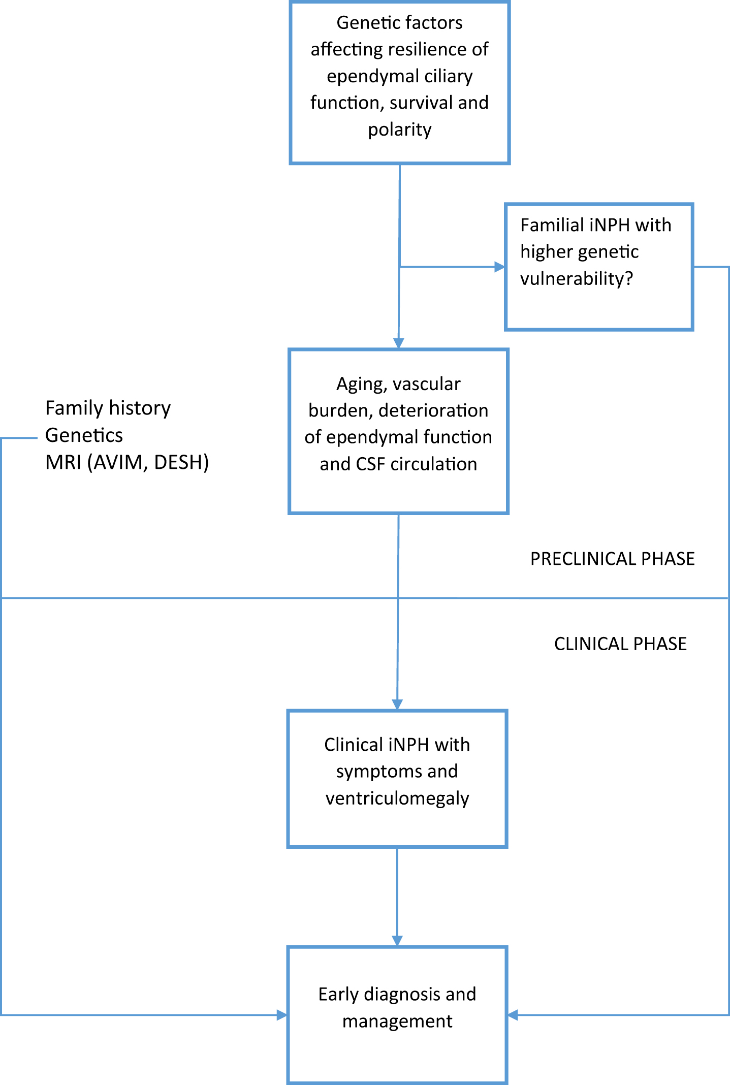
Hypothesized disease course of iNPH. AVIM, asymptomatic ventriculomegaly; DESH, disproportionality between sylvian and suprasylvien subarachnoid spaces.
In conclusion, the modest allelic variation of NME8 should be interpreted cautiously due to the limited sample size. However, the correlation of periventricular changes and the potential role in the cilia function, cell polarization, and cytoskeleton function support the biological link of NME8 with iNPH. Suboptimal reserves in these cellular processes along with environmental burden could increase the vulnerability for iNPH to develop in theelderly.
Footnotes
ACKNOWLEDGMENTS
We would like to acknowledge Marita Parviainen, RN, for the maintenance of KUH NPH Registry and Anniina Savolainen, MSc, for the revision of the English language. The study was partially funded by Emil Aaltonen foundation, Academy of Finland, Kuopio University Hospital (VTR grant V16001, 5252614), Sigrid Juselius Foundation, Cultural Foundation of Northern Savo, VPH Dementia Research Enabled by IT VPH-DARE@IT (no 601055), JPND-CO-FUND program (no 301220), and the Strategic Funding of the University of Eastern Finland.
