






Abstract
Background:
Environmental exposure to toxic metals has been postulated to play a role in the pathophysiological processes of Alzheimer’s disease (AD). However, the circulatory levels of toxic metals in AD patients are not consistent in previous studies.
Objective:
To systematically assess levels of toxic metals (aluminum, mercury, cadmium, lead) in the circulation (blood, serum/plasma) of AD patients and controls.
Methods:
PubMed, Web of Science, Science Direct, Cochrane Library, and the China National Knowledge Infrastructure (CNKI) were systematically searched to identify studies published up to January 1, 2017. Meta-analyses were performed using random-effects models and the pooled standardized mean difference (SMD) were reported with 95% confidence intervals (CI).
Results:
We identified 17, 7, 8, and 10 studies for aluminum, mercury, cadmium, and lead, respectively. Meta-analyses showed significantly elevated circulatory levels of aluminum (SMD = 1.08, 95% CI: 0.66, 1.50), mercury (SMD = 0.55, 95% CI, 0.15, 0.95), and cadmium (SMD = 0.62, 95% CI: 0.12, 1.11), whereas lower levels of lead (SMD = –0.23, 95% CI: –0.38, –0.07) in AD patients than in controls. Publication bias was only observed for aluminum studies, but the “trim and fill” analysis showed that the publication bias did not alter the direction of the effect. Sensitivity analyses showed no studies from the pooled analysis changed the results.
Conclusion:
Compared to controls, circulatory levels of aluminum, mercury, and cadmium are significantly higher but the levels of lead were reduced in AD patients. These findings suggest that elevated aluminum, mercury, and cadmium in the circulation, especially in serum may play a role in the progression of AD.
INTRODUCTION
Alzheimer’s disease (AD) is a progressive neurodegenerative disease manifested by decline of cognitive function, memory, and intellectual ability [1–3]. In the elderly population, AD is the most common dementia representing up to 60% ∼80% of all cases of dementia, and is a major cause of mortality in the developed countries [4]. According to the 2015 World Alzheimer Report Published by Alzheimer’s Disease International (ADI), over 46 million people live with dementia worldwide, with the number of cases being estimated to increase to 131.5 million by 2050 [5]. AD also has a huge impact on economy with an estimated cost of 172 billion dollars per year [4]. Multiple factors may contribute to the etiology of AD. Aging is the primary risk factor for AD, whereas a family history of AD and the presence of the apolipoprotein E (APOE ɛ4) genotype are the inherited genetic determinants known for AD [6]. Other risk factors include environmental and lifestyle factors, which are the avoidable factors [7]. Recent research suggests the importance of epigenetic mechanisms in defining the relationship between environmental exposures and AD [8]. Specifically, environmental toxic metals exposure has been implicated in AD pathogenesis [9]. Humans are exposed to toxic metals through a variety of sources, such as diet and occupational exposures [7]. After absorbing into the body from the gastrointestinal tract, lungs, or through the skin, the toxic metals enter the circulatory system. Thereafter, they may enter the central nervous system from the blood by crossing the blood-brain barrier, or from the blood by crossing the choroid plexus into the cerebrospinal fluid, from which it can diffuse into the central nervous system. Toxic metals could promote the amyloid-β (Aβ) production and the phosphorylation of tau protein (P-tau), which can cause the formation of senile/amyloid plaques and neurofibrillary tangles (NFTs) [10]. Among the various toxic metals, aluminum (Al), cadmium (Cd), lead (Pb), and mercury (Hg) are extensively studied because of their high industrial use and they are all considered as neurotoxic metals. For example, Al has been hypothesized to be an environmental contributor to the pathogenesis of AD since 1960s based on various neurotoxicological, analytical, and epidemiological findings (i.e., aluminum hypothesis) [11, 12]. Hg is a well-known neurotoxic metal and is ubiquitous in the environment [13]. If Hg accumulates in the brain beyond the ability of Selenoprotein P (SelP) to fully buffer its toxicity, Hg can cause oxidative stress and apoptosis in AD [14]. In addition, Pb and Cd cause a reduction in the brain content of acetylcholine, an important neurotransmitter closely related to the pathogenesis of AD [9, 15].
Despite these findings, the hypothesis of toxic metals in AD onset has been the subject of much debate and criticism for several decades. In this context, numerous studies have evaluated the association between the risk of AD and content of these toxic metals in circulatory system (serum/plasma, and blood). For example, several studies have observed higher levels of Al in the circulation of AD patients [16–23], whereas B. Bobba reported lower levels of Al in serum (p < 0.05) [24], and other studies demonstrated no significance of Al level between AD and control [25, 26]. Moreover, several meta-analyses of Al reported inconsistent results [27–29]. In addition, only a few of studies reported significant changes in the levels of Hg, Pb, and Cd. Furthermore, most available data on circulatory toxic metal concentrations is qualitative, and the sample sizes of limited quantitative studies are small. Thus, it is important to systematically review and critically assess available quantitative data on circulatory Al, Hg, Cd, and Pb levels in AD patients compared to controls. The present meta-analyses were conducted to comprehensively and systematically quantify the differences in the levels of toxic metals (Al, Hg, Cd, Pb) in the circulation (serum/plasma, blood) between AD patients and controls.
METHODS
Literature search and selection
PubMed, Web of Science, science direct, Cochrane Library, and the China National Knowledge Infrastructure (CNKI) were systematically searched to identify relevant studies published up to January 1, 2017. Literature search included a combination of keywords and MeSH, including the following terms: “aluminum OR mercury OR cadmium OR lead OR metal OR element” AND “Alzheimer’s OR Alzheimer’s disease OR Alzheimer’s patients OR Dementia” AND “Circulation OR Plasma OR Serum OR Blood”. Additionally, we reviewed the citation lists from each article retrieved for relevant review articles and from the meta-analyses. The literature retrieved was reviewed and the cited references were hand searched for further identification of relevant studies. Acceptable quantitative techniques were included in the analysis as follows: atomic absorption spectroscopy (AAS), inductively coupled plasma mass spectrometry (ICP-MS), or atomic emission spectroscopy (AES), and chromatographic, or spectrophotometric methods(other). Two authors (Lin Xu and Wenchao Zhang) independently screened the title and abstract of records identified in the search and any disagreements between the two reviewers were resolved by consensus involving a third reviewer (Xiulan Zhao).
Selection criteria
This systematic review and meta-analyses included all eligible studies that met the following criteria: 1) Patients met Milan overall dementia assessment (MODA), the National Institute of Neurological and Communicative Disorders and Stroke-Alzheimer’s Disease and Related Disorders Association criteria (NINCDS-ADRDA) [30] or the Diagnostic and Statistical Manual for Mental Disorders (MMSE) [31], DSM-III criteria for AD; 2) Quantitative analytical techniques are acceptable as described above; 3) Original data was available; 4) Study subjects were human adults with AD and control groups; 5) Studies providing a sample size and Al, Hg, Cd, and Pb levels in plasma/serum or blood in both AD patients and control groups.
The exclusion criteria were as follows: 1) Non-quantitative or semi-quantitative analysis; 2) Studies concerning other cognitive disorders: mild cognitive impairment, vascular dementia, or psychogeriatric disease; 3) Studies having inappropriate controls; 4) Studies having no numerical data of toxic metals (Al, Hg, Cd, Pb) levels; 5) Abstracts, case reports, reviews, conference presentations, editorials, and expert opinions; 6) In vivo or experimental studies.
Data extraction and publication quality assessment
All the articles were thoroughly read and evaluated. Data were independently extracted from text, tables, and figures by two authors (L.X. and W. Z.). Extracted data including author’s name, publication year, country, diagnostic criteria of AD, quantitative measurement methods used for analysis of toxic metals, means and SD of toxic metals levels (serum, plasma, blood), sample size of AD and control group, mean age of the participants, percentage of female, and other study characteristics. The publication quality about selection of the study groups, comparability of the groups, and ascertainment of outcome of interest were assessed using the Newcastle Ottawa quality assessment Scale, in which scores for low (0–3), moderate (4–6), and high-quality studies (7–9) were assigned. This scale uses a “star system,” in which included studies are judged on three broad perspectives: selection of the study groups, comparability of the groups, and ascertainment of outcome of interest.
Data analysis
Meta-analyses were performed using R software (version 3.4.0.), package “meta” (version 4.8-1). The standardized mean difference (SMD) and its 95% confidence interval (CI) were used as the summary statistic for the difference of toxic metals (Al, Hg, Cd, Pb) in the circulation (including serum/plasma or whole blood) of AD patients as compared to the controls. The I2 statistic and p-value were used to test statistical heterogeneity of involved studies. A p-value of <0.10 or I2 statistic of >50% indicated a significant heterogeneity. The data were analyzed using the random-effects meta-analyses model. We conducted subgroup analysis to investigate the effect of the study characteristics as possible sources of heterogeneity and meta-regression analysis to assess the effect of continuous variables on the outcomes of the meta-analyses. To test the influence of individual studies on the pooled SMD, a sensitivity analysis was performed by sequentially omitting one single study. Furthermore, publication bias was tested by funnel plot analysis accompanied by Egger’s test. The Trim-and-Fill method was used to explore the impact of studies potentially missing due to publication bias.
RESULTS
Literature search and study characteristics
Figure 1 shows the PRISMA flow diagram for literature search and study selection in this meta-analysis. Publications were found concerning the following toxic metals: Al (17 studies), Hg (7 studies), Cd (8 studies), and Pb (10 studies). The major characteristics of the retrieved publications are presented in Table 1. All studies were case-control studies including AD patients and controls. Of the 25 studies included, 3 studies used MODA criteria, 5 studies used MMSE, 3 studies used DSM-III, and all the others used NINCDS-ADRDA criteria for AD. 11 studies used AAS, 10 studies used ICP-MS, 3 studies used AES, and the other studies used chromatographic or spectrophotometric methods to assess the circulatory levels of toxic metals. The two investigators (L.X and W.Z) used the Newcastle-Ottawa Scale to assess the quality of the studies included in the meta-analysis. The overall quality of studies was good (Supplementary Table 1).
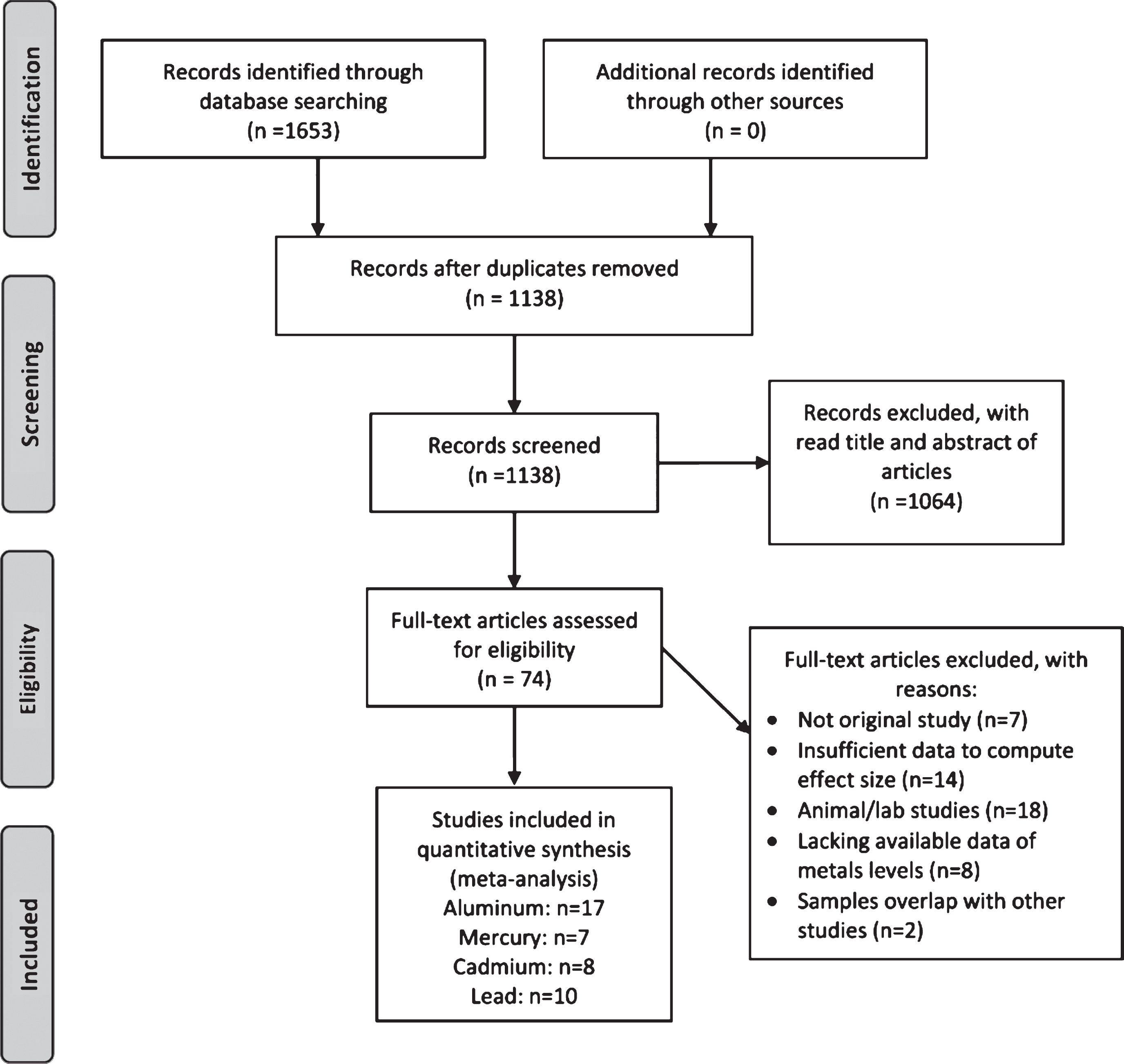
The PRISMA flow diagram for literature search and study selection.
Characteristics of studies included in meta-analyses
AAS, atomic absorption spectroscopy; ICP-MS, inductively coupled plasma mass spectroscopy; AES, atomic emission spectroscopy; Other, chromatographic or spectrophotometric methods; MODA, Milan overall dementia assessment; NINCDS-ADRDA, National Institute of Neurological and Communicative Disorders-Alzheimer’s Disease And Related Disorders Association; DSM-III, Diagnostic and Statistical Manual; NR, not reported; HK, Hong Kong.
Aluminum levels in circulation of AD
Seventeen studies compared the circulatory Al levels in AD patients and control subjects (Fig. 2). The pooled sample size consisted of 1,295 participants: 483 in AD patients and 812 in controls. A significant heterogeneity was found across studies (I2 = 89.6%), the random-effects model was used to compute the pooled effect size. The results indicated that patients with AD had significantly higher circulatory Al levels than control subjects (SMD = 1.08, 95% CI: 0.66, 1.50, z = 5.08, p < 0.001). Serum Al levels in fifteen studies and showed a significant increase with a pooled SMD (95% CI) of 1.17 (0.67, 1.66). Two studies reported whole blood Al levels in AD patients (Fig. 2).
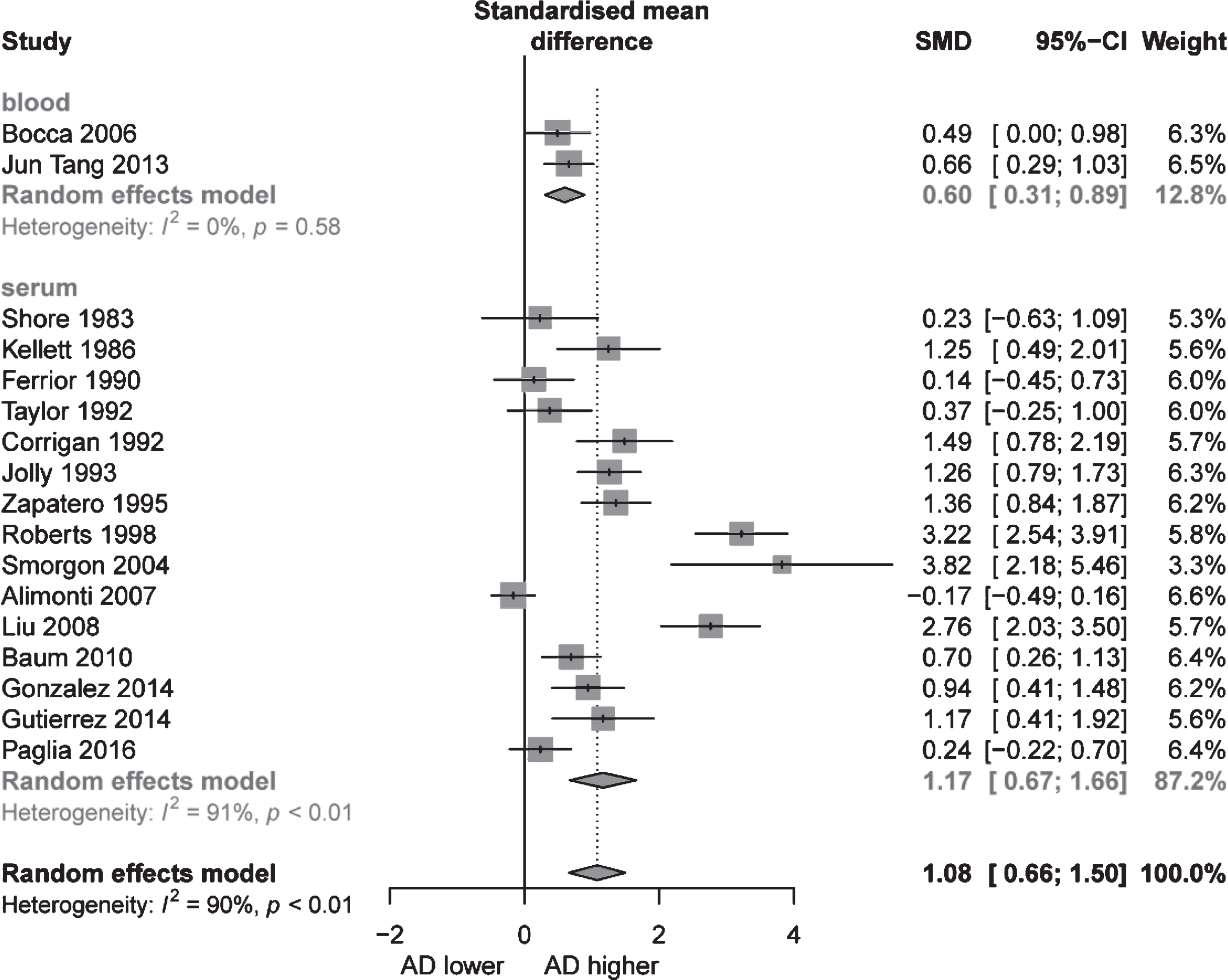
Differences of circulatory aluminum levels in Alzheimer’s disease (AD) patients and control subjects.
The meta-regression analysis was performed with the covariates: place, diagnosis of AD patients, age matched for AD cases and controls, sample test methods, and units. The results showed that place had a significant impact on the heterogeneity among the studies (p < 0.01, Table 2). As shown in Table 3, the subgroup analysis indicated that the methods for measuring Al level was a source of heterogeneity. In sensitivity analysis, leaving-out any individual study produced consistent results and the range of combined SMD identified was stable with no great fluctuations. Funnel plot of Al was asymmetric (Supplementary Figure 1) and publication bias was further observed in the Al meta-analyses as evaluated by the Egger’s test (p < 0.05). The trim-and-fill analysis was used to model the effect of missing studies, which did not alter the direction of the effect but reduced the effect size (adjusted overall combined estimates: SMD = 0.48, 95% CI:0.02, 0.95, z = 2.04, p = 0.04, Fig. 3). The pooled results of Al remained stable with the sensitivity analysis, suggesting main results were not being driven by any single study. Moreover, these results were not attenuated by the exclusion of studies with significant between-group age imbalances.
The meta-regression analysis of studies reporting aluminum in AD
**p < 0.01.
The subgroup analysis of studies reporting aluminum in AD
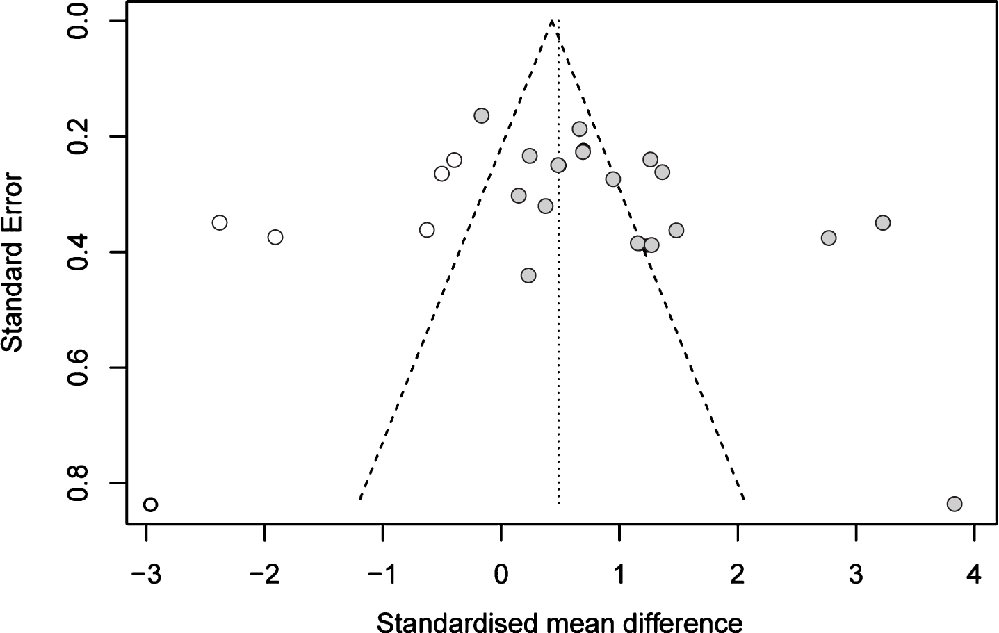
Funnel plot of the original studies included in the analysis (black dots) and the missing studies imputed by the trim-and-fill procedure (white dots).
Mercury levels in circulation of AD
Seven studies were included in pooled analysis of circulatory Hg levels in AD patients compared with controls, including in total of 397 control subjects and 478 AD patients. As displayed in Fig. 4, circulatory Hg levels were significantly higher in patients with AD (SMD = 0.55, 95% CI, 0.15, 0.95; p = 0.0073) than in control subjects. Because a significant heterogeneity was found across studies (I2 = 85.0%, p < 0.01), the random-effects model was used to compute the pooled effect size. Plasma/serum Hg levels in the meta-analyses of 6 studies showed a significant increase with a pooled SMD (95% CI) of 0.57 (0.13, 1.00). One study reported non-significantly elevated whole blood Hg levels in AD patients (Fig. 4).
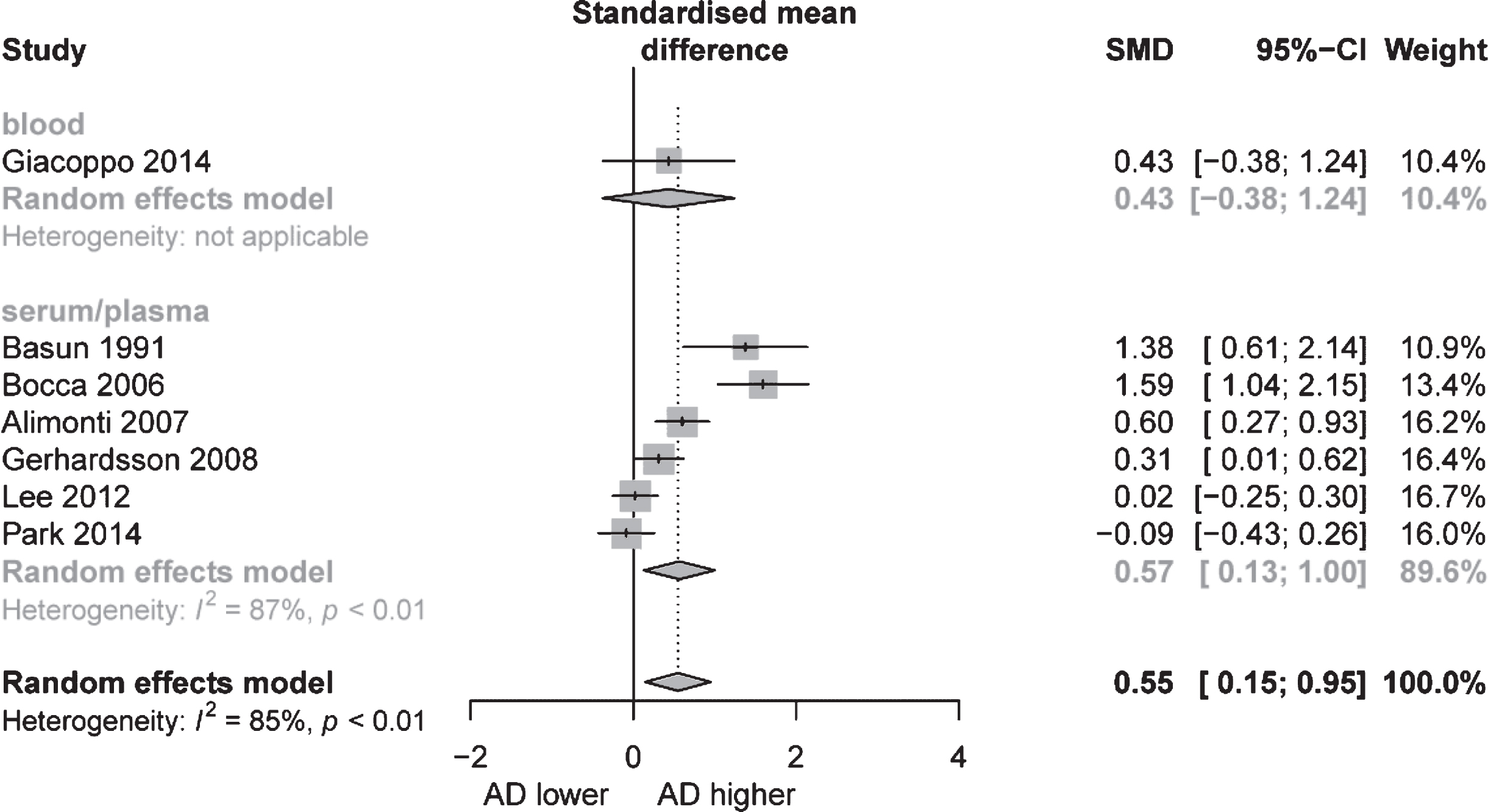
Differences of circulatory mercury levels in Alzheimer’s disease (AD) patients and control subjects.
The meta-regression analysis was performed with the covariates: source and year of publication, age-matching for AD cases and controls, sample test methods and units. Year of publication had significant impact on the heterogeneity among the studies (p = 0.01, Table 4). Sensitivity analyses showed that no studies significantly changed the overall results. Furthermore, despite funnel plot asymmetry (Supplementary Figure 2), no publication bias was observed according to the Egger’s test (p = 0.1176).
The meta-regression analysis of studies reporting mercury in AD
**p < 0.01.
Cadmium levels in circulation of AD
Eight publications included measured pooled circulatory Cd levels in AD and controls, including 405 AD patients and 424 control subjects. As displayed in Fig. 5, circulatory Cd levels were significantly elevated in patients with AD (SMD = 0.62, 95% CI: 0.12, 1.11; p = 0.0144) compared to control subjects. A significant heterogeneity was found across studies (I2 = 90%), thus the random-effects model was used to compute the pooled effect size. Plasma/serum Cd levels in the 6 studies showed a significant increase with a pooled SMD (95% CI) of 0.76 (0.16, 1.37) (Fig. 5). Two studies reported whole blood Cd levels in AD.
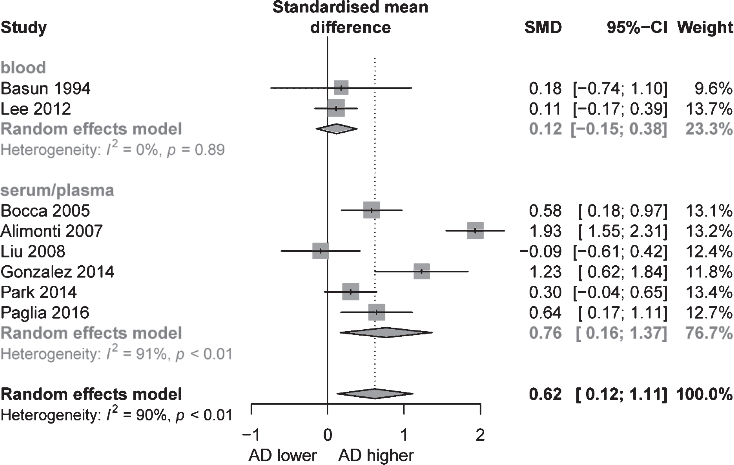
Differences of circulatory cadmium levels in Alzheimer’s disease (AD) patients and control subjects.
A subgroups analysis was conducted to explore possible source of heterogeneity. As shown in Table 5, place of studies in Europe, no-age matching and source of sample were the possible sources of heterogeneity. Sensitivity analyses showed that no studies significantly changed the overall results. Furthermore, despite funnel plot asymmetry (Supplementary Figure 3), no publication bias was observed in the meta-analyses according to the Egger’s test (p = 0.739).
The subgroup and sensitivity analyses of studies reporting cadmium in AD
Lead levels in circulation of AD
Ten publications included measured pooled circulatory Pb levels in AD and controls, including 716 AD patients and 1298 control subjects. The random-effects meta-analyses showed that AD patients had lower Pb levels compared to control subjects (SMD = –0.23, 95% CI: –0.38, –0.07, p = 0.0043; Fig. 6). There was significant heterogeneity among these studies (I2 = 48%, p = 0.04) by a random-effect model. Whole blood Pb level in two studies were not significantly decreased but not significantly in AD patients compared to controls. Plasma/serum Pb levels in eight studies showed a significant decrease with a pooled SMD (95% CI) of –0.19 (–0.37, –0.01) (Fig. 6). Sensitivity analyses showed that no studies significantly changed the overall results. No publication bias was observed in the meta-analyses of Pb according to the Egger’s test (p = 0.4037, Supplementary Figure 4).
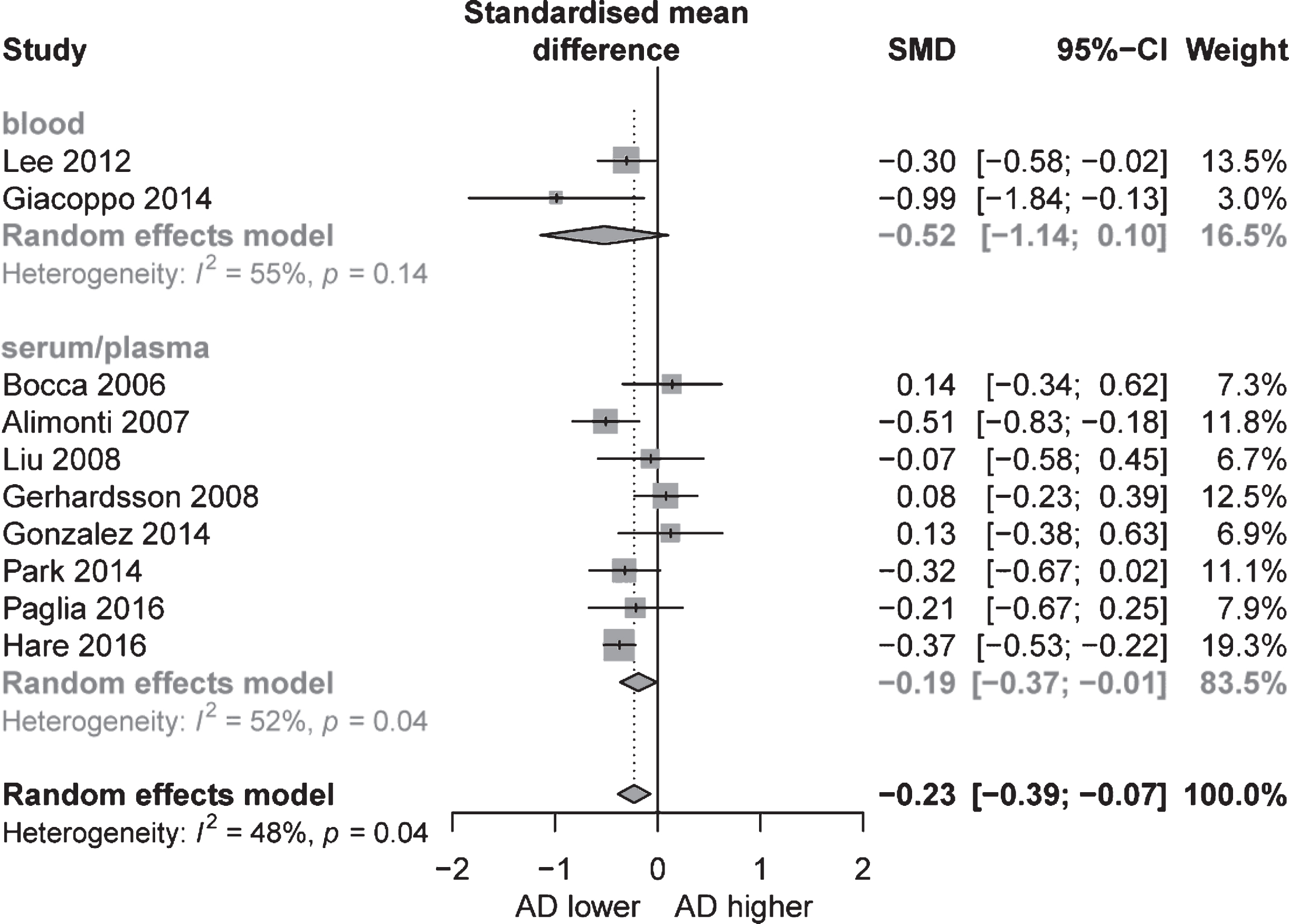
Differences of circulatory lead levels in Alzheimer’s disease (AD) patients and control subjects.
DISCUSSION
Over the past few decades, there has been a growing interest in understanding the metabolism of neurotoxic metals and their impact on AD. However, the circulatory toxic metal levels in AD patients compared to controls in previous studies are not consistent. Our meta-analyses indicated that circulatory Al, Hg, and Cd concentration were significantly elevated in the AD patients compared to controls, while circulatory Pb levels was lower in AD patients.
The relationship between Al and AD has been the subject of scientific debate because the precise mechanism of AD pathogenesis remains unknown. A link between Al and AD was reported by Klatzo et al. in 1965 [32]. In 1973, the first report of increased concentrations of Al in the brains of patients with AD was published [33]. Our meta-analyses demonstrated that circulatory Al levels including seventeen studies with 483 AD cases and 812 controls were significantly increased in AD patients compared to controls. Our findings support the meta-analyses of epidemiological studies that demonstrated the significant association between chronic Al exposure and risk of AD [29]. Neurological effects including impairment of cognitive function and motor dysfunction is associated with occupational exposure to Al in the workplace air [34]. Moreover, most people living in industrialized societies are routinely exposed to bioavailable Al salts in the form of additives-in commercially-prepared foods, alum-clarified drinking water, sunscreens, and other topical applications. Minute amounts of this Al are absorbed into the circulation and then enter the brain by cross the blood-brain barrier. Al progressively accumulates in large pyramidal neurons of the hippocampus, cortex, and other brain regions and may increase the risk of AD [35]. In vivo, Al demonstrates numerous adverse effects relevant to the pathophysiology of AD, including the depletion of microtubules and consequent cortical atrophy [28]. Al is responsible for two main types of toxic damage in cells. As a pro-oxidant, Al causes oxidative damage both on its own and in synergy with iron. Al also competes with, and substitutes for, essential metals-primarily Mg2+, iron and Ca2+ ions—in or on proteins and their co-factors [35]. Al potentiates oxidative damage, which is thought to be an early event in AD [36, 37]. Al can also induce neurofibrillary abnormalities, disruption of axonal transport mechanisms, neurite degeneration, and loss of synapses [38]. Additionally, Al perturbs neuronal Ca2+ homeostasis and inhibits mitochondrial respiration in a complex with amyloid genic Aβ peptide in a triple transgenic mouse model of AD [39].
Despite supporting evidence, the Al hypothesis of AD remains controversial. A number of studies have showed arguments against Al hypothesis. First, some observational studies indicated no association between Al exposure and the risk of AD [34, 40]. Second, it has been argued that neurofibrillary changes in Al-intoxicated animals (Al-NFTs) are different from those in AD patients (AD-NFTs) [41]. However, most of these criticisms were made in the 1990s. Recent immunohistochemical studies have indicated that depositions in the brains of Al-intoxicated animals are stained with the anti-tau antibody [32]. Furthermore, several epidemiological studies reported a high incidence of AD in areas with high Al level in drinking water, including a 15-year follow-up of 1677 elderly subjects [12, 42].
In the present meta-analyses of Hg, we found that the circulatory Hg levels were significantly increased in AD patients compared to controls. Vitro models showed that Hg reproduced all pathological changes observed in AD [14, 44]. APOE ɛ4 allele which is the most important gene risk factor for AD has been reported to be associated with the differential detoxification capacity regarding Hg [43]. Selenoprotein P (SelP), which provides Se for SelP synthesis, is physically associated with both Aβ plaques and NFTs in the AD brain [45]. SelP has high capacity for binding Hg. Accumulation of Hg in the brain, in excess of the ability of SelP to fully buffer its toxicity, can contribute to oxidative stress and apoptosis in AD [46]. Furthermore, Hg potently interferes with neural stem cell development, which may contribute to reduced cortical and hippocampal neuronal density in AD [47]. However, Morris et al. reported an autopsy-based cross-sectional analyses in JAMA which found in nearly 300 brains that there was not an association between mercury levels and brain neuropathology [48]. Thus, further studies needed to research the relationship between blood Hg level and AD.
Cd has garnered considerable attention because of its widespread exposures through occupation, smoking and foods. Cd from polluted soil and water can accumulate in plants and organisms. Tobacco leaves naturally accumulate high amounts of Cd, and as such, smoking greatly increases exposure to Cd [49]. In this study, we demonstrated that circulatory Cd levels were significantly elevated in Asian and European patients with AD compared to control subjects, possibly due to the fact that smoking is prevalent in Asian and European populations. Previous research showed higher Cd concentrations in brain tissues of AD patients than in healthy people [2]. The mechanism underlying the association between Cd concentration and AD has not been elucidated. However, growing evidence indicates that Cd plays a role in the accumulation of Aβ plaques and tau protein [50], which is the main pathological feature of AD [51]. Jiang et al. reported the impact of Cd on the formation of neurofibrillary tangles [52]. Increased blood Cd levels were associated with elevated AD mortality among participants aged 60 years or older in the National Health and Nutrition Examination Survey (NHANES) (1999–2004 cycles) [53]. Similarly, Peng et al. showed that urinary Cd was associated with elevated risk of AD mortality among older adults [54].
From our pooled results, we found that AD patients had lower circulatory Pb levels than healthy controls. To date, while there has been no causative relationship between AD and lead exposure found. studies in the relationship between lead and AD have yielded interesting results. Gerhardsson et al. reported that AD patients had lower Pb level in cerebrospinal fluid than in control subjects [55]. However, Costa et al. suggested that Pb could cause a reduction in the brain content of acetylcholine, which is an important neurotransmitter closely related to the pathogenesis of AD [56]. Duce and colleagues also suggested that Pb could trigger the formation of senile plaques and NFTs to cause failure in neurotransmission and the induction of oxidative stress [57]. Early life exposure to Pb impairs a variety of cognitive, behavioral, and neurochemical processes resulting in a variety of potentially negative outcomes for children exposed to this potent neurotoxicant [58, 59]. However, no specific Pb-associated “behavioral signature” or AD clinical features have been identified [60]. An expert panel on adult Pb toxicity convened by the US Centers for Disease Control concluded that bone Pb levels were the best biomarker of cumulative Pb exposure [8]. Around 95% of the total body burden of lead is in bone. Circulating lead levels was related with acute exogenous exposure and some contribution through remobilization of lead from mineralized storage. Bone lead has a half-life measured in decades, while blood lead has an average turnover of around 30 days [61]. There are potentially many confounding factors that influence lead release from bone into circulation. Thus, further studies are needed to examine both circulating and bone Pb levels in large samples of AD patients and the role of Pb in the development and progress of AD.
Although the present study demonstrates a positive link between circulatory Al, Hg, and Cd levels and AD, our meta-analyses has some limitations. First, studies included in the meta-analyses are heterogeneous. The observed heterogeneity could be attributable to the clinical and methodological variation between studies, such as differences in participant characteristics, classification of AD, sample preparation, and the methods used to analyze toxic metals levels. Second, the number of studies was relatively small. Further studies are required to confirm our findings. Third, the findings of Al from the present study should be interpreted with caution, because publication bias was observed in the Al circulation levels in AD patients. This may be due to the authors and journals being more likely to publish positive results than negative ones. Further investigations are necessary to address circulatory Al levels in AD patients. Fourth, age is a very significant variable in studies of the impact of metals metabolism on AD, we assess the effect of age by used subgroup and meta-regression method which suggested that the pooled effect was not attenuated in the Al analysis by the exclusion of studies with significant between-group age imbalances. However, in the analyses of Hg, Cd, and Pb, the pooled results were affected by the age; this may associate with the small studies included in the Hg, Cd, and Pb analyses. Finally, most studies included in the meta-analyses were conducted in Asian and European AD patients. It is unknown if the findings could be generalized to American and African populations.
In conclusion, the results of our meta-analyses indicate that the circulatory aluminum, mercury, and cadmium levels are significantly increased in AD patients as compared to controls. Given the great patient, family, and socio-economic burden of AD, in the future, steps should be taken to minimize human exposure to these environmental toxic metals to reduce the risk of developing AD. Further studies are needed to examine the role of toxic metals in the development of AD.
