






Abstract
Background:
Natural products are a significantly underutilized source of potential treatments against human disease. Alzheimer’s disease (AD) is a prime example of conditions that could be amenable to such treatments as suggested by recent findings.
Objective:
Aiming to identify novel potentially therapeutic approaches against AD, we assessed the effects of Cichorium spinosum and Sideritis scardica extracts, both distinct components of the Mediterranean diet.
Methods/Results:
After the detailed characterization of the extracts’ composition using LC-HRMS methods, they were evaluated on two AD neuronal cell culture models, namely the AβPP overexpressing SH-SY5Y-AβPP and the hyperphosphorylated tau expressing PC12-htau. Initially their effect on cell viability of SH-SY5Y and PC12 cells was examined, and subsequently their downstream effects on AβPP and tau processing pathways were investigated in the SH-SY5Y-AβPP and PC12-htau cells. We found that the S. scardica and C. spinosum extracts have similar effects on tau, as they both significantly decrease total tau, the activation of the GSK3β, ERK1 and/or ERK2 kinases of tau, as well as tau hyperphosphorylation. Furthermore, both extracts appear to promote AβPP processing through the alpha, non-amyloidogenic pathway, albeit through partly different mechanisms.
Conclusions:
These findings suggest that C. spinosum and S. scardica could have a notable potential in the prevention and/or treatment of AD, and merit further investigations at the in vivo level.
Keywords
INTRODUCTION
Alzheimer’s disease (AD) is a progressive neurodegenerative disease, the most common form of diagnosed dementia (>44 million AD patients worldwide), and the 6th leading cause of death in the USA. By 2050, more than 100 million people are expected to be affected worldwide [1]. Currently, there are no effective treatments, while the FDA approved compounds exhibit short-term benefits and can have serious side effects [2, 3]. Natural products with neuroprotective activities are believed to hold significant promise in preventing or treating AD [4–8].
It is anticipated that some of these products could demonstrate beneficial effects by preventing key AD pathogenetic mechanisms, namely the amyloidogenic and neurofibrillary tangles (NFTs), pathways [9]. The amyloid pathway is implicated in neuronal activity and differentiation, including synapse formation and transmission [10]. Under physiological conditions, the amyloid-β protein precursor (AβPP) is processed by the alpha or beta proteolytic pathways. Through the non-amyloidogenic alpha pathway, AβPP is cleaved by the alpha [11] and gamma secretases to produce soluble AβPP alpha (sAβPPα) and AβPP-C83 [12–14]. The active gamma secretases, PSEN1 and PSEN2, are the products of the respective full-length protein cleavage and amino-/carboxy- terminal fragment (NTF/CTF) heterodimerization [15, 16], and are also encountered in the form of multiprotein complexes [17, 18]. Through the amyloidogenic beta pathway, AβPP is cleaved by beta secretase (BACE1) releasing sAβPPβ to the extracellular space and leaving AβPP-C99 in the plasma membrane. AβPP-C99 can be subsequently processed by gamma secretase into amyloid-β (Aβ) of different lengths (ranging from 37 to 46 amino acids) and AβPP intracellular C-terminal domain (AICD) [12, 19]. Some species of Aβ are particularly toxic causing synaptic failure and neuronal death [20].
The intracellular fibrils and NFTs formed during AD development consist primarily of the hyperphosphorylated and misfolded microtubule-associated protein tau. Normally located at the axons, tau is important for neuronal differentiation and development, maintenance of cellular morphology and polarity, as well as axonal transport of organelles, vesicles, or molecules [21]. Tau is subjected to extensive post-translational modifications, predominantly phosphorylation in >85 sites [22]. The aberrant phosphorylation of tau in AD leads to its dissociation from microtubules, microtubule destabilization, loss of dendritic microtubules and synapses, interruption of axonal transport, plasma membrane degeneration, and eventually neuronal loss [23, 24]. The hyperphosphorylated tau molecules tend to self-assemble intracellularly into filaments forming NFTs. Following the death of tangle-bearing cells, tau filaments are released in the extracellular space as neurotoxic “ghost” tangles stimulating activation of microglial cells and progressive neuronal degeneration [2, 26].
The identification of natural products that could disrupt the amyloid cascade or tau misprocessing and prevent the accumulation of amyloid plaques or NFTs is an area of intense research in the battle against AD. Since dietary habits seem to significantly affect the prevalence of cognitive impairment in a population [27, 28], it has been proposed that the regular intake of certain classes of compounds, such as antioxidants, might act protectively against neuronal cell oxidation and cognitive decline [29, 30]. Natural phenols, such as those encountered in fresh fruits, green vegetables, red wine, and olive oil, are often regarded as health-promoting options for maintaining cognitive health [31–33]. In that sense, the model that combines all of these nutritional elements, the Mediterranean diet, should be thoroughly examined as a potent preventive tool in the battle against neurodegenerative diseases.
The Mediterranean diet constitutes an exemplary model diet with its benefits varying from low rates of heart disease to higher survival rates [34, 35]. In recent years, heightened attention has been drawn to the link between the Mediterranean diet and mental function in older adults [36]. In particular, individuals adhering to a Mediterranean style diet seem to have a reduced risk for developing AD [37] and mild cognitive impairment (MCI), as well as for deterioration from MCI to AD [38]. Recently, a population study in rural Crete, demonstrated a lower prevalence for dementia compared to global data [39]. Consequently, the study of the Cretan/Mediterranean diet, might serve as a rich source of new bioactive natural products.
Among the key components of the Mediterranean diet, the decoction of S. scardica leaves, commonly known as “mountain tea”, represents a daily habit and a traditional remedy. As such, the beneficial properties of plants of the genus Sideritis have been extensively studied. A recent study demonstrated that S. scardica extracts inhibited the uptake of the neurotransmitters serotonin, noradrenaline, and dopamine, which are involved in multiple neurological disorders [40], while other species of the Sideritis genus have been shown to exert antioxidant and anxiolytic-like properties [41], anticholinesterase activity [42, 43], and to improve the spatial learning and memory in mice with Aβ-induced amnesia [44]. Sideritis spp. extracts seem to reduce the amyloid plaque burden in transgenic mice, and significantly ameliorate their memory function [45], while a dietary supplement of S. scardica and selected B vitamins has been found to reduce stress-induced impairment of mental function in young adults [46]. The S. scardica extract has also been found to improve cognition and mental function by modulating the AMPA receptor dependent neurotransmission involved in synaptic plasticity and age-related cognitive decline [47].
Another highly valued and extensively consumed ingredient of the Mediterranean diet, and especially the Cretan diet, is the wild edible greens (chórta) of Crete, C. spinosum, an endemic Mediterranean plant also known as “stamnagkathi” [48, 49], and it is thought to improve liver function [50]. Recently the C. intybus L. member of the Cichorium genus was suggested to improve amnesia and memory process impairment in rats [51]. Importantly, multiple compounds present in C. spinosum extracts, such as aesculetin, cichoric acid, chlorogenic acid, 3,4-dicaffeoylquinic acid, 3,5-dicaffeoylquinic acid, 4,5-dicaffeoylquinic acid, have been demonstrated to play a neuroprotective role [52–56]. Furthermore, the C.spinosum compound quercetin-3-O-glucuronide significantly reduces the generation of Aβ peptides and improves AD-type deficits in hippocampal formation, basal synaptic transmission, and long-term potentiation [57]. Furthermore, both S. scardica and C. spinosum contain high levels of phenolic substances, such as phenolic acids and flavonoids, and they have exhibited potent antioxidant activities in in vitro models [58–61]. They therefore merit further investigation for their potential in modulating the molecular milieu of AD.
In this context, we assessed the effects of these two plants’ natural extracts, distinct components of the Mediterranean diet, in two established in vitro (cell line) models of AD. We demonstrate that treatment with “mountain tea” (S. scardica) or “stamnagkathi” (C. spinosum) extracts modulates multiple steps of the AβPP misprocessing and tau hyperphosphorylation pathways, suggesting a potential preventive and possibly therapeutic potential in AD.
MATERIALS AND METHODS
Plant material extraction
For S. scardica, the dry plant material was extracted with methanol using ultrasounds for 2 h, at room temperature and the extract was concentrated to dryness after filtration. For C. spinosum, the fresh stems and leaves were boiled with distilled water in a ratio of 1 kg plant material / 2 L of water and the obtained decoction was filtered and lyophilized.
UHPLC-ESI(-)-HRMS analysis
Liquid chromatography analysis for S. scardica was performed on an Accela® High-Speed LC System (Thermo Scientific) and for C. spinosum on an Acquity® UPLC System (Waters). For both extracts, detection was carried out on a LTQ-Orbitrap® XL hybrid mass spectrometer equipped with an ESI source (Thermo Scientific), in negative mode. Due to the different polarity of the extracts’ constituents, two different gradient separation methods were used. For S. scardica qualitative analyses, separation was achieved on a Fortis® C18 column (100 mm×2.1 mm, 1.7μm) using a gradient of water containing 0.1% (v/v) formic acid (A) and acetonitrile (B). Elution started with 95% A for 3 min and decreased to 0% A in 21 min. These conditions were maintained for 2 min before reverting to the initial conditions for 7-min of re-equilibration. For C. spinosum analysis, separation was achieved on a Fortis® C18 column (150 mm×2.1 mm, 1.7μm) using the same solvent system. Elution started with 95% A and decreased to 5% in 23 min. These conditions were maintained for 3 min before reverting to initial conditions in 2 min, for a final 3-min re-equilibration. In both cases, the column was maintained at 40°C and the flow rate was set at 0.4 mL/min. 10μL of water extracts at 200μg/mL were injected. MS data were acquired in negative-ion mode, in the full scan of 113–1000 m/z, with a resolution of 30000. Capillary temperature was set at 350°C, whereas source voltage was 2.7 kV in ESI-. Tube lens and capillary voltage were tuned at – 40 V and – 10 V, respectively. Finally, nitrogen was used as sheath gas (40 arbitrary units) and auxiliary gas (10 arbitrary units). For both extracts, a sample was prepared for analysis at a concentration of 0.2 mg/mL in a mixture of the mobile phase.
Cell culture and differentiation
The human neuroblastoma SH-SY5Y and rat pheochromocytoma PC12 cell lines were used as well established neuronal models that can differentiate into neuron-like cells. Furthermore, SH-SY5Y-AβPP cells inducibly over-expressing AβPP695 (a kind gift of Dr S. Efthimiopoulos, Faculty of Biology, National and Kapodistrian University of Athens, Greece) [62], and PC12-htau cells stably transfected with the human tau (htau; 3 R/0 N isoform) transgene and expressing hyperphosphorylated tau (a kind gift of Dr I. Sotiropoulos, Life and Health Sciences Research Institute (ICVS), School of Health Sciences, University of Minho, Portugal)[63], were used as in vitro models of AD. All cells were cultured in Dulbecco’s Modified Eagle’s Medium (DMEM) without L-glutamine, maintained at 37°C in a humidified 5% CO2 environment. For SH-SY5Y cells, the medium was supplemented with 10% (vol/vol) heat-inactivated fetal bovine serum (FBS), 1% antibiotic/antimycotic (10,000 units/mL of penicillin, 10,000μg/mL of streptomycin, 25μg/mL of amphotericin B) and 1% L-alanyl-L-glutamine. For SH-SY5Y-AβPP cells, the SH-SY5Y culture medium contained an additional 100μg/mL of G418 (Gibco, Thermo Fisher Scientific Inc.). For PC12 cells, the DMEM without L-glutamine was supplemented with 5% (v/v) heat-inactivated horse serum (HS) and the cells were plated on collagen-treated flasks/plates [64]. For PC12-htau cells the culture medium contained an additional 100μg/mL of G418 (Gibco, Thermo Fisher Scientific Inc.). Differentiation of SH-SY5Y and SH-SY5Y-AβPP cells was achieved with the addition of all-trans retinal (Sigma-Aldrich Co.) to the culture media, to a final concentration of 10- 5 M for 6 days. For the differentiation of PC12 and PC12-htau cells, 0.75% FBS, 0.75% HS, 100 ng/mL 7 S nerve growth factor (NGF; Invitrogen), 1% antibiotic/antimycotic and 1% L-alanyl-L-glutamine were added to the media for 7 days.
Natural products cell viability assays
Differentiated SH-SY5Y or PC12 cells were exposed to a range of concentrations of either extract (ranging from 0.04μg/mL to 400μg/mL for each natural extract) for 24 h or 72 h. The effect of the exposure to each extract/concentration on cell viability was evaluated with the Water Soluble Tetrazolium Salt -1 assay (WST-1; Takara), a colorimetric test based on the cleavage of WST-1 by mitochondrial dehydrogenase and the measurement of the absorbance of the resulting formazan product at 450 nm in an ELISA reader (Lucy 2; Anthos Labtec Instruments GmbH). Since both extracts were diluted in DMSO, matched concentrations of DMSO were used as control for each product concentration. All experiments were performed at least three times.
Immunoblotting
Cells were lysed in lysis buffer (50 mM Tris, pH 7.5, 150 mM NaCl, 2 mM EDTA, 1% Triton) supplemented with a mixture of protease inhibitors (P8340; Sigma-Aldrich Co.), incubated on ice for 30 min and centrifuged at 13,000 rpm for 5 min. For sAβPPα the culture medium was collected and condensed with centrifugal filter units (Amicon; Millipore) at 4,000 rpm. The protein concentration was determined with the Bradford method [65], using bovine serum albumin to generate a standard curve. All samples were analyzed by SDS-PAGE (Supplementary Table 1). GAPDH and actin were used as loading controls in all cases, except for sAβPPα where the volume of the initial culture media was used in sample normalization and reciprocal sample volume loading. Proteins were transferred to nitrocellulose membranes (Macherey-Nagel GmbH & Co), which were then incubated with primary antibodies (Supplementary Table 1). The nitrocellulose membranes were subsequently washed in 50 mM Tris-HCl, pH 7.5, 150 mM NaCl, and 0.05% Tween 20 and incubated with a peroxidase-conjugated anti-mouse (1:16,000 dilution; Sigma-Aldrich Co.) or anti-rabbit (1:10,000 dilution; BIO-RAD) secondary antibody. Protein signals were detected using electrogenerated chemiluminescence (ECL) reagents according to the manufacturer’s protocol (Thermo Fisher Scientific Inc.). The intensities of the bands of interest from at least three different experiments were quantified using Image J software (version 1.47v; https://imagej.nih.gov/ij/). The measurements for all phosphorylated forms were normalized to the total levels of the corresponding proteins.
Statistical analysis
Descriptive statistics were performed for all experiments and data obtained are presented as mean±standard deviation (SD). A p value≤0.05 was considered statistically significant. For the immunoblotting assays, the integrated densities of the bands obtained were analyzed by Student’s t-test. The ratios of protein isoforms to total protein were assessed by chi-square.
RESULTS
Plant extracts’ UHPLC-HRMS-ESI(-) profiles
HRMS profiles were acquired using ESI, in negative mode. Characterization of the detected metabolites was achieved considering m/z values of the suggested elemental composition with a 5 ppm tolerance from the proposed theoretical mass, as well as RDB equivalent values (Fig. 1). A total of 40 secondary metabolites were identified in the methanolic extract of S. scardica (Supplementary Table 2) and more than 30 compounds in the C. spinosum decoction (Supplementary Table 3). The results of the analysis demonstrate that the S. scardica extract is rich in phenylethanoic glycosides, as well as flavonoids and their glycosylated forms. The major components that were detected under the class of phenylethanoic glycosides, were verbascoside, martynoside, echinacoside, lavandulofolioside, allysonoside, leucosceptoside, forsythoside, samioside, as well as their isomers. Under the class of flavonoids, the main metabolites detected included scutellarein, isoscutellarein, hypolaetin, and apigenin, which were mainly detected in their glycosylated forms, with or without coumaroyl groups. The decoction of C. spinosum appears to be rich in secondary metabolites belonging to different chemical classes, including organic acids, such as citric, malic and cinnamic acid, flavonoid derivatives and sesquiterpene lactones. Caftaric, cichoric and chlorogenic acid are the predominant hydroxycinnamic acids in the extract, whereas in the class of flavonoids, the principal compounds were quercetin and luteolin glucuronides. Interestingly, the extract appears to contain a small number of sesquiterpene lactones such as the sulphonated guianolide, 8-deacetylmatricarin-8-O-sulfate.
UHPLC-HRMS-ESI(-) profiles of: (A) S. scardica methanolic extract, and (B) C. spinosum decoction.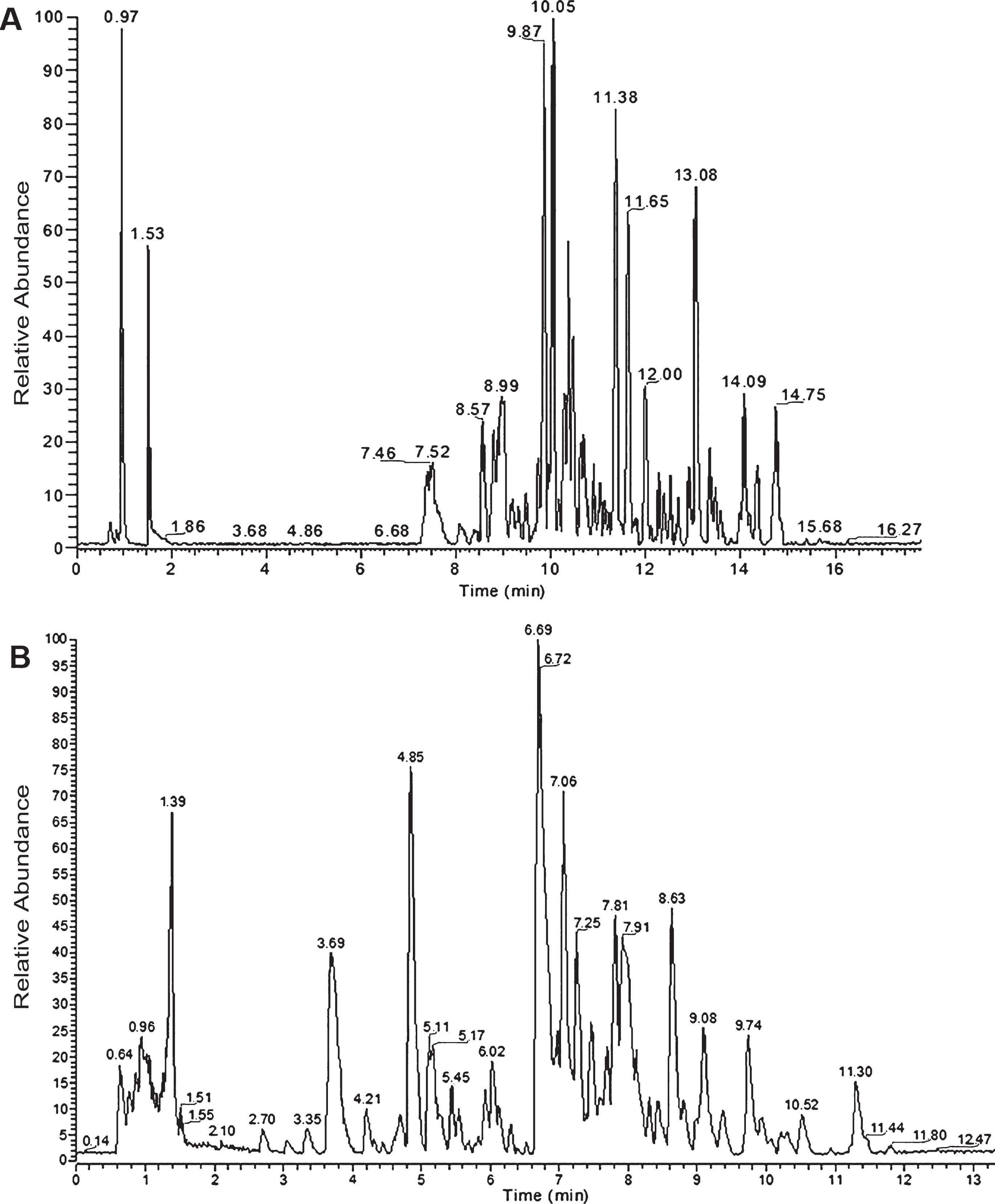
S. scardica and C. spinosum extracts do not compromise viability of SH-SY5Y and PC12 cells
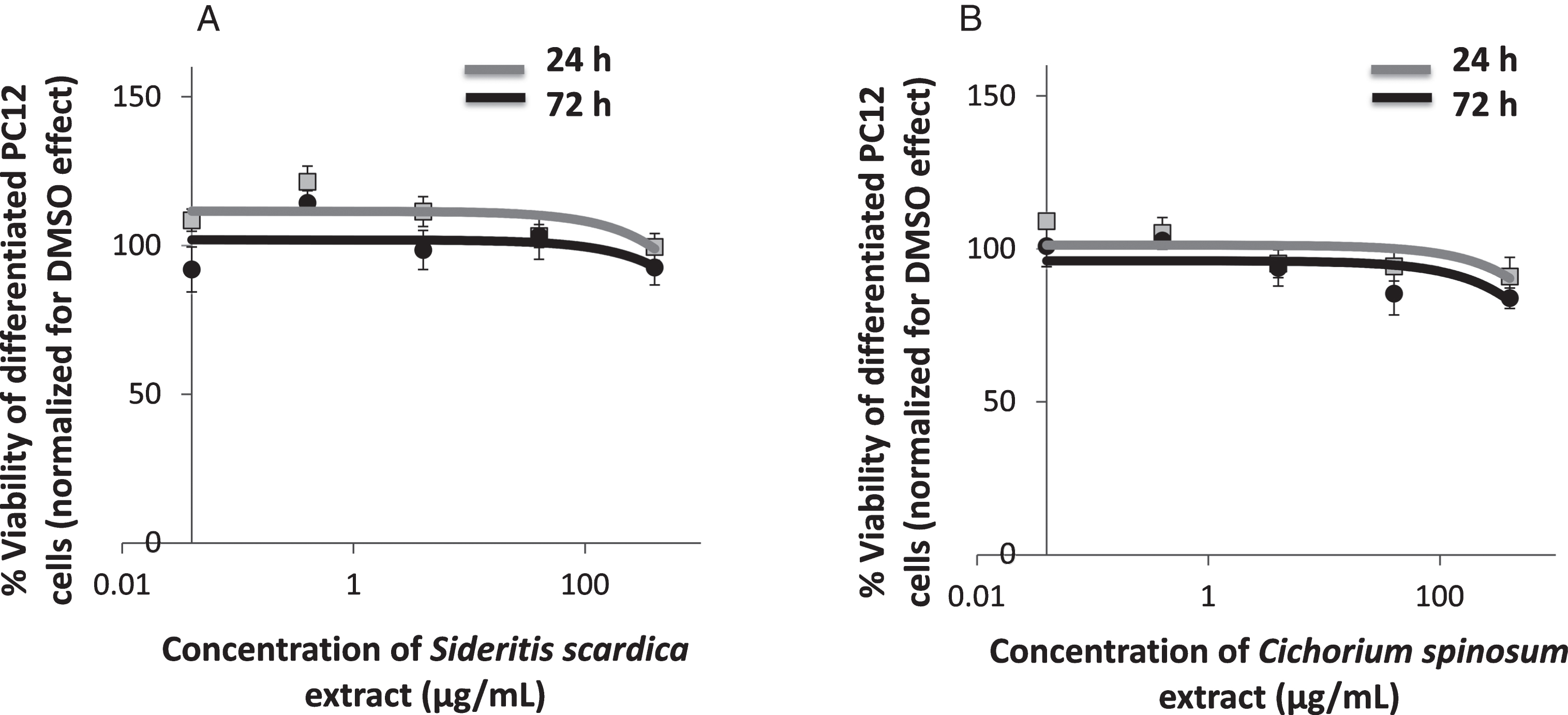
To evaluate the biological tolerance of neuron-like cells for S. scardica and C. spinosum extracts, we exposed them to a range of biologically relevant concentrations for different time periods and measured their effects on cell viability with WST-1 assays [66–68]. In specific, differentiated wild type SH-SY5Y and PC12 cells were exposed to concentrations ranging from 0.04μg/mL to 400μg/mL for each natural extract (Figs. 2 and 3). Both S. scardica and C. spinosum preserved intact the viability of differentiated SH-SY5Y cells across all tested concentrations and incubation times. These results were confirmed by experiments in differentiated PC12 cells.
Diagrammatic presentation of viability of differentiated SH-SY5Y cells at 24 and 72 h of treatment with different concentrations of: (A) S. scardica extract, (B) C. spinosum extract (n = 3, the concentration axis is log scaled). Diagrammatic presentation of viability of differentiated PC12 cells 24 and 72 h of treatment with different concentrations of: (A) S. scardica extract, (B) C. spinosum extract (n = 3, the concentration axis is log scaled).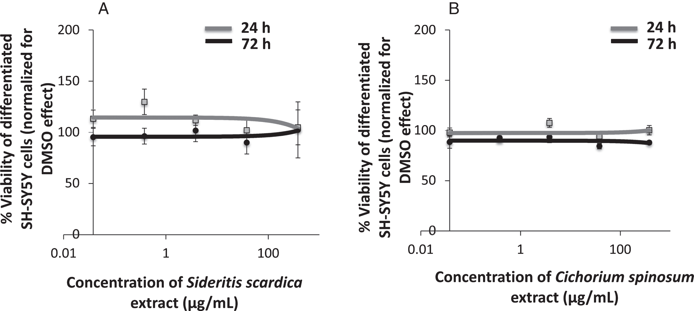
Having determined that the specific S. scardica and C. spinosum extracts do not compromise cell viability, we proceeded to investigate their downstream effects on AβPP processing and tau expression / phosphorylation in two AD neuronal cell culture models (differentiated SH-SY5Y-AβPP and PC12-htau).
Effects on AβPP processing
Since AβPP processing is considered a central pathogenetic mechanism for AD, we proceeded to assess the effect of S. scardica and C. spinosum on the expression of its key molecular players. Differentiated SH-SY5Y-AβPP cells were treated with the maximal non-toxic concentration, as determined above, for 72 h (400μg/mL) and compared against DMSO treatment. AβPP-C83 and sAβPPα were used as markers of the non-amyloidogenic alpha amyloid pathway, while AβPP-C99 and β-secretase (BACE1) as markers of the amyloidogenic Aβ pathway. The levels of γ-secretases (PSEN1 and PSEN2), which are implicated in both the alpha and beta amyloid pathways were also assessed (Fig. 4).
Immunoblotting assessment of AβPP processing components after treatment of differentiated SH-SY5Y-AβPP cells with S. scardica or C. spinosum extracts. A) Immunoblotting detection of cellular AβPP, AβPP-C99, AβPP-C83, sAβPPα, BACE1, PSEN1 complexes, PSEN1, PSEN1-CTF, PSEN2 complexes, PSEN2, PSEN2-CTF, with GAPDH as internal control of protein expression. B) Diagrammatic presentation of the percent change in expression of protein levels following treatment relative to DMSO control. Total AβPP protein levels were based on the sum of the immunoblotting measurements of cellular AβPP, AβPP-C99, AβPP-C83, and sAβPPα. C) Diagrammatic presentation of PSEN1 (top panel) and PSEN2 (bottom panel) processed forms to total protein levels. The size of the pie chart represents the ratio of protein expression to total protein levels compared to DMSO control treatment of cells (*p < 0.05, t-test, n = 3).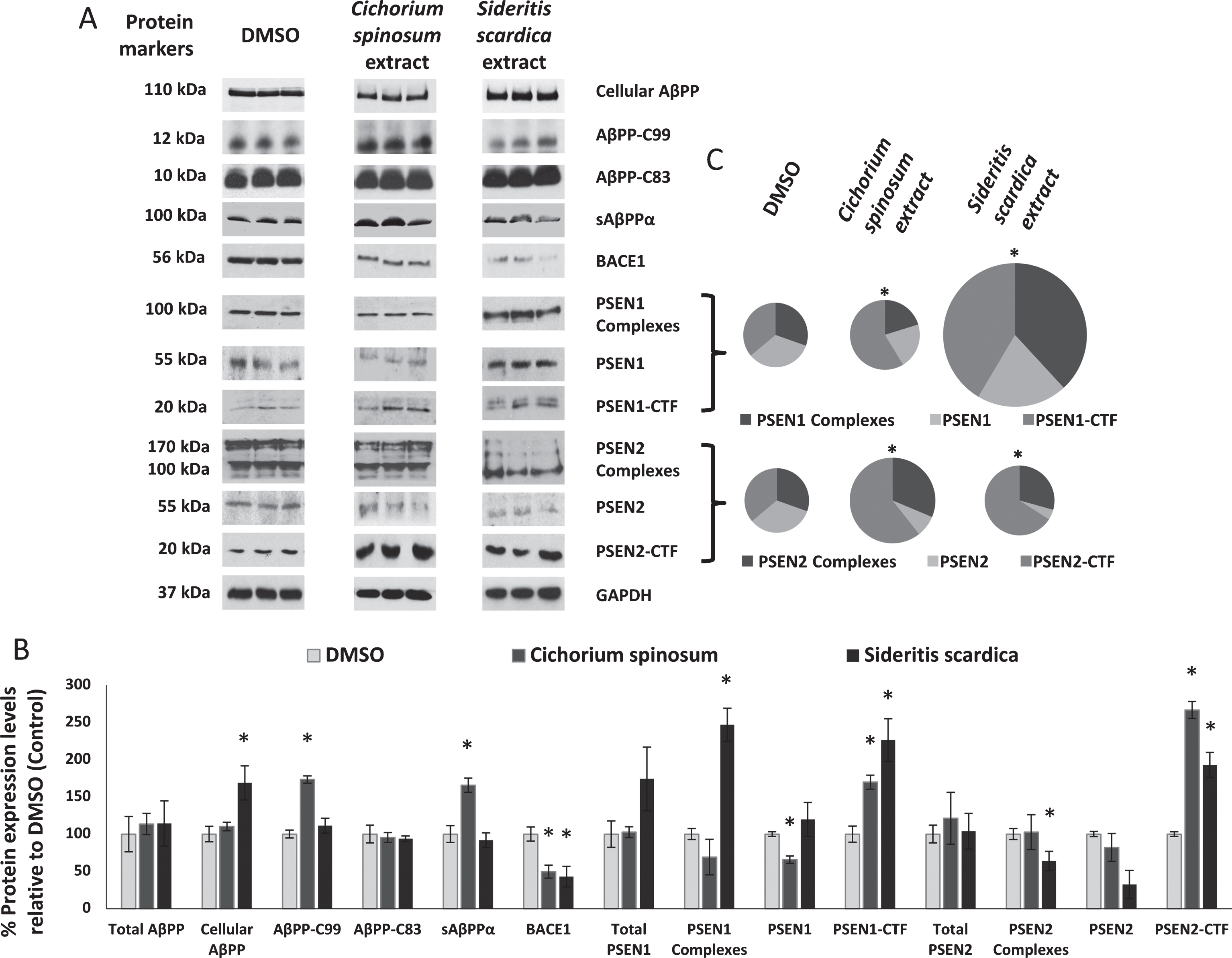
The C. spinosum extract significantly increased AβPP-C99 by 73.3% and sAβPPα by 65.6% , thus significantly altering the ratio of processed AβPP peptides to total AβPP. Furthermore, it increased PSEN1-CTF by 69.7% and PSEN2-CTF by 166.4% , while it reduced BACE1 by 50.4% and PSEN1 by 34.36% , overall affecting the ratio of processed PSEN1 and PSEN2 compared to their respective total protein levels.
The S. scardica extract significantly increased cellular AβPP by 68.7% , PSEN1 by 146.7% , PSEN1-CTF by 126.3% and PSEN2-CTF by 92.7% , while decreasing BACE1 by 57.0% and the PSEN2 complexes by 36.1% . Overall it significantly affected the ratio of processed AβPP peptides to total AβPP, as well as those of PSEN1 and PSEN2.
Effects on tau phosphorylation
Tau hyperphosphorylation is believed to be crucial in AD pathogenesis by promoting the formation of NFTs and ultimately neural loss. Firstly, we confirmed that tau is hyperphosphorylated at Thr231 and Ser199/Ser202 in PC12-htau cells compared to PC12 wt cells (Fig. 5). We then proceeded to assess the effects of S. scardica and C. spinosum using differentiated PC12-htau cells expressing hyperphosphorylated human tau. The endpoints measured included tau phosphorylation (pThr231-tau and pSer199/Ser202-tau), as well as expression and activation of the tau kinases GSK3β and ERK1/2. It should be noted that both GSK3β and ERK1/2 were activated in control PC12-htau cells, in agreement with previous reports on AD (Fig. 6) [22, 70].
Immunoblotting assessment of tau hyperphosphorylation in PC12-htau cells. A) Immunoblotting detection of tau hyperphosphorylation at Thr231 and Ser199/Ser202 in PC12 and PC12-htau cells, with actin as internal control for protein expression. B) Diagrammatic presentation of tau phosphorylation in PC12 and PC12-htau cells (*p < 0.05, t-test, n = 3). Immunoblotting assessment of the tau phosphorylation pathway components after treatment of differentiated PC12-htau cells with S. scardica or C. spinosum extracts. A) Immunoblotting detection of pThr231-tau, pSer199/Ser202-tau, total tau, pSer9-GSK3β, GSK3β, pERK1/2, total ERK1/2, with actin as internal control of protein expression. B) Diagrammatic presentation of quantified protein expression (*p < 0.05, t-test, n = 3).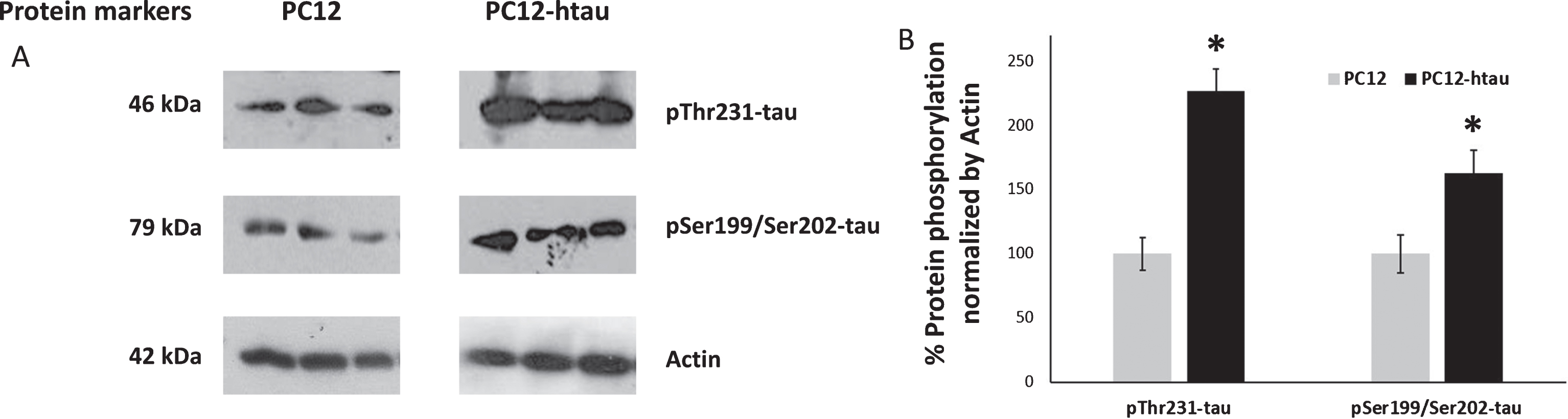
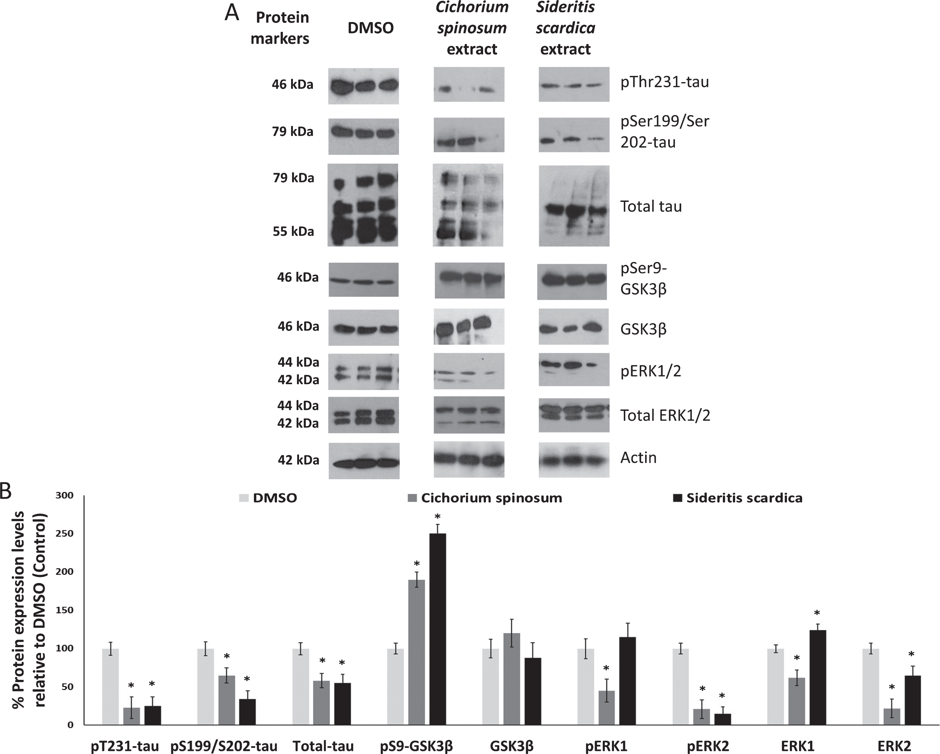
We found that treatment of differentiated PC12-htau cells with the C. spinosum extract decreased total tau (by 42%), the phosphorylation of tau (pThr231 by 77% and pSer199/Ser202 by 35%), ERK1 (by 38%), ERK2 (by 78%), pERK1 (by 55%), and pERK2 (by 79%), while it increased the inactive pSer9-GSK3β (by 90%) compared to the DMSO treatment. The S. scardica extract reduced total tau (by 45%), the phosphorylation of tau (pThr231 by 75% and pSer199/Ser202 by 66%), ERK2 (by 35%) and pERK2 (by 85%), and it increased the levels of inactive pSer9-GSK3β (by 150%) and ERK1 (by 24%). Overall, C. spinosum and S. scardica have similar effects on all investigated components of the tau pathway, with the exception of ERK1 (Fig. 6).
DISCUSSION
AD is characterized by the aggregation of amyloid plaques and the formation of NFTs. In search of new therapeutic approaches against AD we screened Sideritis scardica and Cichorium spinosum extracts, as they are an integral part of the Greek Mediterranean diet, rich sources of polyphenols (such as flavonoids and phenolic acids), and start to emerge as protective agents of memory and cognition [58, 59]. In order to determine the molecular effects of S. scardica and C. spinosum we used two cell culture models of AD and focused on key players of the AβPP and the tau processing pathways, as they constitute promising targets against AD neurodegeneration and disease progression [71–73].
The first step in the evaluation of their therapeutic potential requires the examination of their effects on wild type cell viability. Towards this end, we used an assay based on colorimetric tetrazolium salts cleavage to formazan by enzymes of metabolically active cells [74]. Such assays are recommended because they combine measurement of lethality, proliferation and metabolism, and they are valuable for dose selection [75]. The assays were performed independently for two types of differentiated neuron-like cells, originating from the well-established SH-SY5Y and PC12 cell lines. Both C. spinosum and S. scardica extracts were very well tolerated at the cellular level, maintaining cell viability unaltered. The highest tested biologically relevant and viability favorable doses (400μg/mL) were used for in depth studies at the AβPP and the tau processing pathway levels.
Differentiated neuron-like SH-SY5Y-AβPP over-expressing AβPP695, were used to dissect the effects of the C. spinosum extract on the key steps of the amyloidogenic and non-amyloidogenic AβPP processing pathways. Notably, we observed significantly increased levels of sAβPPα, the product of the non-amyloidogenic AβPP pathway. It has been shown that sAβPPα has neurotrophic, neuroprotective, and synaptotrophic properties, enhances neurite outgrowth, LTP, and memory retention and is involved in the proliferation of neural precursor cells [76–82]. In addition, sAβPPα has been found to act as a neuronal metallotransporter and metallochaperone, regulating metal homeostasis, an important process for Aβ peptides cleavage [77]. The increase in sAβPPα is likely mediated by the observed significant increase in the active components, PSEN1-CTF and PSEN2-CTF, of the respective gamma secretases [83]. Meantime, the levels of PSEN1 were significantly decreased. This could be explained by its increased cleavage to PSEN1-CTF, a decrease in protein expression or a combination of both. Incubation with C. spinosum also led to the reduction of BACE1 levels, the beta secretase mediating Aβ production which is reported increased in the brain of AD patients [84]. Consistently with our findings, cichoric acid, a compound detected in our C. spinosum extract, has been shown to prevent memory impairment and amyloidogenesis, at least in part, through the regulation of BACE1 levels [54]. Despite the reduced levels of BACE1, we observed increase of AβPP-C99. Although the mechanism behind this is unclear, it could potentially be due to accumulation of AβPP-C99 because of its reduced processing to Aβ, as a consequence of the enhanced involvement of gamma secretases in the non-amyloidogenic pathway, as described above. Overall, the C. spinosum extract appears to promote AβPP processing preferentially through the alpha, non-amyloidogenic pathway.
We proceeded to evaluate the effects of C. spinosum on tau hyperphosphorylation, using differentiated neuron-like PC12-htau cells which over-express hyperphosphorylated tau. Notably, a significant decrease of 42% was observed in total tau levels. Tau proteins not only accumulate in the brains of AD patients, but they are also emerging as an informative predictor of a person’s cognitive decline and potential response to treatment [85]. Furthermore, the observed significant decrease in phosphorylated pT231-tau and pSer199/Ser202-tau by 77% and 35% , respectively, suggests a protective effect against phopsho-tau mediated AD pathogenesis.
Since the phosphorylation of tau is primarily regulated by glycogen synthase kinase 3-beta (GSK3β) and ERK1/2, we proceeded to assess both of these pathways [22, 86]. Treatment of the neuron-like PC12-htau cells with C. spinosum led to significant increase of the inactive pSer9-GSK3β, and significant decrease of ERK1/2, as well as its phosphorylated active forms pERK1 and pERK2. Activation of GSK-3β has been associated with the formation of NFTs and the production of Aβ [69], while inhibition of GSK3β through phosphorylation on Serine 9 (pSer9-GSK3β) is neuroprotective [22, 70]. Activation of ERK1/2 increases both tau phosphorylation and abnormal tau deposition in AD, whereas inhibition of the ERK1/2 pathway prevents tau-mediated cell death [87–90]. Consistently with our observations, quercetin-3-O-glucuronide, a compound detected in our C. spinosum extract, has been shown to suppress the phosphorylation of ERK1/2 and significantly reduced the generation of Aβ peptides by primary neuron cultures [57, 91]. Consequently, the C. spinosum extract, appears to have a favorable impact on multiple steps of the tau phosphorylation pathway, suggesting an overall neuroprotective effect.
The S. scardica extract also presented with significant effects on the AβPP processing and tau pathways. In specific, treatment of differentiated SH-SY5Y-AβPP cells led to a significant decrease in BACE1. This is in agreement with the reported down-regulation of BACE1 by apigenin, a compound detected in our S. scardica extract, which has been shown to suppress amyloidogenesis and to ameliorate AD-associated learning and memory impairment [44, 92]. BACE1 has been shown to regulate the levels of full length AβPP [93]. Consistently with these findings, we observed a significant increase in cellular AβPP. Furthermore, the expression of the PSEN1 complexes, PSEN1-CTF and PSEN2-CTF were significantly increased, whereas the PSEN2 complexes’ levels were decreased. These findings, in combination with in vivo studies showing a significant reduction in buffer soluble Aβ42 and decreased amyloid plaque formation (number and size) post S. scardica treatment [45], support the down-regulation of the amyloidogenic pathway.
Importantly, S. scardica appears to also have a significant effect on tau related pathways, similar to this of C. spinosum. In specific, treatment of PC12-htau cells resulted in a significant decrease of pThr231-tau, pSer199/Ser202-tau and pERK2, as well as the increase of the inactive pSer9-GSK3β. These data suggest that S. scardica could be a potential inhibitor of GSK3β and ERK1/2 activation, and consequently of tau phosphorylation, all of which are directly implicated in AD pathogenesis. Indeed, GSK3 is considered a promising therapeutic target for AD, and its inhibition by lithium reduced tau phosphorylation in vivo and lowered the level of tau aggregates [94, 95].
Research on the potential of natural products in preventing or treating specific diseases, including AD, is increasing exponentially [96]. Although there is substantial lack of scientific evidence or clinical trials to support the use of natural products against AD, their market as nutritional supplements is booming [96, 97]. Our findings demonstrate that the C. spinosum and S. scardica extracts cause significant molecular changes that could cumulatively reverse molecular processes associated with amyloidogenesis and tau hyperphosphorylation. Furthermore, our findings support and explain recent epidemiological studies, suggesting the existence of a direct link between adherence to a Mediterranean style diet and low risk for cognitive disease development [37–39]. Recent data from the Hellenic Longitudinal Investigation of Aging and Diet (HELIAD) suggest that adherence to the Mediterranean diet has a neuroprotective effect and is positively associated with better cognitive performance and lower dementia rates in Greek elders (prevalence of dementia in Greece is 4.5% , the lowest in Europe) [98]. In conclusion, the Mediterranean style diet components Cichorium spinosum and Sideritis scardica extracts emerge as promising for the prevention and/or treatment of AD, through the inhibition of AβPP and tau misprocessing pathways leading to AD.
Footnotes
ACKNOWLEDGMENTS
We are grateful to Professor S. Efthymiopoulos, Professor L. Stefanis, Dr. K. Vekrelis, Dr M. Xylouri, Dr N. Koulakiotis, Dr I. Dafnis, and M. Chatzistavraki.
This work has been supported by a “Large Scale Cooperative Project” (TreatAD, 09SYN-21-1003) co-financed by the European Social Fund (ESF) and the General Secretariat for Research and Technology in Greece.
